Abstract
The objective of the study was to evaluate the effects of udder height on upper body kinematics and muscle activity during a simulated attachment task in a parallel parlor set up, and the effects of udder access method (back or side) on the task biomechanics. Twenty males performed the task under conditions that simulated three udder heights and two udder access methods. The muscular load and kinematics during the task confirmed that milking is a physically demanding task. Trunk flexion angle increased with decreasing udder height, and the erector spinae activation was higher when the udder was below shoulder height compared to at or above. Compared to accessing the udder from side of the cow (herringbone parlor style), accessing from behind (Parallel parlor style) was associated with lower trunk flexion, greater shoulder horizontal adduction, lower shoulder elevation, and greater anterior deltoid activation. Milking in herringbone parlor style and with the udder at or above shoulder level may help reduce strain on the trunk/neck.
Keywords: Ergonomics, Dairy farm, Scapular kinematics
1. Introduction
Dairy production in the US has steadily moved toward large-herd milking operations due to the lower production cost (Reinemann, 2002). The shift toward a large-herd model has led to increased physical demand and potentially increased risk of work-related musculoskeletal symptoms (MSS) among milking parlor workers. The estimated prevalence of work-related MSS among dairy milkers is as high as 76% (Douphrate et al., 2012, Douphrate et al., 2014a, Kolstrup, 2012, Dairy Workers, 2014, Douphrate et al., 2013), with shoulder and neck being the most commonly affected areas. It is said that 40–47% of milkers report having shoulder symptoms and 32–47% report having neck symptoms in the past 12 months (Douphrate et al., 2014a, Kolstrup, 2012, Nonnenmann et al., 2008). Long work-hours and biomechanics of milking tasks have been associated with a high prevalence of reported shoulder and neck MSS among milkers (Douphrate et al., 2014b).
In modern milking parlors, cows stand on a raised platform, at a higher level than workers who operate in a milking pit, which enables the workers to perform milking tasks while maintaining a relatively upright posture. Milking routine consists of six tasks; pre-dipping of teats for sanitization, stripping, wiping, milking unit attachment and detachment, and post-dipping of teats for sanitization. Many milkers perform the routine throughout 8–12 h shifts, six days a week for almost 50 weeks per year (Douphrate et al., 2012). Of the six tasks, the attachment task, which requires milkers to reach under the udder to secure a cluster (i.e. device to milk the cow), results in the highest activation level of the arm and forearm muscles, and is regarded as among the most physically demanding (Douphrate et al., 2014a, Pinzke et al., 2001, Douphrate et al., 2016).
The biomechanics of the attachment task has only been described in two previous studies. The first study was conducted in 1986, when stanchion system milking was still being utilized, and milkers operated on the same floor as the cow and had to squat/kneel to perform milking tasks (Arborelius et al., 1986). The second study was conducted more recently by (Jakob et al. 2012). The study examined the effects of udder height on upper body kinematics and muscle activity among professional milkers. This study demonstrated that milking a cow with its udder at the milker’s shoulder height is ideal in minimizing deltoid and upper trapezius muscle activity level (Jakob et al., 2012).
The data collection in the study by Jakob et al. (2012) was conducted in a set up called herringbone parlors where cows are positioned at 30–45° angle to the pit, and thus milkers access udders from side of the cow. Because of better cost and space efficiency, however, herringbone parlors are being gradually replaced by the parallel parlors where the cows are positioned perpendicular to the pit, and thus milkers access the udders through cow’s hind legs. This design requires the milkers to access udders through a narrower space by keeping their elbows close together and making other biomechanical adjustments. However, no study has described upper body kinematics of milking in parallel parlors (Douphrate et al., 2012). Therefore, it is unknown how the difference in body positioning associated with different parlor design influences milkers’ motion and muscle activity.
The objective of the study was to expand on the work conducted by Jakob et al. (2012) to better understand the biomechanics of milking unit attachment task. Specifically, we evaluated the effects of udder height on upper body kinematics and muscle activity during a simulated attachment task in a parallel parlor set up, and the effects of udder access method (back or side), as dictated by the parlor design, on the task biomechanics.
2. Material and methods
2.1. Participants
Data collection took place at the Applied Biomechanics Research Laboratory at the University of Texas at San Antonio. Twenty healthy males (age: 22.2 ± 1.9 years, hand dominance: R/L = 16/4, height: 172.8 ± 5.2 cm, mass: 75.0 ± 8.4 kg, acromion-grip length: 79.0 ± 3.2 cm) agreed to participate in the study. Only males were recruited since approximately 90% of dairy milkers are males (Baker and Chappelle, 2012, Roman-Muniz et al., 2006). No participants had prior experience with cow milking. All participants provided a written informed consent approved by the University of Texas at San Antonio prior to participating in the study.
2.2. Instrumentation
A video-based motion capture system (Vicon Inc., Centennial, CO) with eight T10s cameras was used to capture kinematics at a sampling rate of 100 Hz. Electrical muscle activity of the upper and lower trapezius, neck extensor, erector spinae, and anterior deltoid of the dominant upper extremity was sampled via surface electromyography (EMG) with a differential amplifier (BTS Bioengineering, FreeEMG 300, Brooklyn, NY). The system has an input impedance of 1Gohm, common mode rejection rate greater than 110 dB, and signal-to-noise ratio of 70 db. Muscle activity data were collected at a sampling rate of 1000 Hz. The kinematic data and the EMG data were synchronized electronically.
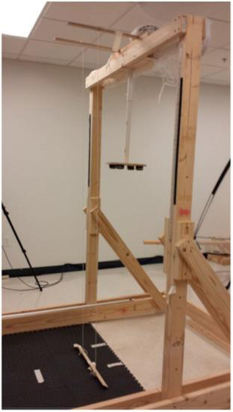
A custom-made simulated udder was constructed for the study (Fig. 1). A PVC pipe with a circular base at the bottom, representing a cow udder, was built into a support frame. The height of the base was adjustable, so that the udder could be moved up or down to the participant’s shoulder height and 15 cm above or below the shoulder height. A milking unit is attached to a cow’s udder, and vacuum pressure enables the harvesting of milk. Magnets were secured to the end of each milking unit teat cup and the base of the simulated udder. Magnetic attraction enabled the attachment of milking unit to the udder. When simulating milking in a parallel parlor design, in which the participants accessed the udder from behind the cow (through the hind legs), two vertical strings were positioned 32 cm apart and 17 cm in front of the udder, based on field-based data from previous studies. Participants were asked to access the udder without touching the strings, which represented cow’s hind legs. The strings were removed when simulating milking in a herringbone parlor, in which the participants accessed the udder from the side of the cow.
Fig. 1.
Custom made Udder.
2.3. Procedures
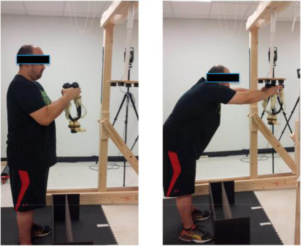
After informed consent, participants proceeded to practice the milking unit attachment task (Fig. 2). The attachment task began with the participant standing in front of the udder holding the milking unit with both hands. On the investigator’s cue, the participant moved the milking unit towards the udder, and attached the back two arms of the cluster to the back two teats followed by the front two arms to the front two teats. This is a common technique used by male workers, since their hands are larger compared to females, which allows them to hold all four teat-cups at once. Although hand-dimension was not measured, none of the participants had trouble holding the cluster. Once the milking unit was attached to the udder, the participant brought his arms back to his side. The participants practiced this task with the udder at their shoulder height for approximately 4–5 min until they were comfortable with the task and were able to repeat the task fluidly and consistently without hesitation or pausing. The investigator explained and demonstrated the tasks before the participants practiced, and provided feedback and correction during practice.
Fig. 2.
Attachment task.
Once participants were familiarized with the task, they removed their shirts to proceed to EMG data collection set up. Researchers identified and marked locations for electrode placement for anterior deltoid, neck extensors, upper trapezius, lower trapezius, and erector spinae on the participant’s dominant side. Skin was lightly abraded with gauze and cleaned using alcohol wipes to minimize impedance (Hermens et al., 2000). Two 24 mm diameter Ag/AgCI disposable surface electrodes (Covidien, Minneapolis, MN) were placed parallel to the muscle fibers on locations specified in the literature (Cram, 1998), with an inter-electrode distance of 2 cm.
After electrodes were secured using pre-wrap and adhesive tapes (BSN medical, Luxembourg), participants performed maximal voluntary isometric contractions (MVC) trials. The standardized procedures to conduct manual muscle testing (Boettcher et al., 2008) were utilized for MVC trials. Instead of using manual resistance, however, we asked the participants to contract the muscle being tested against an unyielding strap, in order to limit movement and ensure that participants performed a maximal isometric muscle contraction. The participants held the contraction for 5 s for each trial, and the trials were repeated three times for each muscle.
The MVC for the anterior deltoid was performed in a seated position. Participants flexed their shoulder 90° with a strap attached to the wrist. The participants performed a maximum effort isometric shoulder flexion against the strap (Boettcher et al., 2008). The MVC for upper trapezius was also performed in a seated position. with a strap placed over the acromion process of the dominant shoulder. The participants performed a maximum effort isometric shoulder shrug against the strap placed over the acromion process and anchored to the floor (Boettcher et al., 2008). The MVC for the erector spinae was performed while the participants lay prone with the head and upper part of the chest (above xyphoid process) unsupported, and arms crossed in front of the chest. The participants’ legs and torso were secured to the treatment table with harnesses. The participants performed a maximum effort isometric back extension against the strap that was placed around the chest/upper back and anchored to the floor (Park et al., 2014). The MVC test for neck extensor required the participants to lay prone on the treatment table with their head unsupported. The participants’ torso was secured to the treatment table with harnesses. Participants performed a maximum isometric neck extension against the strap that was placed around their head, and was anchored to the floor. Participants remained prone on the treatment table for the MVC for the lower trapezius. The participants abducted their arm approximately 150° and hanging off the table. The participants attempted to retract the scapula against the strap that was wrapped around the participants’ wrist, and was anchored to the floor (Park et al., 2014, De Mey et al., 2013). The muscle activity data during the MVC trials were later used to normalize muscle activity during the attachment task.
After MVC trials, investigators placed the reflective markers on participants’ anatomical bony landmarks to capture kinematics. Markers were secured using double-sided tape, pre-wrap, and athletic tape. Specifically, markers were placed over the sternal notch, xyphoid process, spinous process of 7th cervical spine, anterior arm, medial epicondyle, lateral epicondyle, radial styloid process, and ulnar styloid process. Additionally, a cluster of three markers attached onto a rigid base was placed over the spinous process of the 8th thoracic spine.
Once markers were secured, participants proceeded to the milking unit attachment task. The attachment task was performed in a set up that simulated the parallel parlor (udder accessed from behind the cow) under three udder height conditions; udder at shoulder height (SHL-P), 15 cm below shoulder height (LOW-P), and 15 cm above shoulder height (HIGH-P). In these conditions, participants were required to access the cow through the vertical strings that simulated cow’s hind legs. Additionally, the task was performed with an udder at shoulder height in a set up that simulated milking in a herringbone parlor, where the udders are accessed from the side of the cow (SHL-H). The order of conditions was counterbalanced, and participants performed five repetitions of the attachment task under each condition. The kinematics and the muscle activity data were recorded during all trials.
2.4. Data processing
The marker coordinate data were filtered using a 4th order Butterworth filter with a cut off frequency of 10 Hz. The filtered coordinate data were used to estimate joint centers and to define body segments. The anatomical coordinate system of the trunk, arm, and forearm were defined based on recommendations from the International Society of Biomechanics (ISB) (Wu et al., 2005). The trunk flexion angle was calculated as the angle of the longitudinal axis of the trunk relative to vertical. Shoulder angles were calculated as the orientation of arm relative to thorax using an Euler angle sequence of plane of elevation (horizontal adduction/abduction), elevation, and internal/external rotation (Wu et al., 2005). Elbow angle was calculated as the angle between the longitudinal axis of the arm and forearm in the sagittal plane of the arm (Wu et al., 2005). The frame that the participants finished attaching all arms of the cluster to the udder (task completion) was visually identified from the video data. The kinematic variables at the time of task completion were used for data analysis.
Myoelectric activity was first filtered with a zero-lag 4th order bandpass filter with a cutoff frequency of 10–350 Hz, and then filtered with a 60 Hz notch filter (width = 1 Hz) to minimize noise. After subtracting the average EMG activity of each muscle at rest (baseline), the data were smoothed using a 50 ms moving root mean square window. Based on a visual examination of raw EMG signal at rest, we identified that the data from erector spinae muscle from one participant was visibly contaminated by heartbeat. Therefore, the data were removed from analysis. The average muscle activation level during the MVC was calculated from the middle 3 s of the 5-s trial. The average activation level for each muscle during the attachment task was calculated using the data collected while the participants’ hand (marker placed on participant’s head of the 3rd metacarpal) was within 30 cm of the udder in the horizontal plane. Once the mean muscle activity level for each muscle was calculated, values were normalized to the muscle activity during the MVC trials. Kinematic and EMG variables were calculated as the mean of five trials performed in each condition. All data processing was performed using Matlab R2015a (The MathWorks Inc., Natick, Massachusetts).
2.5. Data analysis
The effects of udder height (LOW-P vs. SHL-P vs. HIGH-P) on the upper body kinematics and muscle activation level were examined using separate 1-way repeated measures analysis of variance (ANOVA) followed by the Bonferroni post hoc tests. The effects of udder access methods (SHL-P vs. SHL-H) on the upper body kinematics and muscle activation level were examined using paired sample t-tests. A priori alpha level of 0.05 was used to determine statistical significance. Bonferroni adjustment was used for post-hoc analysis to compare variables between three height conditions (= 0.05/3 comparisons).
3. Results
The udder height had significant effect on trunk flexion angle (F1,38 = 102.7, p ≤ 0.001), elbow flexion angle (F1,38 = 10.0, p ≤ 0.001), neck extensor activation level (F1,37 = 5.3, p = 0.014), and erector spinae activation level (F1,38 = 18.0, p ≤ 0.001) (Table 1). The udder height did not have significant effect on any of the other variables. Specifically, participants flexed their trunk more with lower udder height (p < 0.001). The participant’s elbow was in greater flexion in LOW-P condition compared to SHL-P (p = 0.008) and HIGH-P (p ≤ 0.001) conditions. The erector spinae activation level was higher in LOW-P compared to SHL-P (p ≤ 0.001) and HIGH-P (p ≤ 0.001) conditions, and higher in SHL-P condition than for HIGH-P condition (p = 0.016). Post hoc comparisons did not show any significant differences in neck extensor activation level between the udder height conditions.
Table 1.
Effects of udder height on kinematics and muscle activation level during attachment.
| Low | Shoulder height | High | F | p | |
|---|---|---|---|---|---|
| Kinematics (°) | |||||
| Trunk flexion a,b,c | 51.3 ± 11.3 | 37.2 ± 11.9 | 29.5 ± 12.7 | 102.7 | <0.001* |
| Shoulder horizontal adduction | 87.3 ± 8.5 | 87.7 ± 9.6 | 87.0 ± 8.4 | 0.2 | 0.725 |
| Shoulder elevation | 92.0 ± 14.1 | 89.0 ± 13.8 | 93.0 ± 15.0 | 3.2 | 0.064 |
| Scapular internal rotation | 47.1 ± 13.0 | 47.9 ± 12.0 | 48.2 ± 11.9 | 0.6 | 0.572 |
| Scapular upward rotation | 38.4 ± 12.4 | 35.9 ± 13.7 | 38.7 ± 11.8 | 1.9 | 0.165 |
| Scapular anterior tilt | 0.7 ± 10.8 | 1.2 ± 12.0 | 0.6 ± 10.6 | 0.1 | 0.876 |
| Clavicle elevation | 22.7 ± 6.6 | 21.2 ± 8.7 | 21.7 ± 6.9 | 1.1 | 0.338 |
| Clavicle protraction | −30.3 ± 10.3 | −29.0 ± 13.1 | −30.9 ± 12.2 | 0.8 | 0.445 |
| Elbow flexion a,b | 58.0 ± 22.4 | 47.0 ± 18.2 | 43.4 ± 16.0 | 10.0 | <0.001* |
| Muscle activation level (%MVIC) | |||||
| Anterior deltoid | 44.0 ± 11.2 | 44.7 ± 11.1 | 47.7 ± 9.0 | 2.7 | 0.083 |
| Upper trapezius | 42.9 ± 21.8 | 36.1 ± 20.9 | 40.0 ± 17.6 | 2.7 | 0.081 |
| Lower trapezius | 5.3 ± 2.1 | 4.9 ± 2.3 | 4.7 ± 2.0 | 2.1 | 0.140 |
| Neck extensor | 50.6 ± 38.5 | 40.1 ± 34.8 | 46.0 ± 41.0 | 5.3 | 0.014* |
| Erector spinae a,b,c | 38.8 ± 18.2 | 31.2 ± 14.3 | 26.1 ± 9.6 | 18.2 | <0.001* |
Statistically significant main effect for udder height at α < 0.05.
Significant difference between low and normal conditions.
Significant difference between low and high conditions.
Significant difference between normal and high conditions at a Bonferroni adjusted alpha level of 0.016 (= 0.05/3 comparisons).
Compared to accessing the udder from side of the cow (SHL-H), accessing the udder from behind the cow by keeping the arms close together (SHL-P) was associated with more upright trunk position (smaller trunk flexion angle) (t19 = −3.2, p = 0.005), greater shoulder horizontal adduction angle (t19 = 4.3, p ≤ 0.001), lower shoulder elevation angle (t19 = −4.5, p ≤ 0.001), and greater anterior deltoid activation level (t19 = 6.0, p ≤ 0.001) (Table 2). The udder access method did not have significant effect on any of the other variables.
Table 2.
Pair-wise comparisons of kinematics and muscle activity level between conditions that simulate milking from behind the cow (SHL-P) and from the side (SHL-H).
| Mean difference (= Back - Side) | T19 | ||
|---|---|---|---|
| Kinematics (°) | |||
| Trunk flexion | −3.4 ± 4.7 | −3.2 | 0.005* |
| Shoulder horizontal adduction | 11.3 ± 11.9 | 4.3 | <0.001* |
| Shoulder elevation | −6.0 ± 5.9 | −4.5 | <0.001* |
| Scapular internal rotation | 1.1 ± 7.8 | 0.6 | 0.543 |
| Scapular upward rotation | 2.2 ± 6.5 | 1.5 | 0.144 |
| Scapular anterior tilt | 3.3 ± 12.5 | 1.2 | 0.249 |
| Clavicle elevation | −1.0 ± 6.2 | −0.7 | 0.469 |
| Clavicle protraction | −2.0 ± 6.3 | −1.4 | 0.901 |
| Elbow flexion | −3.8 ± 7.6 | −2.2 | 0.039* |
| Muscle activation level (%MVIC) | |||
| Anterior deltoid | 12.3 ± 9.1 | 6.0 | <0.001* |
| Upper trapezius | −0.6 ± 19.6 | −0.1 | 0.890 |
| Lower trapezius | −0.8 ± 2.8 | −1.3 | 0.194 |
| Neck extensor | 3.6 ± 11.1 | 1.4 | 0.170 |
| Erector spinae | −1.5 ± 5.0 | −1.3 | 0.204 |
Statistically significant at α = 0.05.
4. Discussion
Attaching the cluster to the cow’s udder is a physically demanding task, especially when performed repetitively throughout the long work shift. This is highlighted by the data demonstrating that the anterior deltoid, upper trapezius, erector spinae, and neck extensors were highly activated during the task. A work by Jonsson et al. (Jonsson, 1978) indicates that for work performed for over an hour, the average muscle activation level should not exceed 10%. and peak muscle activation level should not exceed 50% of MVC. While we cannot speak to the average muscle activation level during work, because we only looked at average during isolated segments of work, we can say that the peak muscle activation level of the anterior deltoid, upper trapezius, and neck extensors at least approached or exceeded 50% during the task, considering that the average activation level ranged from 36.1 to 50.6% MVC. Especially considering that attachment tasks are performed repetitively throughout the day, along with the other physically demanding tasks, our data supports the claim that milking, which includes the attachment task is a physically demanding task which exceeds recommended thresholds (reference Jonsson, 1978).
In addition to the high muscle load, exposure to repetitive arm elevation has been identified as a risk factor for shoulder MSS in manual laborers (Punnett et al., 2000, Svendsen et al., 2004, Hanvold et al., 2015). Peak shoulder elevation angle that was reached during the milking task was around 90°. Having to elevate the arm to 90° for every cow that needs to be milked, predisposes the milkers to shoulder MSS. Maintaining the arm elevation above 90° for more than 10% of the work shift has been linked to in increased risk of chronic or recurrent shoulder disorders (Punnett et al., 2000), and the greater time spent with arm maintained above 60° for more than 5 s has been correlated to shoulder pain (Svendsen et al., 2004, Hanvold et al., 2015).
Considering that the milking task is very demanding on the workers, it is important to investigate the ways to help minimize the physical load. The effect of udder height on upper body kinematics and muscle activities during the attachment task has been evaluated previously in the herringbone parlor setting (Jakob et al., 2012). The study concluded that the udder height should be at the milker’s shoulder level, based on the observation that having an udder above the shoulder resulted in higher shoulder elevation angle and higher activation level of the deltoid and trapezius muscles compared to having the udder at the shoulder height (Jakob et al., 2012). However, since the herringbone parlors are gradually being replaced by the parallel and rotary parlors, the objectives of the current study was to evaluate the effects of udder height on upper body kinematics and muscle activities in the simulated parallel parlor setting, and to compare the biomechanics of attachment task performed using the udder access methods associated with parallel parlors (from the side) vs. herringbone parlors (from behind).
In contrast to the previous study (Jakob et al., 2012), we observed no differences in shoulder elevation angles or muscle activation level of the deltoid or trapezius muscles between having the udder above or at the shoulder level. The discrepancy between the studies may be due to the difference in the udder access method, since the pervious study used the herringbone parlor design where the udder is accessed from the side of the cow, while we simulated the parallel parlor design where the udder is accessed from behind the cow. However, the difference can also due to the fact that Jakob et al. (2012) reported mean shoulder elevation angles during the task, while we reported peak elevation angles. We chose to report the peak angle because the shoulder elevation angle peaks as the milking unit is being attached to the udder, which is the most biomechanically demanding part of the task. Furthermore, difference in the participants’ experience level in the milking task may have attributed to the discrepancy. The participants in the study by Jakob et al. (2012). were professional milkers, while participants in our study were college-aged students with no previous milking experience.
In both the current study and the study by Jakob et al. (2012), the lower udder height resulted in greater forward trunk flexion. This is not surprising, since accessing the lower udder requires greater trunk flexion. Our study also demonstrated that the lower udder condition results in higher erector spinae activation levels. This increase in muscle activity may be explained by the anterior shift in the center of gravity from the increased forward trunk flexion. Furthermore, the participants kept their elbows more flexed when milking at the low condition, which would require them to shift their weight forward to reach the udder. Neck extensor muscle activity level was also higher in low and high udder conditions. Higher erector spinae and neck extensor muscle activity levels are an indication of higher demand on these muscles, which may lead to early onset of fatigue and the development of MSS in the neck and low back region (Howarth et al., 2015, Lisinski, 2000). The erector spinae activation level was unaffected by the udder height in the study by Jakob et al. (2012). This difference may be due to the difference in the parlor design that was simulated in two studies or the experience level of the participants.
Overall, the udder height had no effect on the participant’s kinematics or muscle activity at the shoulder. This indicates that the demand on shoulders may be unaffected by the udder height, and that most of the adjustment to the task was made at the trunk/neck. On the other hand, the higher udder height resulted in less trunk flexion and demand on the back extensor muscles. Given these observations, having the udder at or above the shoulder level may lessen the physical load on the lower back. For those workers with pre-existing low back MSS, having the udder slightly higher than the shoulder level, and thereby decreasing trunk flexion may be recommended.
The cow’s udder height can vary based on the cow’s height and age. Therefore, providing instruction/training to milkers to use their lower extremity joints so that the udder is at slightly above their shoulder level may be recommended. This may reduce the need to flex the trunk and extend the neck, thereby decreasing muscular strain on the trunk. However, feasibility of implementing such intervention needs to be examined. On the other hand, since there is a variation in the cow height, the workers modify their movement to attach clusters to cows. This variability in udder height may keep the milkers from working in the same position over a long time, thus reducing physical demands by increasing task variability.
The second objective of the study was to compare the effects of udder access methods used in the herringbone vs. parallel parlors on task biomechanics. In herringbone style parlor, the udder is accessed from the side of the cow. In contrast, in parallel style parlor, the udder is accessed from behind the cow. Therefore, parallel parlors require milkers to reach through a narrow space between the legs, requiring them to keep their elbows closer together. This requirement adds to the task constraints and precision needed to perform the task.
We observed that having a narrow access to the udder resulted in greater shoulder horizontal adduction, lower trunk flexion, lower shoulder elevation, and higher anterior deltoid activation level. The higher activation level of the anterior deltoid (44.7 vs. 32.4%MVC) is likely associated with the greater shoulder horizontal adduction, and may result in the early onset of fatigue over the work-shift. The prevalence of neck and back MSS tends to be higher for the workers in parallel parlor compared to the herringbone design parlor (Douphrate et al., 2014b). Although parallel parlors are becoming more popular due to cost effectiveness, our data suggest that they may increase the physical demand on milkers, and possibly contribute to slightly higher prevalence of MSS. Further research is needed to confirm this hypothesis. This study only investigated muscle activation level and postures associated with the task. It is possible that other exposures such as repetition, long work hours, and limited task variability may contribute to the development of MSS.
4.1. Limitations
There are a few limitations that need to be acknowledged. First, the data collection for this study took place in a research laboratory, and not in an actual milking parlor. However, data collection in the laboratory allowed greater control the task performed by participants. Secondly, participants were not experienced milkers. Therefore, replication of the study with experienced milkers may be needed. However, our participants were given ample opportunity to practice tasks prior to performing each test condition. While we examined the superficial shoulder muscles, we were unable to collect muscle activity data from the rotator cuff muscles that play an important role in shoulder function. The activity of the rotator cuff muscles should be examined in the future.
5. Conclusion
Our results confirm that milking is a physically demanding task that requires muscular load that exceeds recommended muscle activation thresholds, and involves repetitive elevation of the arms that is associated with increased injury risk. Our data suggests that milking with the udder at or above the shoulder level may help reduce strain on the trunk/neck when milking from behind the cow. The muscular load on the anterior deltoid was lower when milking the cow from the side (herringbone parlor), than from the back (parallel parlor). However, further study is needed to determine if the mitigation of muscular load from using higher udder position or side access to the udder would make a clinically significant impact on musculoskeletal health among milkers.
References
- Arborelius UP, Ekholm J, Nisell R, Nemeth G, Svensson O. Shoulder load during machine milking. An electromyographic and biomechanical study Ergonomics, 29 (12) (1986), pp. 1591–1607 [DOI] [PubMed] [Google Scholar]
- Baker D, Chappelle D. Health status and needs of Latino dairy farmworkers in Vermont J. agromedicine., 17 (3) (2012), pp. 277–287 [DOI] [PubMed] [Google Scholar]
- Boettcher CE, Ginn KA, Cathers I. Standard maximum isometric voluntary contraction tests for normalizing shoulder muscle EMG J. Orthop. Res. Official Publ. Orthop. Res. Soc., 26 (12) (2008), pp. 1591–1597 [DOI] [PubMed] [Google Scholar]
- Cram JR Introduction to Surface Electromyography Aspen Publichers (1998) [Google Scholar]
- Dairy Workers. 2014; http://www.ncfh.org/docs/fs-DairyWorkers.pdf. Accessed 27 August 2014, 2014.
- De Mey K, Danneels L, Cagnie B, Van den Bosch L, Flier J, Cools AM Kinetic chain influences on upper and lower trapezius muscle activation during eight variations of a scapular retraction exercise in overhead athletes J. Sci. Med. sport/Sports Med. Aust., 16 (1) (2013), pp. 65–70 [DOI] [PubMed] [Google Scholar]
- Douphrate DI, Fethke NB, Nonnenmann MW, Rosecrance JC, Reynolds SJ Full shift arm inclinometry among dairy parlor workers: a feasibility study in a challenging work environment Appl. Ergon., 43 (3) (2012), pp. 604–613 [DOI] [PubMed] [Google Scholar]
- Douphrate DI, Lunner Kolstrup C, Nonnenmann MW, Jakob M, Pinzke S. Ergonomics in modern dairy practice: a review of current issues and research needs J. agromedicine., 18 (3) (2013), pp. 198–209 [DOI] [PubMed] [Google Scholar]
- Douphrate DI, Gimeno D, Nonnenmann MW, Hagevoort R, Rosas-Goulart C, Rosecrance JC Prevalence of work-related musculoskeletal symptoms among US large-herd dairy parlor workers Am. J. industrial Med, 57 (3) (2014), pp. 370–379 [DOI] [PMC free article] [PubMed] [Google Scholar]
- Douphrate DI, Gimeno D, Nonnenmann MW, Hagevoort R, Rosas-Goulart C, Rosecrance JC Prevalence of work-related musculoskeletal symptoms among US large-herd dairy parlor workers Am. J. Industrial Med, 57 (3) (2014), pp. 370–379 [DOI] [PMC free article] [PubMed] [Google Scholar]
- Douphrate D, Fethke N, Nonnenmann M, Rodriguez A, Hagevoort R, Gimeno D. Ruiz de Porras Full-shift and task-specific assessment of muscle activity among large herd dairy parlor workers Ergonomics (17 November 2016), pp. 1–39, 10.1080/00140139.2016.1262464 [DOI] [Google Scholar]
- Hanvold TN, Wærsted M, Mengshoel AM, Bjertness E, Veiersted KB Work with prolonged arm elevation as a risk factor for shoulder pain: a longitudinal study among young adults Appl. Ergon, 47 (2015), pp. 43–51 [DOI] [PubMed] [Google Scholar]
- Hermens HJ, Freriks B, Disselhorst-Klug C, Rau G. Development of recommendations for SEMG sensors and sensor placement procedures J. Electromyogr. Kinesiol. Official J. Int. Soc. Electrophysiol. Kinesiol, 10 (5) (2000), pp. 361–374 [DOI] [PubMed] [Google Scholar]
- Howarth SJ, Grondin DE, La Delfa NJ, Cox J, Potvin JR Working position influences the biomechanical demands on the lower back during dental hygiene Ergonomics (2015), pp. 1–11 [DOI] [PubMed] [Google Scholar]
- Jakob M, Liebers F, Behrendt S. The effects of working height and manipulated weights on subjective strain, body posture and muscular activity of milking parlor operatives–laboratory study Appl. Ergon., 43 (4) (Jul 2012), pp. 753–761 [DOI] [PubMed] [Google Scholar]
- Jonsson B Kinesiology: with special reference to electromyographic kinesiology Electroencephalogr. Clin. Neurophysiol. Suppl., 34 (1978), pp. 417–428 [PubMed] [Google Scholar]
- Kolstrup CL Work-related musculoskeletal discomfort of dairy farmers and employed workers J. Occup. Med. Toxicol. Lond. Engl, 7 (1) (2012), p. 23. [DOI] [PMC free article] [PubMed] [Google Scholar]
- Lisinski P Surface EMG in chronic low back pain Eur. Spine J., 9 (6) (2000), pp. 559–562 [DOI] [PMC free article] [PubMed] [Google Scholar]
- Nonnenmann MW, Anton D, Gerr F, Merlino L, Donham K. Musculoskeletal symptoms of the neck and upper extremities among Iowa dairy farmers Am. J. industrial Med, 51 (6) (2008), pp. 443–451 [DOI] [PubMed] [Google Scholar]
- Park SY, Yoo WG, An DH, Oh JS, Lee JH, Choi BR Comparison of isometric exercises for activating latissimus dorsi against the upper body weight J. Electromyogr. Kinesiol., 25 (1) (2015. Feb), pp. 47–52, 10.1016/j.jelekin.2014.09.001 [DOI] [PubMed] [Google Scholar]
- Pinzke S, Stal M, Hansson GA Physical workload on upper extremities in various operations during machine milking Ann. Agric. Environ. Med. AAEM, 8 (1) (2001), pp. 63–70 [PubMed] [Google Scholar]
- Punnett L, Fine LJ, Keyserling WM, Herrin GD, Chaffin DB Shoulder disorders and postural stress in automobile assembly work Scand. J. work, Environ. health. (2000), pp. 283–291 [DOI] [PubMed] [Google Scholar]
- Reinemann D. Evolution of Automated Milking in the USA. Paper Presented at: First North American Conference on Robotic Milking March 20–22, 2002 (2002) Toronto, Ontario, Canada [Google Scholar]
- Roman-Muniz IN, Van Metre DC, Garry FB, Reynolds SJ, Wailes WR, Keefe TJ Training methods and association with worker injury on Colorado dairies: a survey J. agromedicine., 11 (2) (2006), pp. 19–26 [DOI] [PubMed] [Google Scholar]
- Svendsen SW, Bonde JP, Mathiassen SE, Stengaard-Pedersen K, Frich L. Work related shoulder disorders: quantitative exposure-response relations with reference to arm posture Occup. Environ. Med, 61 (10) (2004), pp. 844–853 [DOI] [PMC free article] [PubMed] [Google Scholar]
- Wu G, van der Helm FC, Veeger HE, et al. ISB recommendation on definitions of joint coordinate systems of various joints for the reporting of human joint motion–Part II: shoulder, elbow, wrist and hand J. Biomech., 38 (5) (2005), pp. 981–992 [DOI] [PubMed] [Google Scholar]


