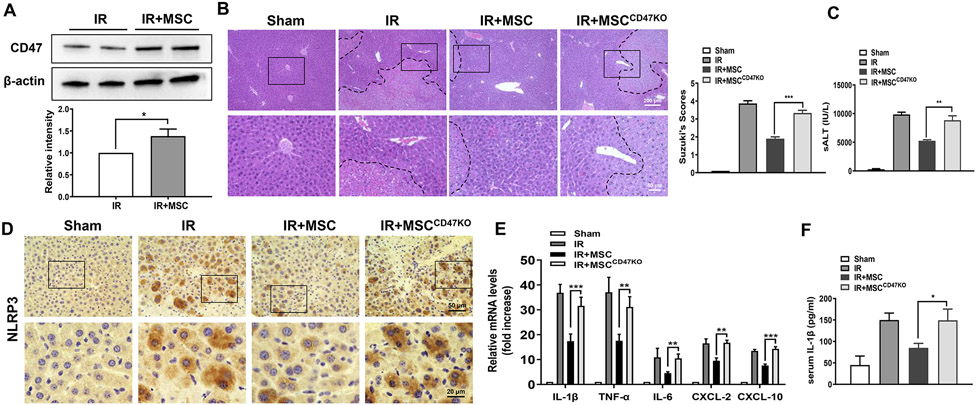
Fig. 1. Disruption of CD47 in MSCs exacerbates IR-induced liver injury and augments proinflammatory mediators.
WT mice were adoptive transferred MSCs or CD47-deficient MSCs (1x10⁶) 24h prior to liver ischemic insult. (A) The CD47 expression was detected by Western blot assay in ischemic livers. The representative of three experiments. (B) Representative histological staining (H&E) of ischemic liver tissue (n=4-6 mice/group) and Suzuki's histological score. Scale bars, 200μm and 50μm. (C) Liver function was evaluated by serum ALT levels (IU/L) (n=4-6 samples/group). (D) Immunohistochemistry staining of NLRP3 in ischemic livers (n=4-6 mice/group). Scale bars, 50μm and 20μm. (E) qRT-PCR analysis of IL-1β, TNF-α, IL-6, CXCL-2, and CXCL-10 in ischemic livers (n=3-4 samples/group). (F) ELISA analysis of serum IL-1β levels (n=3-4 samples/group). All data represent the mean±SD. *p<0.05, **p<0.01, ***p<0.001.

