PLOS ONE logoLink to PLOS ONE
. 2022 Sep 1;17(9):e0273544. doi: 10.1371/journal.pone.0273544

Impact of number and type of identified antigen on transplant-free survival in hypersensitivity pneumonitis

Margaret Kypreos 1, Kiran Batra 2, Craig S Glazer 1, Traci N Adams 1,*
Editor: Vipa Thanachartwet3
PMCID: PMC9436128  PMID: 36048790

Abstract

Background

Identification of inciting antigen can affect diagnostic confidence, quality of life, and prognosis in patients with HP. It is unknown whether the number and type of antigen affect results of diagnostic testing or prognosis, whether antigen identified by clinical history alone affects prognosis, and whether feather exposure is associated with outcomes similar to those of other antigens.

Methods

To evaluate whether the number or type of antigen identified by clinical history alone affects clinical outcomes, we evaluated a retrospective cohort of patients with a high or definite probability of HP based on recent guidelines.

Results

In our retrospective cohort, 136 patients met high or definite probability of HP and were included in the analysis. Median transplant-free survival was better in patients with antigen identified on clinical history alone than patients without identified antigen. Feather exposure was associated with improved TFS compared to patients without antigen identified; there was no difference in TFS between patients with feather exposure and either mold or live bird exposure. Mold antigen was associated with increased risk of fibrotic HP compared to avian antigen. Among patients with identified antigen, the number and type of antigen did not affect TFS.

Discussion

Our study suggests that clinical history is adequate for providing prognostic information to patients with HP and classifying the diagnostic probability of HP according to recent guidelines. Feather exposure should be considered an inciting antigen in patients with ILD.

Introduction

Hypersensitivity pneumonitis (HP) is a group of granulomatous, interstitial, bronchiolar, and alveolar-filling pulmonary diseases caused by repeated exposure and sensitization to a variety of organic and chemical antigens [1]. Inciting antigens are typically microbial particulate matter such as mold or hot tubs, plant or animal proteins such as birds or feathers, or chemicals such as isocyanates [2]. Exposures may occur in the home, at work, or in recreational activities [3].

Identification of an inciting antigen is important for several reasons. Identifying potential antigens increases diagnostic confidence and is associated with improved quality of life and better prognosis [35]. Identification of antigen confirmed by an industrial hygienist or serum precipitating antibodies has been previously associated with improved survival in HP [4]. However, it remains unclear whether the number or type of identified antigens affects survival, radiographic or histopathologic findings and whether antigen identified on clinical history without confirmation by an industrial hygienist or the presence of serum precipitating antibodies is associated with survival in chronic HP.

To evaluate whether the number or type of antigen identified by clinical history alone affects clinical outcomes, we evaluated a retrospective cohort of patients with a high or definite probability of HP based on recent guidelines [3]. We also evaluated characteristics of high-resolution computed tomography (HRCT), transbronchial biopsy (Tbbx), bronchoalveolar lavage (BAL), and surgical lung biopsy (SLB) in these patients to determine whether the number or type of antigen affects diagnostic findings and potentially contributes to lead time bias. We hypothesized that identification of antigen exposure by the treating physician would be associated with improved transplant-free survival (TFS) but that the type of antigen or number of antigens would not be associated with TFS.

Methods

We conducted a retrospective cohort study of all patients with a high or definite diagnosis of chronic HP based on recent guidelines [3]. We derived the HP cohort from the UT Southwestern pulmonary clinic Epic registry, which includes all patients seen in pulmonary clinic with a diagnosis of interstitial lung disease. Clinical data extracted from the medical record included age, gender, baseline pulmonary function testing, antigen exposure, BAL lymphocyte percentage, TBBx results, HRCT results, SLB results, survival, and transplant-free survival. We identified antigen exposure through a detailed history from a ILD specialist rather than a template questionnaire. An antigen was counted as avian if the patient was regularly exposed to a live bird or feather products. Mold exposure could be in the home or office or related to farming and was considered significant if the patient was regularly exposed to visible mold or regularly using a composte heap. An occupational medicine specialist (CSG) reviewed the exposure history in cases where it was unclear whether the exposure was significant enough to potentially lead to sensitization. Patients were classified as having 2 separate antigens if the category of identified antigen was different; for example, a feather comforter and a pet bird would be counted as avian antigen only, whereas home mold and a pet bird would count as 2 antigens. We defined a diagnostic BAL as a lymphocyte percentage greater than 30 [6]. HRCTs were reviewed by a thoracic radiologist (KB) who was blinded to the clinical diagnosis. We defined HRCT results as indeterminate, compatible, and typical HP based on recent guidelines [3]. The HRCT was defined as fibrotic or inflammatory based on the presence or absence of reticulations and traction bronchiectasis [3]. We defined TBBx and SLB results as typical, probable, or indeterminate for HP based on recent guidelines [3]. Based on the review of antigen exposure, HRCT, BAL, TBBx, and SLB, patients were classified as HP excluded, low probability HP, moderate probability HP, high probability of HP, or HP based on recent guidelines. Only those with high or definite probability of HP were included in the study.

Continuous variables were expressed as means and standard deviations and comparisons were made using Student’s t test. Categorical variables were expressed using counts and percentages; comparisons were made using Chi-squared test or Fisher’s exact test, where appropriate. We used the Kaplan-Meier method to display and the log-rank test to compare survival curves. The association between antigen exposure and transplant-free survival were assessed using univariable and multivariable Cox proportional hazards regression. Known predictors of survival including age, previous smoking, presence of antigen, FVC % predicted, DLCO % predicted, and presence of fibrosis were included in a multivariable model [4].

The primary outcome of this study was transplant-free survival for patients with and without identified antigen exposure, defined as time from diagnosis of interstitial lung disease (ILD) to death or transplant. This study was approved by the Institutional Review Board at University of Texas Southwestern Medical Center (IRB approval protocol number STU-2021-0598), and consent was waived for the study.

Results

In our retrospective cohort of 1157 patients with ILD, 136 patients met high or definite probability of HP and were included in the analysis. Demographic characteristics of the retrospective cohort are shown in Table 1. Mean age was 63 years, and 84% of the patients were non-Hispanic white. A potentially fibrogenic exposure was found in 84.6% of the cohort; of these, 88 patients (64.7%) had 1 antigen identified and 27 (19.9%) had more than 1 antigen identified. Baseline FVC in our cohort was 67.4% predicted, and baseline DLCO was 50.7% predicted. Sixty-eight percent of patients had an SLB performed for diagnosis, and 50% underwent bronchoscopy with Tbbx. All patients included in the study had a BAL, TBBx, and/or SLB for confirmation of diagnosis based on ATS criteria for high or definite HP. Median time from ILD diagnosis to death or transplant was 11.7 years.

Table 1. Demographic characteristics of retrospective cohort.

All HP (N = 136) No antigen (N = 21) 1 antigen (N = 88) ≥2 antigens (N = 27)
Mean age (SD) 63.0 (10.9) 63.2 (7.7) 61.9 (11.5) 61 (10.5)
Male, No. (%) 58 (42.6) 11 (52.4) 36 (40.9) 10 (37.0)
Ethnicity, No. (%)
    White 115 (84.6) 14 (66.7) 78 (88.6) 23 (85.2)
    Black 3 (2.2) 1 (4.8) 1 (11.4) 1 (1.1)
    Hispanic or Latino 8 (5.9) 2 (9.5) 5 (5.7) 1 (1.1)
    Asian 6 (4.4) 4 (1.9) 0 (0.0) 2 (7.4)
    Other 0 (0.0) 0 (0.0) 0 (0.0) 0 (0.0)
    Unknown 4 (2.9) 0 (0.0) 4 (4.5) 0 (0.0)
Ever Smoker, No. (%) 54 (39.7) 7 (33.3) 34 (38.6) 13 (48.1)
Pack years, median (IQR) 15 (6.4–24) 21 (8–23) 16.3 (8.1–24.5) 10 (4–24)
Antigen identified, No. (%)* 115 (84.6) 0 (0.0) 88 (100) 27 (100)
    Avian 72 (52.9) 0 (0.0) 45 (51.1) 27 (100)
    Mold 58 (42.6) 0 (0.0) 34 (38.6) 24 (88.9)
    Other** 13 (9.6) 0 (0.0) 9 (10.2) 4 (14.8)
Baseline Lung Function, mean (SD), N
    FVC % predicted 67.4 (19.3),134 63.3 (20), 20 68.7 (19.0),88 66.0 (19.0),26
    DLCO % predicted 50.7 (17.4), 134 51.0 (22.5), 20 50.2 (17.0),88 52.2(13.6),26
HRCT Available for Scoring, No. (%) 136 (100) 21 (100) 88 (100) 27 (100)
Lung Biopsy Performed, No. (%)***
    Surgical Biopsy 93 (68.4) 17 (81.0) 58 (65.9) 18 (66.7)
    Transbronchial 68 (50) 6 (28.5) 45 (51.1) 17 (63.0)
    Biopsy
Bronchoalveolar Lavage**** 57 (41.9) 6 (28.5) 39 (44.3) 12 (44.4)
Follow Up Time in years, median (IQR) 3.2 (1.8–5.5) 1.83 (1.0–3.2) 3.0 (1.9–5.5) 2.8 (1.2–4.9)
Clinical Outcomes
    Death, N (%) 15 (11.0) 4 (19.0) 8 (9.1) 3 (11.1)
    Transplant, N (%) 22 (16.2) 5 (23.8) 13 (14.8) 4 (14.8)
    Transplant-free 11.1 4.89 12.8 11.2
    survival, median
    years

*1 patient had 3 antigens identified; the remainder had 2 antigens identified; antigen exposure was identified by history

**Other antigens included isocyanate exposure and fish tank exposure

***30 patients had both SLB and Tbbx

****49 patients had both BAL and TBBx

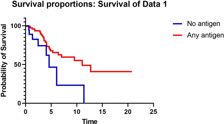
TFS was significantly better in patients with antigen identified by detailed clinical history compared to those without identified antigen (median TFS 11.1 vs 4.9 years, respectively p = 0.003) (Fig 1). The presence of antigen was associated with TFS in a univariable model (HR 0.38, 95% CI 0.16–0.72, p = 0.005) of 136 patients. In a multivariable model with 134 patients (2 had missing DLCO baseline data and were excluded) adjusted for age, previous smoking, FVC % predicted, DLCO % predicted, and presence of fibrosis, identified antigen was associated with improved TFS (HR 0.39, 95% CI 0.17–0.89, p = 0.025) (Table 2). Because emphysema has been variably reported to impact mortality, the presence of emphysema, which was present in 25 patients (18.4%) in the cohort, was evaluated in a univariable model but did not impact TFS (HR 1.2, 95% CI 0.53–2.61, p = 0.59) so emphysema was not included in the multivariable model.

Fig 1. Kaplan-Meier curve comparing transplant-free survival in years in patients with no antigen identified vs at least 1 antigen identified.

Fig 1

Table 2. Variables included in the Cox proportional hazards survival model.

Variable HR for death or transplant 95% CI P value
Age 1.011 0.98–1.05 0.54
Previous smoking 1.03 0.45–2.34 0.95
Identified antigen 0.39 0.17–0.89 0.025
FVC % predicted 0.98 0.96–1.01 0.22
DLCO % predicted 0.97 0.94–1.0 0.025
Presence of fibrosis 5.6 0.71–40.3 0.10

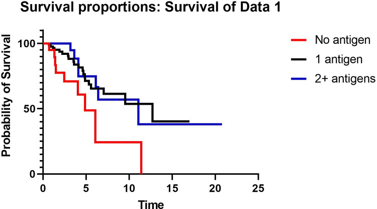
To evaluate whether the number of antigens identified was associated with outcome, TFS was compared between patients with no antigen, 1 antigen, and 2 or more antigens. TFS was significantly worse in patients with no antigen identified compared to those with either 1 antigen identified or 2 or more antigens identified by history, but TFS did not differ between patients with 1 antigen identified and those with 2 antigens identified (Fig 2). When estimating outcome risk, in a univariable model, the number of antigens was associated with TFS (HR 0.29, HR 0.31–0.95, p = 0.03), but in a multivariable model adjusted for age, smoking, FVC % predicted, DLCO % predicted, and fibrosis, the number of antigens was not associated with transplant-free survival (HR 0.31, 95% CI 0.31–1.05, p = 0.07).

Fig 2. Kaplan-Meier curve comparing transplant-free survival in years in patients with no antigen identified, 1 antigen identified, or 2 or more antigens identified.

Fig 2

HRCT results were similar between patients with no identified antigen, 1 antigen, and 2 antigens (Table 3). There was no difference in the proportion of patients with indeterminate, compatible, and typical HP HRCT scans between groups. Sixteen percent of the cohort had inflammatory HP, and the proportion did not differ between groups (9.5% for no antigen, 18.2% for 1 antigen, and 14.5% for 2 or more antigens, p = 0.61). The proportion of patients with mosaicism, nodules, ground glass, and upper lobe predominance was not different between groups. When patients with no antigen were compared to those with any antigen identified, there was no difference in the proportion of patients with indeterminate, compatible, or typical HP scans, the proportion of patients with fibrotic vs inflammatory scans, or any of the inconsistent with UIP features. Histopathologic findings were also similar between patients with no identified antigen, 1 antigen, and 2 antigens (Table 4). There was no difference in the probability of HP based on Tbbx or SLB between patients with any antigen identified vs no antigen (p = 0.72 for Tbbx and p = 0.68 for SLB), and the proportion of patients undergoing Tbbx or SLB was also similar between groups.

Table 3. Radiographic and histopathologic features of retrospective cohort.

Features No antigen (N = 21) 1 antigen (N = 88) 2+ antigen (N = 27) P value
HRCT pattern, N (%) 0.13
    Definite UIP 1 (4.8) 2 (2.3) 0 (0)
    Possible UIP 0 (0.0) 10 (11.4) 0 (0)
    Inconsistent with UIP 20 (95.2) 76 (86.4) 27 (100)
Type of HP 0.61
    Inflammatory 2 (9.5) 16 (18.2) 4 (14.5)
    Fibrotic 19 (90.5) 72 (81.8) 23 (85.2)
HRCT honeycombing, N (%) 5 (23.8) 20 (22.7) 8 (29.6) 0.76
Probability of HP based on HRCT, N (%)* 0.39
    Indeterminate 7 (33.3) 23 (26.1) 3 (11.1)
    Compatible 2 (9.5) 12 (13.6) 5 (18.5)
    Typical 12 (57.1) 53 (60.2) 20 (74.1)
Inconsistent features
    Mid/upper lung predominant fibrosis 7 (33.3) 33 (37.5) 6 (22.2) 0.34
    Peribronchovascular predominance 13 (61.9) 36 (40.0) 11 (40.7) 0.20
    Extensive ground glass > reticulations 11 (52.4) 51 (58.0) 16 (59.2) 0.88
    Micronodules, No. (%) 4 (19.0) 19 (21.6) 7 (25.9) 0.84
    Mosaic attenuation in ≥ 3 lobes 14 (66.7) 62 (70.5) 19 (70.4) 0.94
    Cysts, No. (%) 2 (9.5) 10 (11.4) 0 (0) 0.19
    Consolidation, No. (%) 1 (4.8) 9 (10.2) 0 (0) 0.18

Table 4. Transbronchial biopsy and SLB characteristics of retrospective cohort.

Histopathology features No antigen (N = 21) 1 antigen (N = 88) 2+ antigen (N = 27) P value
BAL performed 6 (28.6) 39 (44.3) 12 (44.4) 0.40
    BAL lymph > 30 5 (83.3) 19 (21.6) 5 (18.5) 0.22
Tbbx performed 6 (28.6) 45 (51.1) 17 (63.0) 0.06
TBBx Findings
    Indeterminate HP 3 (50.0) 17 (37.8) 8 (47.1) 0.72
    Probable HP 0 (0) 0 (0) 0 (0)
    Typical HP 3 (50.0) 28 (62.2) 9 (52.9)
SLB performed 17 (81.0) 58 (65.9) 18 (66.7) 0.40
SLB HP classification 0.68
    Indeterminate HP 0 (0) 3 (5.2) 0 (0)
    Probable HP 6 (35.3) 17 (29.3) 7 (38.9)
    Typical HP 11 (64.7) 38 (65.5) 11 (61.1)
SLB Findings
    Poorly formed granulomas 12 (70.6) 37 (63.8) 11 (61.1) 0.83
    Airway-centered fibrosis 16 (94.1) 48 (54.5) 18 (100) 0.10
    Chronic fibrosing interstitial pneumonia 15 (88.2) 51 (87.9) 18 (100) 0.30
    Cellular interstitial pneumonia 1 (5.9) 7 (12.1) 0 (0) 0.25
    Cellular bronchiolitis 1 (5.9) 6 (10.3) 0 (0) 0.33

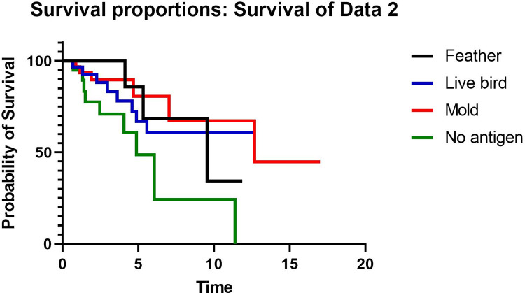
To evaluate whether the type of antigen was associated with outcomes, TFS was compared between patients with no antigen identified (N = 21), feather antigen (N = 16), live bird antigen (N = 29), and mold antigen (N = 34). Patients with more than one identified antigen were not included this portion of the analysis in order to isolate the effect of each antigen. TFS was worst for patients with no identified antigen compared to those with feather, live bird, or mold antigen (Fig 3). When estimating outcome risk, patients with feather antigen exposure showed a decreased risk for death or transplant compared to patients with no antigen identified (HR 0.30, 95% CI 0.10–0.96, p = 0.043). Patients with feather antigen exposure had no difference in the risk of death or transplant compared to patients with live bird (HR 1.4, 95% CI 0.40–5.07, p = 0.59) or mold exposure (HR 0.70, 95% CI 0.15–3.2, p = 0.65). When patients with both live bird and feather exposure (all avian antigen) were compared to patients with mold antigen in a univariable model, there was no difference in TFS (HR 0.47, 95% CI 0.42–2.6, p = 0.91). Patients with avian antigen exposure were more likely than those with mold antigen exposure to have inflammatory HP (p = 0.014) and an HRCT that is inconsistent with UIP pattern (p = 0.01) (Table 5). The probability of HP based on HRCT, BAL lymphocyte percentage, and baseline PFTs were the same between groups. Patients with avian antigen exposure were more likely to undergo TBBx than patients with mold antigen, but among those who underwent TBBx the findings were not different between groups. The proportion of patients who underwent SLB and the probability of HP based on SLB results was similar between those with avian vs mold antigen.

Fig 3. Kaplan-meier curve comparing transplant-free survival in years in patients with no antigen, feather, live bird, or mold.

Fig 3

Table 5. Radiographic and histopathologic features by avian vs mold antigen.

Avian antigen (N = 45) Mold antigen (N = 34) P-value
HRCT pattern 0.01
    Definite UIP 1 (2.2) 0 (0)
    Possible UIP 1 (2.2) 8 (23.5)
    Inconsistent with UIP 43 (95.6) 26 (76.5)
Type of HP 0.005
    Inflammatory 12 (26.7) 1 (2.9)
    Fibrotic 33 (73.3) 33 (97.1)
HRCT honeycombing 7 (15.6) 10 (29.4) 0.14
Probability of HP based on 0.17
    HRCT
    Indeterminate 8 (17.8) 12 (35.3)
    Compatible 6 (13.3) 5 (14.7)
    Typical 31 (68.9) 17 (50)
Inconsistent Features, N (%)
    Mid/upper lung predominance 18 (40) 12 (35.3) 0.67
    Peribronchovascular 21 (46.7) 12 (35.3) 0.22
    Extensive ground glass 27 (60) 18 (52.9) 0.53
    Micronodules 13 (28.9) 5 (14.7) 0.14
    Mosaic attenuation ≥ 3 lobes 35 (77.8) 21 (61.8) 0.12
    Cysts 5 (11.1) 3 (8.8) 0.74
    Consolidation 6 (13.3) 2 (5.9) 0.28
BAL performed 22 (48.9) 15 (44.1) 0.67
    BAL lymph > 30 11 (50) 6 (40) 0.55
    BAL lymph mean (SD) 35 (22.5) 34.73 (31.1) 0.98
Tbbx performed 29 (64.4) 14 (41.2) 0.04
Tbbx pathology
    Indeterminate HP 12 (41.4) 4 (28.6) 0.42
    Probable HP 0 (0) 0 (0)
    Typical HP 17 (58.6) 10 (71.4)
SLB performed 28 (62.2) 22 (64.7) 0.82
SLB findings 0.99
    Indeterminate HP 1 (3.6) 1 (4.5)
    Probable HP 9 (32.1) 7 (31.2)
    Typical HP 18 (64.3) 14 (63.4)
Baseline FVC mean (SD) 69.1 (18.1) 69.1 (21.1) 1.00
Baseline DLCO mean (SD) 52.7 (16.9) 48.5 (16.8) 0.28

Discussion

In this study, we examined the effect of number and type of antigen on transplant-free survival, radiographic, and histopathologic findings in a cohort of patients with a confident diagnosis of HP. Our study has several notable findings. 1) Identification of antigen by clinical history alone was associated with improved TFS. 2) Identification of a feather exposure was associated with improved TFS compared to unidentified antigen. 3) Among patients with identified antigen, the number and type of antigen did not affect transplant-free survival. 4) Patients with mold exposure were more likely to have fibrosis on HRCT than patients with avian antigen exposure. 5) Emphysema is not a predictor of TFS in HP.

Our findings are similar to a prior study, which showed that identification of antigen confirmed by an industrial hygienist or serum precipitating antibodies was associated with improved survival in patients with biopsy-proven HP [4]. This may be explained by both a higher rate of fibrotic HP in patients without identified antigen and by reduced rate of FVC decline in patients who remove antigen compared to those who remain in exposure [712]. However, our study adds to the literature by demonstrating that the diagnostic evaluation, including radiographic and histopathologic probability of HP, do not differ between patients based on the identification of antigen. Our study indirectly supports the importance of antigen removal by eliminating lead-time bias as a potentially contributing factor for improved survival in patients with identified antigen. We routinely recommend antigen remediation for all patients with identified antigen but are unable to rigorously assess whether that occurred in this retrospective study.

Our findings are also unique in that we demonstrated survival benefit with antigen identification alone without confirmation by an industrial hygienist or serum precipitating antibodies in patients with a confident diagnosis of HP and that we included patients with feather exposure alone [4, 10]. The use of an industrial hygienist may be limited by cost, availability, and expertise and lack of standardization of sample collection and analysis [13]. Further, while a positive serum precipitating antibody test may be informative that sensitization has occurred, available commercial tests have variable methods of measurement, different antigens, and nonuniform thresholds for positivity, which limit their negative predictive value [13]. Our study demonstrates that a thorough history for antigen exposure has prognostic value for patients with HP without the need for confirmatory testing. We agree with prior expert consensus statements that exposure history should be structured, standardized, and comprehensive [13], and several questionnaires have been previously published to guide exposure assessment [2, 14]. We suggest that exposure questionnaires also ascertain down exposure, as identification of down exposure in our study was associated with improved survival compared to patients with no antigen identified. While this study is the first to note survival association with down exposure compared to no identified antigen, it fits with prior HP studies that have included down as an inciting antigen and with numerous case series which have also suggested an association between down exposure and the development of HP [1518].

The impact of the characteristics of antigen exposure, including type of antigen, intensity, and duration, on the development of hypersensitivity pneumonitis remains poorly understood [7]. Only a small percentage of people with antigen exposure will go on to develop HP, ranging from 8–540,000 per 100,000 per year among farmers and 6000–21000 per 100,000 per year in pigeon breeders [1925]. What accounts for the difference in prevalence of disease in exposed patients and whether that translates to any meaningful difference in disease phenotype or outcome remains unclear. Our findings help to resolve conflicting data in the literature with regard to the effect of the type of antigen on mortality and fibrotic phenotype in HP. Two prior studies suggested that patients with avian antigen exposure have a better survival than patients with other types of exposure [4, 10], while other studies found that the type of antigen did not affect mortality [7, 26, 27]. We did not find an association between antigen type and TFS but did demonstrate that mold antigen leads to a higher proportion of fibrotic HP compared to avian antigen, a finding that has been shown in the literature and in our multivariate analysis of our cohort to lead to higher mortality [4, 10, 26]. Conversely, a prior study from the Mayo Clinic revealed that patients with avian antigen exposure were more likely to have fibrosis on HRCT than those without avian antigen, but this did not correspond to a change in mortality [26], while other studies did not find an association between antigen type and fibrotic phenotype [7, 27, 28]. Our study required high or definite diagnosis of HP, whereas the Mayo Clinic study only required histopathologic confirmation if antigen was not identified, which could have led to a lower confidence diagnosis and may account for different results.

Finally, our study elucidates the prevalence of emphysema in HP and its impact on TFS. Emphysema is a predictor of mortality in the literature in patients without other underlying lung disease [29], but the role that emphysema plays in mortality in patients with ILD remains unclear. In 2005, Cottin et al described the syndrome of combined pulmonary fibrosis and emphysema (CPFE), which is characterized by upper-lobe predominant emphysema and lower-lobe predominant fibrosis with preserved lung volumes and severely diminished DLCO [30]. Studies comparing the mortality between CPFE and idiopathic pulmonary fibrosis have yielded conflicting results [31, 32]. However, in patients with scleroderma, mortality is higher in patients with both ILD and emphysema compared to patients with ILD without emphysema [33]. Our study adds to the literature by being the first to look for an association between emphysema and TFS in patients with HP, and like the prior studies of CPFE, we did not find an association between TFS and emphysema. It is possible that a confounder such as pulmonary hypertension, which is present in both CPFE patients and patients in scleroderma, may limit our ability to detect an association between emphysema and TFS.

Strengths of our study include the large cohort of patients with a highly confident diagnosis of HP, the identification of antigen by history alone, and the inclusion of down products as inciting antigen. The use of clinical history alone to identify antigen exposure increases the generalizability of our study. Further, we have limited selection bias by using published guidelines to define our HP cohort [3]. Finally, by analyzing feather exposure compared to no identifying antigen, we have strengthened the association between down exposure and the development of HP.

There are limitations to this study that should be acknowledged. The study is retrospective, and we were unable to determine for all patients if and when an antigen was removed. In addition, given the retrospective nature of the study, we could not accurately assess the influence of treatment at the time of HRCT or biopsy on the radiographic or histopathologic findings and could not control for lead-time bias other than adjustment for FVC and DLCO. We lacked statistical power to analyze fibrotic and non-fibrotic HP separately, though we did adjust for fibrosis in the multivariable model.

In summary, the identification of antigen by a clinician taking a detailed history is associated with improved transplant-free survival in HP but does not affect radiographic or histopathologic findings. Among those with identified antigen, the number of antigens does not affect transplant-free survival, radiographic, or histopathologic findings. The type of antigen does not influence transplant-free survival, but patients with mold antigen exposure are more likely to have fibrotic HP compared to those with avian antigen exposure. Patients with feather exposure had comparable transplant-free survival to those with live bird exposure but better transplant-free survival than patients without identified antigen. We suggest that clinical history is adequate for providing prognostic information to patients with HP and classifying the diagnostic probability of HP according to recent guidelines. Appropriate history taking and identification of antigen exposure is associated with prognosis of patients with ILD.

Supporting information

S1 Data

(XLSX)

Data Availability

All relevant data are within the paper and its Supporting Information files.

Funding Statement

The author(s) received no specific funding for this work.

References

  • 1.Glazer CS, Rose CS, Lynch DA. Clinical and radiologic manifestations of hypersensitivity pneumonitis. J Thorac Imaging. 2002;17(4):261–72. doi: 10.1097/00005382-200210000-00003 [DOI] [PubMed] [Google Scholar]
  • 2.Barnes H, Morisset J, Molyneaux P, Westall G, Glaspole I, Collard HR, et al. A Systematically Derived Exposure Assessment Instrument for Chronic Hypersensitivity Pneumonitis. Chest. 2020;157(6):1506–12. doi: 10.1016/j.chest.2019.12.018 [DOI] [PMC free article] [PubMed] [Google Scholar]
  • 3.Raghu G, Remy-Jardin M, Ryerson CJ, Myers JL, Kreuter M, Vasakova M, et al. Diagnosis of Hypersensitivity Pneumonitis in Adults. An Official ATS/JRS/ALAT Clinical Practice Guideline. Am J Respir Crit Care Med. 2020;202(3):e36–e69. doi: 10.1164/rccm.202005-2032ST [DOI] [PMC free article] [PubMed] [Google Scholar]
  • 4.Fernandez Perez ER, Swigris JJ, Forssen AV, Tourin O, Solomon JJ, Huie TJ, et al. Identifying an inciting antigen is associated with improved survival in patients with chronic hypersensitivity pneumonitis. Chest. 2013;144(5):1644–51. doi: 10.1378/chest.12-2685 [DOI] [PMC free article] [PubMed] [Google Scholar]
  • 5.Barber CM, Wiggans RE, Fishwick D. Impaired quality of life in chronic hypersensitivity pneumonitis. Chest. 2015;147(6):e230. doi: 10.1378/chest.15-0198 [DOI] [PubMed] [Google Scholar]
  • 6.Adams TN, Newton CA, Batra K, Abu-Hijleh M, Barbera T, Torrealba J, et al. Utility of Bronchoalveolar Lavage and Transbronchial Biopsy in Patients with Hypersensitivity Pneumonitis. Lung. 2018;196(5):617–22. doi: 10.1007/s00408-018-0139-1 [DOI] [PMC free article] [PubMed] [Google Scholar]
  • 7.Barnes H, Lu J, Glaspole I, Collard HR, Johannson KA. Exposures and associations with clinical phenotypes in hypersensitivity pneumonitis: A scoping review. Respir Med. 2021;184:106444. doi: 10.1016/j.rmed.2021.106444 [DOI] [PubMed] [Google Scholar]
  • 8.Braun SR, doPico GA, Tsiatis A, Horvath E, Dickie HA, Rankin J. Farmer’s lung disease: long-term clinical and physiologic outcome. Am Rev Respir Dis. 1979;119(2):185–91. doi: 10.1164/arrd.1979.119.2.185 [DOI] [PubMed] [Google Scholar]
  • 9.Selman M, Pardo A, King TE Jr., Hypersensitivity pneumonitis: insights in diagnosis and pathobiology. Am J Respir Crit Care Med. 2012;186(4):314–24. [DOI] [PubMed] [Google Scholar]
  • 10.De Sadeleer LJ, Hermans F, De Dycker E, Yserbyt J, Verschakelen JA, Verbeken EK, et al. Effects of Corticosteroid Treatment and Antigen Avoidance in a Large Hypersensitivity Pneumonitis Cohort: A Single-Centre Cohort Study. J Clin Med. 2018;8(1). [DOI] [PMC free article] [PubMed] [Google Scholar]
  • 11.Tsutsui T, Miyazaki Y, Kuramochi J, Uchida K, Eishi Y, Inase N. The amount of avian antigen in household dust predicts the prognosis of chronic bird-related hypersensitivity pneumonitis. Ann Am Thorac Soc. 2015;12(7):1013–21. doi: 10.1513/AnnalsATS.201412-569OC [DOI] [PubMed] [Google Scholar]
  • 12.Sema M, Miyazaki Y, Tsutsui T, Tomita M, Eishi Y, Inase N. Environmental levels of avian antigen are relevant to the progression of chronic hypersensitivity pneumonitis during antigen avoidance. Immun Inflamm Dis. 2018;6(1):154–62. doi: 10.1002/iid3.202 [DOI] [PMC free article] [PubMed] [Google Scholar]
  • 13.Johannson KA, Barnes H, Bellanger AP, Dalphin JC, Fernandez Perez ER, Flaherty KR, et al. Exposure Assessment Tools for Hypersensitivity Pneumonitis. An Official American Thoracic Society Workshop Report. Ann Am Thorac Soc. 2020;17(12):1501–9. doi: 10.1513/AnnalsATS.202008-942ST [DOI] [PMC free article] [PubMed] [Google Scholar]
  • 14.Vasakova M, Morell F, Walsh S, Leslie K, Raghu G. Hypersensitivity Pneumonitis: Perspectives in Diagnosis and Management. Am J Respir Crit Care Med. 2017;196(6):680–9. doi: 10.1164/rccm.201611-2201PP [DOI] [PubMed] [Google Scholar]
  • 15.Hanak V, Golbin JM, Ryu JH. Causes and presenting features in 85 consecutive patients with hypersensitivity pneumonitis. Mayo Clin Proc. 2007;82(7):812–6. doi: 10.4065/82.7.812 [DOI] [PubMed] [Google Scholar]
  • 16.Wang P, Jones KD, Urisman A, Elicker BM, Urbania T, Johannson KA, et al. Pathologic Findings and Prognosis in a Large Prospective Cohort of Chronic Hypersensitivity Pneumonitis. Chest. 2017;152(3):502–9. doi: 10.1016/j.chest.2017.02.011 [DOI] [PubMed] [Google Scholar]
  • 17.Inase N, Ohtani Y, Sumi Y, Umino T, Usui Y, Miyake S, et al. A clinical study of hypersensitivity pneumonitis presumably caused by feather duvets. Ann Allergy Asthma Immunol. 2006;96(1):98–104. doi: 10.1016/S1081-1206(10)61047-2 [DOI] [PubMed] [Google Scholar]
  • 18.Jacobs MR, Andrews CP, Ramirez RM, Jacobs RL. Frequency of goose and duck down causation of hypersensitivity pneumonitis within an 80-patient cohort. Ann Allergy Asthma Immunol. 2019;123(2):201–7. doi: 10.1016/j.anai.2019.04.023 [DOI] [PubMed] [Google Scholar]
  • 19.Salvaggio JE. The identification of hypersensitivity pneumonitis. Hosp Pract (1995). 1995;30(5):57–62, 5–6. doi: 10.1080/21548331.1995.11443199 [DOI] [PubMed] [Google Scholar]
  • 20.Dalphin JC, Debieuvre D, Pernet D, Maheu MF, Polio JC, Toson B, et al. Prevalence and risk factors for chronic bronchitis and farmer’s lung in French dairy farmers. Br J Ind Med. 1993;50(10):941–4. doi: 10.1136/oem.50.10.941 [DOI] [PMC free article] [PubMed] [Google Scholar]
  • 21.Arya A, Roychoudhury K, Bredin CP. Farmer’s lung is now in decline. Ir Med J. 2006;99(7):203–5. [PubMed] [Google Scholar]
  • 22.Grant IW, Blyth W, Wardrop VE, Gordon RM, Pearson JC, Mair A. Prevalence of farmer’s lung in Scotland: a pilot survey. Br Med J. 1972;1(5799):530–4. doi: 10.1136/bmj.1.5799.530 [DOI] [PMC free article] [PubMed] [Google Scholar]
  • 23.Christensen LT, Schmidt CD, Robbins L. Pigeon breeders’ disease—a prevalence study and review. Clin Allergy. 1975;5(4):417–30. doi: 10.1111/j.1365-2222.1975.tb01881.x [DOI] [PubMed] [Google Scholar]
  • 24.Hendrick DJ, Faux JA, Marshall R. Budgerigar-fancier’s lung: the commonest variety of allergic alveolitis in Britain. Br Med J. 1978;2(6130):81–4. doi: 10.1136/bmj.2.6130.81 [DOI] [PMC free article] [PubMed] [Google Scholar]
  • 25.Terho EO, Vohlonen I, Husman K. Prevalence and incidence of chronic bronchitis and farmer’s lung with respect to socioeconomic factors. Eur J Respir Dis Suppl. 1987;152:29–36. [PubMed] [Google Scholar]
  • 26.Hanak V, Golbin JM, Hartman TE, Ryu JH. High-resolution CT findings of parenchymal fibrosis correlate with prognosis in hypersensitivity pneumonitis. Chest. 2008;134(1):133–8. doi: 10.1378/chest.07-3005 [DOI] [PubMed] [Google Scholar]
  • 27.Vourlekis JS, Schwarz MI, Cherniack RM, Curran-Everett D, Cool CD, Tuder RM, et al. The effect of pulmonary fibrosis on survival in patients with hypersensitivity pneumonitis. Am J Med. 2004;116(10):662–8. doi: 10.1016/j.amjmed.2003.12.030 [DOI] [PubMed] [Google Scholar]
  • 28.Adams TN, Newton CA, Glazer CS. Role of Antigen Type in Survival in Chronic Hypersensitivity Pneumonitis. Lung. 2019;197(1):113–4. doi: 10.1007/s00408-018-0187-6 [DOI] [PMC free article] [PubMed] [Google Scholar]
  • 29.Johannessen A, Skorge TD, Bottai M, Grydeland TB, Nilsen RM, Coxson H, et al. Mortality by level of emphysema and airway wall thickness. Am J Respir Crit Care Med. 2013;187(6):602–8. doi: 10.1164/rccm.201209-1722OC [DOI] [PubMed] [Google Scholar]
  • 30.Cottin V, Nunes H, Brillet PY, Delaval P, Devouassoux G, Tillie-Leblond I, et al. Combined pulmonary fibrosis and emphysema: a distinct underrecognised entity. Eur Respir J. 2005;26(4):586–93. doi: 10.1183/09031936.05.00021005 [DOI] [PubMed] [Google Scholar]
  • 31.Ryerson CJ, Hartman T, Elicker BM, Ley B, Lee JS, Abbritti M, et al. Clinical features and outcomes in combined pulmonary fibrosis and emphysema in idiopathic pulmonary fibrosis. Chest. 2013;144(1):234–40. doi: 10.1378/chest.12-2403 [DOI] [PubMed] [Google Scholar]
  • 32.Mejia M, Carrillo G, Rojas-Serrano J, Estrada A, Suarez T, Alonso D, et al. Idiopathic pulmonary fibrosis and emphysema: decreased survival associated with severe pulmonary arterial hypertension. Chest. 2009;136(1):10–5. doi: 10.1378/chest.08-2306 [DOI] [PubMed] [Google Scholar]
  • 33.Champtiaux N, Cottin V, Chassagnon G, Chaigne B, Valeyre D, Nunes H, et al. Combined pulmonary fibrosis and emphysema in systemic sclerosis: A syndrome associated with heavy morbidity and mortality. Semin Arthritis Rheum. 2019;49(1):98–104. doi: 10.1016/j.semarthrit.2018.10.011 [DOI] [PubMed] [Google Scholar]

Decision Letter 0

Vipa Thanachartwet

1 Aug 2022

PONE-D-22-19345Impact of number and type of identified antigen on transplant-free survival in hypersensitivity pneumonitisPLOS ONE

Dear Dr. Traci Adams,

Thank you for submitting your manuscript to PLOS ONE. After careful consideration, we feel that it has merit but does not fully meet PLOS ONE’s publication criteria as it currently stands. Therefore, we invite you to submit a revised version of the manuscript that addresses the points raised during the review process. We appreciate the interesting study. However, there are some important points that are required to define clearly. Please carefully respond to the reviewers’ comments and suggestions. Please submit your revised manuscript by Sep 12 2022 11:59 PM. If you will need more time than this to complete your revisions, please reply to this message or contact the journal office at plosone@plos.org. When you're ready to submit your revision, log on to https://www.editorialmanager.com/pone/ and select the 'Submissions Needing Revision' folder to locate your manuscript file.

Please include the following items when submitting your revised manuscript:

  • A rebuttal letter that responds to each point raised by the academic editor and reviewer(s). You should upload this letter as a separate file labeled 'Response to Reviewers'.

  • A marked-up copy of your manuscript that highlights changes made to the original version. You should upload this as a separate file labeled 'Revised Manuscript with Track Changes'.

  • An unmarked version of your revised paper without tracked changes. You should upload this as a separate file labeled 'Manuscript'.

If you would like to make changes to your financial disclosure, please include your updated statement in your cover letter. Guidelines for resubmitting your figure files are available below the reviewer comments at the end of this letter.

If applicable, we recommend that you deposit your laboratory protocols in protocols.io to enhance the reproducibility of your results. Protocols.io assigns your protocol its own identifier (DOI) so that it can be cited independently in the future. For instructions see: https://journals.plos.org/plosone/s/submission-guidelines#loc-laboratory-protocols. Additionally, PLOS ONE offers an option for publishing peer-reviewed Lab Protocol articles, which describe protocols hosted on protocols.io. Read more information on sharing protocols at https://plos.org/protocols?utm_medium=editorial-email&utm_source=authorletters&utm_campaign=protocols.

We look forward to receiving your revised manuscript.

Kind regards,

Vipa Thanachartwet, M.D.

Academic Editor

PLOS ONE

Journal Requirements:

When submitting your revision, we need you to address these additional requirements.

1. Please ensure that your manuscript meets PLOS ONE's style requirements, including those for file naming. The PLOS ONE style templates can be found at 

https://journals.plos.org/plosone/s/file?id=wjVg/PLOSOne_formatting_sample_main_body.pdf and 

https://journals.plos.org/plosone/s/file?id=ba62/PLOSOne_formatting_sample_title_authors_affiliations.pdf

2. Please include your full ethics statement in the ‘Methods’ section of your manuscript file. In your statement, please include the full name of the IRB or ethics committee who approved or waived your study, as well as whether or not you obtained informed written or verbal consent. If consent was waived for your study, please include this information in your statement as well. 

[Note: HTML markup is below. Please do not edit.]

Reviewers' comments:

Reviewer's Responses to Questions

Comments to the Author

1. Is the manuscript technically sound, and do the data support the conclusions?

The manuscript must describe a technically sound piece of scientific research with data that supports the conclusions. Experiments must have been conducted rigorously, with appropriate controls, replication, and sample sizes. The conclusions must be drawn appropriately based on the data presented.

Reviewer #1: Yes

Reviewer #2: Partly

**********

2. Has the statistical analysis been performed appropriately and rigorously?

Reviewer #1: Yes

Reviewer #2: No

**********

3. Have the authors made all data underlying the findings in their manuscript fully available?

The PLOS Data policy requires authors to make all data underlying the findings described in their manuscript fully available without restriction, with rare exception (please refer to the Data Availability Statement in the manuscript PDF file). The data should be provided as part of the manuscript or its supporting information, or deposited to a public repository. For example, in addition to summary statistics, the data points behind means, medians and variance measures should be available. If there are restrictions on publicly sharing data—e.g. participant privacy or use of data from a third party—those must be specified.

Reviewer #1: Yes

Reviewer #2: Yes

**********

4. Is the manuscript presented in an intelligible fashion and written in standard English?

PLOS ONE does not copyedit accepted manuscripts, so the language in submitted articles must be clear, correct, and unambiguous. Any typographical or grammatical errors should be corrected at revision, so please note any specific errors here.

Reviewer #1: Yes

Reviewer #2: Yes

**********

5. Review Comments to the Author

Please use the space provided to explain your answers to the questions above. You may also include additional comments for the author, including concerns about dual publication, research ethics, or publication ethics. (Please upload your review as an attachment if it exceeds 20,000 characters)

Reviewer #1: Kypreos et al. investigate the relationship between antigen type and transplant-free survival (TFS) in patients with hypersensitivity pneumonitis. The authors determined that identification of inciting antigen led to improved TFS. While TFS was not different in patients with mold or feather exposure, mold antigen was associated with increased risk of fibrotic HP. Additionally, in patients with an identified antigen, the number of antigens or the type of antigen did not affect TFS. The authors present interesting data, summarized below are my concerns.

1. Does the type of antigen exposure correlate with lung function (FVC or DLCO)?

2. Does the type of antigen exposure correlate with BAL lymphocyte percentage?

Reviewer #2: The study sought to determine if patients with HP with identifiable exposure antigens had better transplant free survival. This was a secondary database analysis that tested a hypothesis that had been previously confirmed by a more rigorous method of determining antigen exposure. The authors argued that the contribution of this study was that clinical history alone could be sufficient. However, the description in the methods including patient population, exposure history and statistical analysis was inadequate to allow for assessment of the validity of the results. Specific comments are outlined below.

1) How were the 136 cases of HP identified? Did these cases have HP diagnosis already or some numbers of patients with ILD diagnosis were matched with the guidelines and only those with high confidence or definitive HP were selected? Were they identified because they all had typical CT findings of small airway disease? A detailed description on how the cohort was established is critical to avoid any case selection bias. A flow chart on the patient selection process would help.

2) How were the antigen exposures identified and confirmed to be relevant? Based on review of the clinic notes, serum IgG tests, or other environmental tests (like indoor air quality test)? If the information was obtained from clinic notes, were these notes from pulmonologists/allergists? It is well known that adequate history-taking in HP requires a high index of suspicion, time, and content expertise. Differences in approach and content among clinicians lead to varying reliability.

3) HRCT was read by one of the co-authors. Why were the original CT reports not used? Potential bias could be introduced if the readers know the patient has a high pre-test probability of HP.

4) The guidelines were not published until 2020. Many of these patients likely were diagnosed with HP before 2020. What was the time frame from which the patients came from?

5) There were another HP guidelines published by CHEST in 2020. The guidelines prioritize environmental exposure in the algorithm. Were the results the same if the CHEST guidelines were used?

6) Page 5. "The primary outcome of this study was transplant-free survival... defined as time from diagnosis of interstitial lung disease (ILD) to death or transplant". The clinical course of HP is highly variable, especially between non-fibrotic and fibrotic HP. How did you account for lead time bias, especially for fibrotic HP? Also was the worse survival in HP patients without identifiable exposure due to fibrotic HP patients since many more had no identifiable exposure compared to non-fibrotic HP (Table 3)? If the analysis were done separately for fibrotic and non-fibrotic HP, would the same results be expected?

7) Line 80. Please describe how mold exposure was considered significant and relevant, since molds are ubiquitous in the environment.

8) Table 1. Please define "sensitizing antigens". Were they from history, IgG test or other methods?

9) Line 94. In the model, how were the missing values handled, since not all patients had PFT, lung biopsy etc?

10) In Table 1, in the 37 death and transplant, how many were fibrotic HP and how many were non-fibrotic HP? It was a bit of a surprise that in Table 2, presence of fibrosis was not a factor.

11) In Table 1 under antigen identified, what does "other" indicate?

12) Table 3. In the ATS guidelines, BAL and lung biopsy play a significant role in determining the diagnostic confidence. So without BAL or lung pathology, it is difficult to reach high confidence or definitive diagnosis of HP, especially for non-fibrotic HP. This could potentially introduce selection bias. This is a significant issue that needs to be addressed. Perhaps additional analysis should be performed only in patients who had lung biopsy and BAL.

**********

6. PLOS authors have the option to publish the peer review history of their article (what does this mean?). If published, this will include your full peer review and any attached files.

If you choose “no”, your identity will remain anonymous but your review may still be made public.

Do you want your identity to be public for this peer review? For information about this choice, including consent withdrawal, please see our Privacy Policy.

Reviewer #1: No

Reviewer #2: No

**********

[NOTE: If reviewer comments were submitted as an attachment file, they will be attached to this email and accessible via the submission site. Please log into your account, locate the manuscript record, and check for the action link "View Attachments". If this link does not appear, there are no attachment files.]

While revising your submission, please upload your figure files to the Preflight Analysis and Conversion Engine (PACE) digital diagnostic tool, https://pacev2.apexcovantage.com/. PACE helps ensure that figures meet PLOS requirements. To use PACE, you must first register as a user. Registration is free. Then, login and navigate to the UPLOAD tab, where you will find detailed instructions on how to use the tool. If you encounter any issues or have any questions when using PACE, please email PLOS at figures@plos.org. Please note that Supporting Information files do not need this step.

PLoS One. 2022 Sep 1;17(9):e0273544. doi: 10.1371/journal.pone.0273544.r002

Author response to Decision Letter 0


3 Aug 2022

Reviewer #1: Kypreos et al. investigate the relationship between antigen type and transplant-free survival (TFS) in patients with hypersensitivity pneumonitis. The authors determined that identification of inciting antigen led to improved TFS. While TFS was not different in patients with mold or feather exposure, mold antigen was associated with increased risk of fibrotic HP. Additionally, in patients with an identified antigen, the number of antigens or the type of antigen did not affect TFS. The authors present interesting data, summarized below are my concerns.

1. Does the type of antigen exposure correlate with lung function (FVC or DLCO)?

The type of exposure does not correlate with FVC or DLCO. We have added a sentence in the results section to note this and have included PFT data data in Table 5.

“The probability of HP based on HRCT, BAL lymphocyte percentage, and baseline PFTs were the same between groups.”

2. Does the type of antigen exposure correlate with BAL lymphocyte percentage?

The type of exposure does not correlate with BAL lymphocyte percentage. We have added a sentence in the results section to note this and have included BAL data in Table 5.

“The probability of HP based on HRCT, BAL lymphocyte percentage, and baseline PFTs were the same between groups.”

Reviewer #2: The study sought to determine if patients with HP with identifiable exposure antigens had better transplant free survival. This was a secondary database analysis that tested a hypothesis that had been previously confirmed by a more rigorous method of determining antigen exposure. The authors argued that the contribution of this study was that clinical history alone could be sufficient. However, the description in the methods including patient population, exposure history and statistical analysis was inadequate to allow for assessment of the validity of the results. Specific comments are outlined below.

1) How were the 136 cases of HP identified? Did these cases have HP diagnosis already or some numbers of patients with ILD diagnosis were matched with the guidelines and only those with high confidence or definitive HP were selected? Were they identified because they all had typical CT findings of small airway disease? A detailed description on how the cohort was established is critical to avoid any case selection bias. A flow chart on the patient selection process would help.

We identified the cases from our retrospective cohort of patients diagnosed with interstitial lung disease from 2005-2021. The cohort is derived from a database in our electronic medical record, which includes all patients with a diagnosis of interstitial lung disease (or any subtype of ILD including IPF, HP, etc) on the problem list who had been seen in the pulmonary clinic at UT Southwestern. Our cohort contains 1157 patients. Of these, 92 had moderate probability of HP by ATS criteria and were excluded. 136 patients had a high or definite probability of HP by ATS criteria and were included. Each of these patient’s medical records was reviewed, and our thoracic radiologist entered interpretations of each of the HRCT scans into a spreadsheet without knowledge of the diagnosis because she opens the imaging software only, rather than the full medical record where the diagnosis might be visible. Pathology findings, demographics, and antigen exposure were also entered into a spreadsheet, though we were not able to be blind to the diagnosis when entering data from the medical record. Once all available data was present for review, we used the objective data to determine whether the patient met ATS criteria and used a multidisciplinary discussion to confirm a diagnosis of HP if one had not been conducted previously. Most, but not all, of these patients had a prior diagnosis of HP in the medical record. We chose to only include those with a high or definite confidence level because to reach this level of confidence, ancillary information including a BAL or lung biopsy are needed, and we thought that inclusion of patients with moderate probability could introduce significant selection bias as those patients are not required to undergo bronchoscopy or lung biopsy.

We did not include a flow chart (though we can if the reviewer needs this), but have included the following statements in the manuscript:

“We derived the HP cohort from the UT Southwestern pulmonary clinic Epic registry, which includes all patients seen in pulmonary clinic with a diagnosis of interstitial lung disease.”

“Based on the review of antigen exposure, HRCT, BAL, TBBx, and SLB, patients were classified as HP excluded, low probability HP, moderate probability HP, high probability of HP, or HP based on recent guidelines. Only those with high or definite probability of HP were included in the study.”

“In our retrospective cohort of 1157 patients with ILD, 136 patients met high or definite probability of HP and were included in the analysis.”

2) How were the antigen exposures identified and confirmed to be relevant? Based on review of the clinic notes, serum IgG tests, or other environmental tests (like indoor air quality test)? If the information was obtained from clinic notes, were these notes from pulmonologists/allergists? It is well known that adequate history-taking in HP requires a high index of suspicion, time, and content expertise. Differences in approach and content among clinicians lead to varying reliability.

Each of these patients had been seen by an interstitial lung disease specialist at UT Southwestern. Our providers take a detailed exposure history and typically provide commentary about whether this exposure is considered significant. The majority of our providers use the following list of questions, which we did not include because it has not uniformly been used by our providers across the 16 years of the registry.

Type of home

Pier and beam or concrete slab

Crawl space inspection (wet, dry, mold, no mold)

Water damage

Visible mold in home

Type of air conditioning unit

Visible mold in air conditioning unit

Use of humidifier, pool, hot tub, sauna, CPAP/bipap

Bird exposure

Feather products in the home

Hobbies (gardening, metal/wood working, wind instrument)

We agree that it can be difficult to determine whether an exposure is relevant in some cases, as there is not a defined threshold that puts a patient at risk for HP. When there was any discrepancy about the exposure, Craig Glazer, who completed an occupational lung disease fellowship at National Jewish, reviewed the exposure to determine whether it was significant. We have added the following sentences to the Methods section to address this:

“Mold exposure could be in the home or office or related to farming and was considered significant if the patient was regularly exposed to visible mold or regularly using a composte heap. An occupational medicine specialist (CSG) reviewed the exposure history in cases where it was unclear whether the exposure was significant enough to potentially lead to sensitization.”

3) HRCT was read by one of the co-authors. Why were the original CT reports not used? Potential bias could be introduced if the readers know the patient has a high pre-test probability of HP.

We have only one thoracic radiologist at UT Southwestern, who has been involved in a number of studies and whose work has very closely correlated with radiologists at other centers including UC Davis, University of Chicago, and UCSF in prior studies. She read the scans without knowledge of diagnosis and did not know the exposure history or clinical diagnosis. This led to less variability than using the original CT reports, which may have been read by a radiologist without expertise in thoracic imaging.

We have included a statement about this in the methods section: “HRCTs were reviewed by a thoracic radiologist (KB) who was blinded to the clinical diagnosis.”

4) The guidelines were not published until 2020. Many of these patients likely were diagnosed with HP before 2020. What was the time frame from which the patients came from?

Our electronic medical record began in 2005, so patients were included who were seen in our ILD clinic after 2005 through 2020. Our assignment of diagnosis was retrospective using the ATS guidelines as outlined under question 1. We included the following in the methods section.

“Based on the review of antigen exposure, HRCT, BAL, TBBx, and SLB, patients were classified as HP excluded, low probability HP, moderate probability HP, high probability of HP, or HP based on recent guidelines. Only those with high or definite probability of HP were included in the study.”

5) There were another HP guidelines published by CHEST in 2020. The guidelines prioritize environmental exposure in the algorithm. Were the results the same if the CHEST guidelines were used?

The Chest criteria do not overlap entirely with ATS criteria, as they classify antigen exposure as unidentified, indeterminate, and identified. Further, Chest guidelines allow for a confident diagnosis based on exposure and HRCT alone, which would have led to additional patients being included that are not included in high/definite classification of ATS criteria. However, patients who meet a high or definite confidence of HP diagnosis in the ATS guidelines also meet a “provisional high confidence” or “HP” category of diagnosis in the Chest guidelines, so all of our patients would have been included by Chest criteria as well. We felt that having a cohort that is better-defined with either BAL or biopsy as confirmation would lead to clearer conclusions than including patients who were defined only by antigen exposure and CT scan. We agree that inclusion of those patients who did not have bronchoscopy or lung biopsy could lead to selection bias.

We included the following statement in the results section: “All patients included in the study had a BAL, TBBx, and/or SLB for confirmation of diagnosis based on ATS criteria for high or definite HP.”

6) Page 5. "The primary outcome of this study was transplant-free survival... defined as time from diagnosis of interstitial lung disease (ILD) to death or transplant". The clinical course of HP is highly variable, especially between non-fibrotic and fibrotic HP. How did you account for lead time bias, especially for fibrotic HP? Also was the worse survival in HP patients without identifiable exposure due to fibrotic HP patients since many more had no identifiable exposure compared to non-fibrotic HP (Table 3)? If the analysis were done separately for fibrotic and non-fibrotic HP, would the same results be expected?

We are not able to account for lead-time bias which is consistent with other retrospective cohort studies in ILD, including the initial study by Fernandes-Perez. We try to adjust for that by adjusting for FVC and DLCO at the time of ILD diagnosis, but there is not a way to directly adjust for lead-time bias using available data from the medical record. We added the following statement to the discussion: “In addition, given the retrospective nature of the study, we could not accurately assess the influence of treatment at the time of HRCT or biopsy on the radiographic or histopathologic findings and could not control for lead-time bias other than adjustment for FVC and DLCO.”

If we did a separate analysis for fibrotic and non-fibrotic HP, we would be unlikely to have statistical power to demonstrate any conclusions given the number of variables we need to control for. We did control for fibrotic disease in our model, which is the best we can do to account for the differential finding. We included the following statement in the discussion: “We lacked statistical power to analyze fibrotic and non-fibrotic HP separately, though we did adjust for fibrosis in the multivariable model.”

7) Line 80. Please describe how mold exposure was considered significant and relevant, since molds are ubiquitous in the environment.

Molds are ubiquitous, but they only grow in moist environments. We count a mold exposure as significant if the patient was regularly exposed to visible mold or regularly using composte. Industrial hygienists would typically examine for visible mold and quantify indoor vs outdoor air quality, and if there is either visible mold or the counts are 10x indoors what they are outdoors that would count as a significant exposure. We used visible mold as a significant and relevant exposure. Any questionable exposures were reviewed by Craig Glazer. We added the following sentences to the methods section: “Mold exposure could be in the home or office or related to farming and was considered significant if the patient was regularly exposed to visible mold or regularly using a composte heap. An occupational medicine specialist (CSG) reviewed the exposure history in cases where it was unclear whether the exposure was significant enough to potentially lead to sensitization.”

8) Table 1. Please define "sensitizing antigens". Were they from history, IgG test or other methods?

We have clarified that these were antigens from history in the table. We also removed “Sensitizing antigen” from the text to avoid confusion and substituted: “A potentially fibrogenic exposure”

9) Line 94. In the model, how were the missing values handled, since not all patients had PFT, lung biopsy etc?

All variables in the univariate and multivariate analysis had complete data except for 2 patients who were missing DLCO. For univariable analysis, any patient missing data is excluded. In the multivariable analyses, any patient that is missing any of the included variables is excluded. We have added a statement in the manuscript about how many patients were included in this analysis. “The presence of antigen was associated with TFS in a univariable model (HR 0.38, 95% CI 0.16-0.72, p=0.005) of 136 patients. In a multivariable model with 134 patients (2 had missing DLCO baseline data and were excluded) adjusted for age, previous smoking, FVC % predicted, DLCO % predicted, and presence of fibrosis, identified antigen was associated with improved TFS (HR 0.39, 95% CI 0.17-0.89, p=0.025) (Table 2).”

10) In Table 1, in the 37 death and transplant, how many were fibrotic HP and how many were non-fibrotic HP? It was a bit of a surprise that in Table 2, presence of fibrosis was not a factor.

We will clarify this in the manuscript. Only 1 patient of the 37 patients who died was nonfibrotic. We suspect that it is the low number of nonfibrotic patients or the association between nonfibrotic disease and other included variables that led to the lack of statistical significance in the multivariable model. We have included the following statement in the discussion section: “We lacked statistical power to analyze fibrotic and non-fibrotic HP separately, though we did adjust for fibrosis in the multivariable model.”

11) In Table 1 under antigen identified, what does "other" indicate?

Other indicated antigen other than avian or mold. Our “other” cohort consisted of isocyanate exposure and fish tank exposure. We have included a note about this under Table 1: **Other antigens included isocyanate exposure and fish tank exposure

12) Table 3. In the ATS guidelines, BAL and lung biopsy play a significant role in determining the diagnostic confidence. So without BAL or lung pathology, it is difficult to reach high confidence or definitive diagnosis of HP, especially for non-fibrotic HP. This could potentially introduce selection bias. This is a significant issue that needs to be addressed. Perhaps additional analysis should be performed only in patients who had lung biopsy and BAL.

We only included patients with a high or definite confidence of HP, so all patients did undergo bronchoscopy or lung biopsy. The goal of including only these patients, rather than those who met a diagnosis of HP with moderate confidence by exposure + HRCT, is to minimize selection bias. We absolutely agree that including patients who did not have bronchoscopy or biopsy demonstrated HP would introduce selection bias. We included this statement in the results section to clarify: “All patients included in the study had a BAL, TBBx, and/or SLB for confirmation of diagnosis based on ATS criteria for high or definite HP.”

Attachment

Submitted filename: Response to reviewers.docx

Decision Letter 1

Vipa Thanachartwet

9 Aug 2022

PONE-D-22-19345R1Impact of number and type of identified antigen on transplant-free survival in hypersensitivity pneumonitisPLOS ONE

Dear Dr. Adams,

Thank you for submitting your manuscript to PLOS ONE. After careful consideration, we feel that it has merit but does not fully meet PLOS ONE’s publication criteria as it currently stands. Therefore, we invite you to submit a revised version of the manuscript that addresses the points raised during the review process.

We appreciate your efforts for the study and the authors have made a careful revision to the manuscript. However, there are some minor points that are required to define clearly. Please carefully respond to the reviewer’ comments and suggestions. 

Please submit your revised manuscript by Sep 23 2022 11:59PM. If you will need more time than this to complete your revisions, please reply to this message or contact the journal office at plosone@plos.org. When you're ready to submit your revision, log on to https://www.editorialmanager.com/pone/ and select the 'Submissions Needing Revision' folder to locate your manuscript file.

Please include the following items when submitting your revised manuscript:

  • A rebuttal letter that responds to each point raised by the academic editor and reviewer(s). You should upload this letter as a separate file labeled 'Response to Reviewers'.

  • A marked-up copy of your manuscript that highlights changes made to the original version. You should upload this as a separate file labeled 'Revised Manuscript with Track Changes'.

  • An unmarked version of your revised paper without tracked changes. You should upload this as a separate file labeled 'Manuscript'.

If you would like to make changes to your financial disclosure, please include your updated statement in your cover letter. Guidelines for resubmitting your figure files are available below the reviewer comments at the end of this letter.

If applicable, we recommend that you deposit your laboratory protocols in protocols.io to enhance the reproducibility of your results. Protocols.io assigns your protocol its own identifier (DOI) so that it can be cited independently in the future. For instructions see: https://journals.plos.org/plosone/s/submission-guidelines#loc-laboratory-protocols. Additionally, PLOS ONE offers an option for publishing peer-reviewed Lab Protocol articles, which describe protocols hosted on protocols.io. Read more information on sharing protocols at https://plos.org/protocols?utm_medium=editorial-email&utm_source=authorletters&utm_campaign=protocols.

We look forward to receiving your revised manuscript.

Kind regards,

Vipa Thanachartwet, M.D.

Academic Editor

PLOS ONE

Journal Requirements:

Please review your reference list to ensure that it is complete and correct. If you have cited papers that have been retracted, please include the rationale for doing so in the manuscript text, or remove these references and replace them with relevant current references. Any changes to the reference list should be mentioned in the rebuttal letter that accompanies your revised manuscript. If you need to cite a retracted article, indicate the article’s retracted status in the References list and also include a citation and full reference for the retraction notice.

[Note: HTML markup is below. Please do not edit.]

Reviewers' comments:

Reviewer's Responses to Questions

Comments to the Author

1. If the authors have adequately addressed your comments raised in a previous round of review and you feel that this manuscript is now acceptable for publication, you may indicate that here to bypass the “Comments to the Author” section, enter your conflict of interest statement in the “Confidential to Editor” section, and submit your "Accept" recommendation.

Reviewer #1: All comments have been addressed

Reviewer #2: (No Response)

**********

2. Is the manuscript technically sound, and do the data support the conclusions?

The manuscript must describe a technically sound piece of scientific research with data that supports the conclusions. Experiments must have been conducted rigorously, with appropriate controls, replication, and sample sizes. The conclusions must be drawn appropriately based on the data presented.

Reviewer #1: Yes

Reviewer #2: Yes

**********

3. Has the statistical analysis been performed appropriately and rigorously?

Reviewer #1: Yes

Reviewer #2: Yes

**********

4. Have the authors made all data underlying the findings in their manuscript fully available?

The PLOS Data policy requires authors to make all data underlying the findings described in their manuscript fully available without restriction, with rare exception (please refer to the Data Availability Statement in the manuscript PDF file). The data should be provided as part of the manuscript or its supporting information, or deposited to a public repository. For example, in addition to summary statistics, the data points behind means, medians and variance measures should be available. If there are restrictions on publicly sharing data—e.g. participant privacy or use of data from a third party—those must be specified.

Reviewer #1: Yes

Reviewer #2: Yes

**********

5. Is the manuscript presented in an intelligible fashion and written in standard English?

PLOS ONE does not copyedit accepted manuscripts, so the language in submitted articles must be clear, correct, and unambiguous. Any typographical or grammatical errors should be corrected at revision, so please note any specific errors here.

Reviewer #1: Yes

Reviewer #2: Yes

**********

6. Review Comments to the Author

Please use the space provided to explain your answers to the questions above. You may also include additional comments for the author, including concerns about dual publication, research ethics, or publication ethics. (Please upload your review as an attachment if it exceeds 20,000 characters)

Reviewer #1: The authors have addressed all of this reviewer's concerns. The manuscript is greatly improved. Great job!

Reviewer #2: The manuscript is now more clear. The response is satisfactory. Just a few remaining points.

1) Your response indicates that all patients included in the study had a BAL, TBBx, and/or SLB. I assume there were patients with BAL but no Tbbx? If so, please update Table 1 to include total number of patients with BAL (with or without Tbbx) in each column since BAL is a separate criterion in the diagnostic algorithm.

2) Please add IRB approval protocol number.

**********

7. PLOS authors have the option to publish the peer review history of their article (what does this mean?). If published, this will include your full peer review and any attached files.

If you choose “no”, your identity will remain anonymous but your review may still be made public.

Do you want your identity to be public for this peer review? For information about this choice, including consent withdrawal, please see our Privacy Policy.

Reviewer #1: No

Reviewer #2: No

**********

[NOTE: If reviewer comments were submitted as an attachment file, they will be attached to this email and accessible via the submission site. Please log into your account, locate the manuscript record, and check for the action link "View Attachments". If this link does not appear, there are no attachment files.]

While revising your submission, please upload your figure files to the Preflight Analysis and Conversion Engine (PACE) digital diagnostic tool, https://pacev2.apexcovantage.com/. PACE helps ensure that figures meet PLOS requirements. To use PACE, you must first register as a user. Registration is free. Then, login and navigate to the UPLOAD tab, where you will find detailed instructions on how to use the tool. If you encounter any issues or have any questions when using PACE, please email PLOS at figures@plos.org. Please note that Supporting Information files do not need this step.

PLoS One. 2022 Sep 1;17(9):e0273544. doi: 10.1371/journal.pone.0273544.r004

Author response to Decision Letter 1


9 Aug 2022

Reviewer #1: The authors have addressed all of this reviewer's concerns. The manuscript is greatly improved. Great job!

We appreciate the reviewer feedback.

Reviewer #2: The manuscript is now more clear. The response is satisfactory. Just a few remaining points.

1) Your response indicates that all patients included in the study had a BAL, TBBx, and/or SLB. I assume there were patients with BAL but no Tbbx? If so, please update Table 1 to include total number of patients with BAL (with or without Tbbx) in each column since BAL is a separate criterion in the diagnostic algorithm.

We have added the total number of patients with BAL (N=57, 49 of whom also had TBBx) to table 1 as requested.

We also created a note underneath the table with the following text to improve transparency with regard to diagnostic workup in our cohort.

***30 patients had both SLB and Tbbx

****49 patients had both BAL and TBBx

2) Please add IRB approval protocol number

We have added the IRB approval number.

“This study was approved by the Institutional Review Board at University of Texas Southwestern Medical Center (IRB approval protocol number STU-2021-0598), and consent was waived for the study.”

Attachment

Submitted filename: Response to reviewers 2.docx

Decision Letter 2

Vipa Thanachartwet

11 Aug 2022

Impact of number and type of identified antigen on transplant-free survival in hypersensitivity pneumonitis

PONE-D-22-19345R2

Dear Dr. Traci Adams,

We’re pleased to inform you that your manuscript has been judged scientifically suitable for publication and will be formally accepted for publication once it meets all outstanding technical requirements.

Within one week, you’ll receive an e-mail detailing the required amendments. When these have been addressed, you’ll receive a formal acceptance letter and your manuscript will be scheduled for publication.

An invoice for payment will follow shortly after the formal acceptance. To ensure an efficient process, please log into Editorial Manager at http://www.editorialmanager.com/pone/, click the 'Update My Information' link at the top of the page, and double check that your user information is up-to-date. If you have any billing related questions, please contact our Author Billing department directly at authorbilling@plos.org.

If your institution or institutions have a press office, please notify them about your upcoming paper to help maximize its impact. If they’ll be preparing press materials, please inform our press team as soon as possible -- no later than 48 hours after receiving the formal acceptance. Your manuscript will remain under strict press embargo until 2 pm Eastern Time on the date of publication. For more information, please contact onepress@plos.org.

Kind regards,

Vipa Thanachartwet, M.D.

Academic Editor

PLOS ONE

Additional Editor Comments (optional):

All issues were revised according to the reviewers' comments and suggestions.

Reviewers' comments:

Acceptance letter

Vipa Thanachartwet

23 Aug 2022

PONE-D-22-19345R2

Impact of number and type of identified antigen on transplant-free survival in hypersensitivity pneumonitis

Dear Dr. Adams:

I'm pleased to inform you that your manuscript has been deemed suitable for publication in PLOS ONE. Congratulations! Your manuscript is now with our production department.

If your institution or institutions have a press office, please let them know about your upcoming paper now to help maximize its impact. If they'll be preparing press materials, please inform our press team within the next 48 hours. Your manuscript will remain under strict press embargo until 2 pm Eastern Time on the date of publication. For more information please contact onepress@plos.org.

If we can help with anything else, please email us at plosone@plos.org.

Thank you for submitting your work to PLOS ONE and supporting open access.

Kind regards,

PLOS ONE Editorial Office Staff

on behalf of

Associate Professor Vipa Thanachartwet

Academic Editor

PLOS ONE

Associated Data

    This section collects any data citations, data availability statements, or supplementary materials included in this article.

    Supplementary Materials

    S1 Data

    (XLSX)

    Attachment

    Submitted filename: Response to reviewers.docx

    Attachment

    Submitted filename: Response to reviewers 2.docx

    Data Availability Statement

    All relevant data are within the paper and its Supporting Information files.


    Articles from PLoS ONE are provided here courtesy of PLOS

    RESOURCES