Main text
(Molecular Therapy: Nucleic Acids 28, 17–31; June 2022)
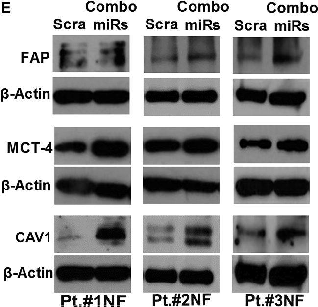
In the originally published version of this article, Figure 4E included incorrect β-actin blots in row 6 (Pt. #2) and row 4 (Pt. #3) and an incorrect MCT4 blot in row 3 (Pt. #3). The authors have replaced the incorrect blots in the revised version of Figure 4E. For additional clarification, the authors added text to the Figure 4E legend to disclose the reuse of β-actin blots that were derived from the same gels. These errors have been corrected online, and the authors regret these errors.
Figure 4. Differentially expressed miRNAs in fibroblasts upon exposure toMDA-MB-231 cell-derived exosomes (original).
(E) Western blot showing overexpression of FAP, Caveolin-1, and MCT4 proteins in NFs (pt. #1, #2, and #3) transfected with combo miRs compared with control (scrambled [Scra]) after 72 h
Figure 4. Differentially expressed miRNAs in fibroblasts upon exposure toMDA-MB-231 cell-derived exosomes (corrected).
(E) Western blot showing overexpression of FAP, Caveolin-1, and MCT4 proteins in NFs (pt. #1, #2, and #3) transfected with combo miRs compared with control (scrambled [Scra]) after 72 h. FAP and CAV-1 blots (pt. #1 and pt. #3) are from the same gel, so they have the same β-actin normalization. MCT-4 (pt.#2) and MMP3(pt.#2 [Fig.5E]) blots are from the same gel, so they have the same β-actin normalization. CAV1 (pt.#2) and p-FAK (Y576/577)/FAK (pt.#2 [Figure 6E]) blots are from the same gel, so they have the same β-actin normalization.


