Abstract
Breast cancer is the most common female tumors arising worldwide, and genetic and epigenetic events are constantly accumulated in breast tumorigenesis. The melastatin-related transient receptor potential 7 channel (TRPM7) is a nonselective cation channel, mainly maintaining Zn2+, Ca2+ and Mg2+ homeostasis. It is also involved in regulating proliferation and migration in various cancers including breast cancer. However, epigenetic alterations (such as promoter methylation) of TRPM7 and their correlation with clinical outcomes in breast cancer patients remain largely unclear. In this study, we found that TRPM7 was highly expressed in the luminal A subtype of breast cancers but no other subtypes compared with GTEx (Genotype-Tissue Expression Rad) or normal samples by analyzing the TCGA database. Correspondingly, TRPM7 was methylated in 42.7% (93 of 219) of breast cancers. Further studies found that promoter methylation of TRPM7 were significantly associated with better clinical outcomes in breast cancer patients, especially in the Luminal A subtype. Besides, methylated TRPM7 was correlated with less number of metastatic lymph nodes and longer local failure free survival time in this subtype. In summary, our data indicate that promoter methylation of TRPM7 may predict poor prognosis in patients with luminal A breast cancer.
Supplementary Information
The online version contains supplementary material available at 10.1186/s12885-022-10038-z.
Keywords: Breast cancer, TRPM7, Promotor methylation, Methylation-Specific PCR (MSP), Clinical outcomes
Introduction
Breast cancer (BC) is one of the leading cause cancers that affect women health all around the world [1].It is thus vital to gain a better understanding of the molecular mechanisms underlying the development of this disease [2]. In recent years, many researchers have been devoted to find and validate molecular alterations to serve as a prognostic and predictive biomarker. One of the validated and widely used multi-gene signature tests is the 21 genes, which is commonly applied for predicting the breast cancer outcomes [3, 4]. In addition, there are also studies demonstrating that epigenetic alterations, such as promoter hypermethylation or hypomethylation, can lead to aberrant gene expression in tumor cells [5, 6]. What’s more, the extension and functional importance of metabolic alterations and the associated genes also become a hot topic study in numerous cancers.
It is the fact that mammary microcalcifications occur in 30% to 50% of breast cancer patients, which are frequently associated with poor patient survival [7, 8]. Moreover, serum calcium (Ca2+) has been proved to be associated with the risk of breast cancer (9). Besides, calcium signals play important roles in tumorigenesis by interacting with and modulating tumor microenvironment [9, 10]. In breast cancer cells, intracellular Ca2+ is maintained via two classes of Ca2 + channels [11]. The transient receptor potential (TRP) super family of ion channels contains about thirty members, forming a non-selective cation permeable channels and is organized into seven subgroups according to their sequence homology [12, 13]. The transient receptor potential cation channel melastatin-subfamily (TRPM), composed of eight members from TRPM1 to TRPM8, is involved in various physiological functions including carcinogenesis [12].
The transient receptor potential cation channel melastatin-subfamily, member 7 (TRPM7, ChaK1, TRP-PLIK andLTRPC7), is ubiquitously expressed in many tissues [14, 15]. This protein is made up of 6 transmembrane domains and a COOH-terminal α-kinase domain [16]. The TRPM7 ion channel is involved in various physiological and pharmacological processes both with its channel activity and its kinase activity [15, 17]. The crucial role of TRPM7 in the balance of Zn(2 +), Mg(2 +), Ca(2 +) implicated in many metabolic processes and signalling pathways [18–21]. Through its varied physiological roles, TRPM7 is of major importance in much human pathology in various cancers, including breast cancer [22–24]. For example, in prostate cancer, the upregulation of TRPM7 enhanced the migration and invasion potential of cancer cells by promoting epithelial–mesenchymal transition (EMT) process [25, 26]. Similarly, knocking down TRPM7 in pancreatic cancer cells inhibited cancer cell migration [27]. For gastric cancer, several studies suggest that TRPM7 participates in the survival of human gastric adenocarcinoma cells by acting as detoxifiers [28–30].Also, there are studies suggesting that TRPM7 expression is significantly higher in invasive breast ductal carcinomas compared with control subjects, and demonstrating that TRPM7 is involve in regulating breast cancer cell proliferation, apoptosis, EMT, migration, invasion and microcalcification [31, 32]. Given that approximately 15%-20% of breast cancer patients diagnosed with triple negative breast cancer (TNBC) in the United States and lack appropriate targeted therapeutic drugs, chemical modulators of the TRPM7 channel might potentially be used in therapeutic applications [33, 34].
Considering that most previous studies focus on the expression of TRPM7 and its role in promoting malignant transformation of numerous cancers, the aims of the present study are to investigate the methylation status of TRPM7 in a cohort of breast cancer patients and its association with their clinicopathological features, better understanding the pathogenesis of breast cancer.
Patients and methods
Patients
With the approval of our institutional review board and human ethics committee, where required, a total of 219 paraffin-embedded breast cancer tissues were randomly obtained at the First Affiliated Hospital of Xi’an Jiaotong University. All individuals signed informed consent to participate in the study and familial cases of breast cancer were excluded. Tumor staging was performed according to the tumor, node, and metastasis (TNM) classification. In addition, patients who showed the breast and/or ovarian cancers in the first- and second-degree relatives were excluded from the study. Tumor samples were obtained following surgical resection and their pathological features were examined on macro dissection of the samples. All samples were histologically examined by a senior pathologist at Department of Pathology of the Hospital based on World Health Organization (WHO) criteria. The demographic and clinical features of the study population are summarized in Table 1.
Table 1.
Clinicopathological characteristics of breast cancer patients (n = 219)
| Characteristics | Percentage of Patients (n/n) |
Methylation frequency of TRPM7(n/n) |
|---|---|---|
| Age,years | ||
| Mean | 51.5 | n/a |
| SD | 11.5 | n/a |
| WHO grade | ||
| I | 3.2(7/219) | 28.6(2/7) |
| II | 74.4(163/219) | 45.4(74/163) |
| III | 22.4(49/219) | 34.7(17/49) |
| Molecular Subtype | ||
| Luminal A | 35.6(78/219) | 33.3(26/78) |
| Luminal B | 32.0(70/219) | 45.7(32/70) |
| Her2+ | 14.6(32/219) | 46.9(15/32) |
| Basal like | 17.8(39/219) | 51.3(20/39) |
| ER | ||
| - | 37.0(81/219) | 37.0(30/81) |
| + | 63.0 (138/219) | 45.6(63/138) |
| PR | ||
| - | 43.8(96/219) | 38.5(37/96) |
| + | 56.2(123/219) | 45.5(56/123) |
| Her2 | ||
| - | 78.1(171/219) | 45.0(77/171) |
| + | 21.9(48/219) | 33.3(16/48) |
| Lymph node metastasis (LNM) | ||
| No | 69.8(153/219) | 56.2(86/153) |
| Yes | 30.2(66/219) | 10.6(7/66)* |
| Radiotherapy | ||
| No | 51.6(113/219) | 54.0(61/113) |
| Yes | 48.4(106/219) | 30.2(32/106) |
| Chemotherapy | ||
| No | 14.2(31/219) | 54.8(17/31) |
| Yes | 85.8(188/219) | 40.4(76/188) |
| Recurrence | ||
| No | 84.9(186/219) | 44.6(83/186) |
| Yes | 15.1(33/219) | 30.3(10/33) * |
| Survival status | ||
| Alive | 82.6(181/219) | 45.3(82/181) |
| Dead | 17.4(38/219) | 28.9(11/38)* |
*stand for p <0.05
DNA extraction and bisulfite modification
As shown in our previous study [35]. All tissues sections were reviewed by board certified pathologists to ensure that ≥ 50% of the cells used for DNA purification were neoplastic. The tissues were first treated with xylene for 12 h at room temperature to remove the paraffin, and were then subjected to digestion with 1% sodium dodecylsulfate (SDS) and proteinase K at 48 °C for 48 to 72 h with the addition of several spiking aliquots of concentrated proteinase K to facilitate digestion. Next, genomic DNA was isolated from the digested tissues followed by a standard phenol–chloroform extraction and ethanol precipitation protocol. The samples were then stored at -80 °C until use. Quantity and quality of the extracted DNA were evaluated by a spectrophotometer (Nanodrop 2000, Thermo Scientific, USA). About 1 μg of genomic DNA was then treated with bisulfite to convert unmethylated cytosine to uracil prior to a methylation-specific PCR (MSP) using Epi Tect Bisulfite Kit (Qiagen, Germany) according to the manufacturer’s instructions.
MSP assay
The bisulfite-modified DNA was amplified using specific methylated or unmethylated primersas follow: 5’-TCGGTATAGGTTAGGTTTAGTTAGC-3’ (forward) and 5’-AAAATAATAAAATATTCACGCCGTA-3’ (reverse) for methylated gene; 5’-TGGTATAGGTTAGGTTTAGTTAGTGG-3’ (forward) and 5’-AAAATAATAAAATATTCACACCATA-3’ (reverse) for unmethylated gene. As shown in our previous study [35]. PCR amplification was then carried out in the buffer containing 16.6 mM ammonium sulfate, 67 mM Tris base, 2.5 mM MgCl2, 10 mM 2-mercaptoethanol, 0.1% DMSO, 0.2 mM each of dATP, dCTP, dGTP and dTTP, 600 nM each of forward and reverse primers and 0.6 unit Platinum Taq polymerase. Each sample was run in triplicate. The condition for TRPM7 amplification was 10 min at 95 °C, followed by amplification cycles including 30 s at 95 °C, 30 secs at 60 °C and 30 s at 72 °C for the extension, in addition to final elongation of 10 min at 72 °C. Normal leukocyte DNA was methylated in vitro with Sss I methylase (New England Biolabs, Beverly, MA) to generate completely methylated DNA as a positive control. After PCR, electrophoresis was performed on a 1.5% agarose gel.
Statistical analysis
Data were analyzed using SPSS 16.0 (SPSS Inc. Chicago, USA). As shown in our previous study [35].The Mann–Whitney U test and Kruskal–Wallis test were performed for numerical data and the Chi-square test was used to analyze the relationship between parameter data. Multivariate models were then developed that adjusted for the most important covariates, including age, tumor size, differentiation and lymph node metastasis. Survival length was determined from the day of primary tumor surgery to the day of death or last clinical follow-up. The Kaplan–Meier method was used for survival analysis grouping with methylation status of TRPM7. Differences between curves were analyzed using the log-rank test. Multivariate Cox regression analysis was used to evaluate the effect of gene methylation on survival of independently of the number of lymph node metastasis, tumor invasion and differentiation. Differences were considered statistically significant if P < 0.05.
Results
Different expression of TRPM7 is correlated with molecular subtype in breast cancers
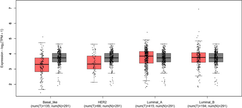
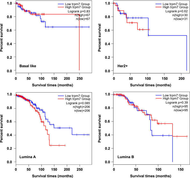
Previous studies have indicated that high level of TRPM7 proteins is correlated with shorter survival time in breast cancer after surgery. By analyzing mRNA level of TRPM7 in TCGA database using the online tools GEPIA (http://gepia2.cancer-pku.cn) (Fig. 1), we found that the expression of TRPM7 differed from the different subtype of breast cancer tissues. Although not significantly, the expression of TRPM7 in the Lumina A and Luminal B subtypes was higher, and was lower in the Basal-like and Her2 positive subtypes than either GTEx or normal samples (data from TCGA database). Next, we also found that high expression of TRPM7 was related to poor patient survival in the Lumina A subtype, although it did not reach statistical significance (Fig. 2). These observations, taken together, support that TRPM7 might act as an oncogene, and suggest that it may be a predictor for poor survival in patients with Lumina A breast cancers.
Fig. 1.
Expression of TRPM7 in breast cancer and normal tissues. In TCGA database, compared with GTEx (Genotype-Tissue Expression Rad), the expression of TRPM7 was lower in Basal like and Her2 + breast cancers. However, the expression of TRPM7 was higher in the Lumina A and Luminal B subtypes
Fig. 2.
High level of TRPM7 leads to poor prognosis of breast cancer patients. Expression of TRPM7 gene causes shorter survival times after surgery especially in the Lumina A subtype
Association of promoter methylation of TRPM7 with clinicopathological features in patients with breast cancers
Considering that the regulatory effect of promoter methylation on gene expression in human cancers [27, 28], we next examined promoter methylation of TRPM7 using MSP approach in a cohort of breast cancers and control subjects. As shown in Table 1, the methylated rate of TRPM7 was 42.7% (93/219) in the whole cohort. In consonance with TCGA database shown high expression of TRPM7 in the Lumina A subtype, we found the methylation frequency of TRPM7 was much lower in Lumina A subtype (33.3%) than that in others subtypes (51.3% in Basal-like, 46.9% in Her2+, and 45.7% in Luminal B). Moreover, we also found methylation frequency of TRPM7 was negatively correlated with lymph node metastasis, disease recurrence and cancer related death. However, no significant difference was seen between TRPM7 methylation with TNM, therapy strategies and hormone receptors.
We also examined the relationship of TRPM7 methylation status with clinicopathological characteristics using logistic regression analysis. The univariate analyses showed that TRPM7 methylation status was significantly associated with molecular subtypes (OR = 1.27; 95% CI = 1.00–1.63; P = 0.05), lymph node metastasis (OR = 0.09; 95% CI = 0.04–0.22; P = 0.001), and radiotherapy (OR = 0.37; 95% CI = 0.21–0.64; P = 0.001) (Table 2). Besides, although the difference did not reach statistical significance, the cancer related death also trended to be associated with TRPM7 methylation (OR = 0.49; 95% CI = 0.23–1.05; P = 0.06). In order to assess the independent association of gene methylation with molecular subtypes (including Basal like, Her2 positive, Luminal A and Luminal B), lymph node metastasis, endocrine therapy, radiotherapy, chemotherapy, relapse and cancer-related death, we conducted multiple multivariable logistic regression analysis. Also shown in Table 2 and Figure S1, TRPM7 methylation remained negatively associated with lymph node metastasis (OR = 0.09; 95% CI = 0.03–0.27; P = 0.001) and cancer related death (OR = 0.20; 95% CI = 0.03–1.21; p = 0.07).
Table 2.
TRPM7methylation in breast cancer: univariate and multivariate models with clinicopathological characteristics
| Characteristics | Univariate | Multivariate | ||
|---|---|---|---|---|
| ORa (95% CI) | P | ORa (95% CI) | P | |
| Ageb | 1.04 (0.84–1.28) | 0.75 | / | / |
| WHO gradec | 0.66 (0.37–1.18) | 0.16 | / | / |
| Molecular subtyped | 1.27 (1.00–1.63) | 0.05 | 1.34 (0.94–1.90) | 0.10 |
| LNMe | 0.09 (0.04–0.22) | 0.001 | 0.09 (0.03–0.27) | 0.001 |
| ER | 1.43 (0.81–2.50) | 0.21 | ||
| PR | 1.33 (0.77–2.29) | 0.30 | ||
| Her2 | 0.61 (0.31–1.20) | 0.15 | ||
| Endocrine therapy | 1.43 (0.81–2.50) | 0.21 | 1.45 (0.76–2.76) | 0.26 |
| Radiotherapy | 0.37 (0.21–0.64) | 0.001 | 0.94 (0.36–2.44) | 0.90 |
| Chemotherapy | 0.56 (0.26–1.20) | 0.14 | 0.09 (0.31–1.97) | 0.61 |
| Relapse | 0.63 (0.29–1.38) | 0.25 | 3.25 (0.49–21.58) | 0.22 |
| Survival statusf | 0.49 (0.23–1.05) | 0.06 | 0.20 (0.03–1.21) | 0.07 |
aOR: odds ratio with 95% confidence interval; bAge (per 10 years); cWHO grade (I, II, III and IV); dMolecular subtype (Luminal A, Luminal B, Her2 positive and Basal like); eLymph node metastasis; fSurvival status (alive vs. dead)
Next, we further examined the relationship of TRPM7 methylation with clinicopathological characteristics in patients with different molecular subtypes using logistic regression. As shown in Table 3, TRPM7 methylation was negatively associated with lymph node metastasis and radiotherapy in patients with Her2 positive (Figure S2) and Lumina A subtypes (Figure S3). In the Lumina B subtype (Figure S4), we similarly found that TRPM7 methylation was negatively associated with lymph node metastasis and radiotherapy, while positively associated with endocrine therapy. In the Basal like subtype, we failed to find the above relationships. Notably, our analysis showed that TRPM7 methylation trended to have a negative correlation with cancer-related death only in the Lumina A subtype (Table 3 and Figure S3).We then conducted multiple multivariable logistic regression analysis in Lumina A and Lumina B subtypes, and expectedly found that TRPM7methylationwas negatively associated with lymph node metastasis in Lumina A subtype, while positively associated with endocrine therapy in Lumina B subtype (Table 4).These findings further support the above conclusions.
Table 3.
Univariate analysis of TRPM7 methylation with clinicopathological characteristics in different subtypes of breast cancers
| Characteristics | Basal like | Her2 positive | Luminal A | Luminal B | ||||
|---|---|---|---|---|---|---|---|---|
| ORa (95% CI) | P | ORa (95% CI) | P | ORa (95% CI) | P | ORa (95% CI) | P | |
| Ageb | 0.98 (0.61–1.58) | 0.93 | 0.78 (0.42–1.46) | 0.44 | 0.80 (0.54–1.21) | 0.30 | 1.41 (0.98–2.05) | 0.07 |
| WHO gradec | 0.59 (0.15–2.34) | 0.45 | 0.32 (0.07–1.44) | 0.14 | 1.19 (0.44–3.27) | 0.73 | 0.47 (0.15–1.45) | 0.19 |
| LNMd | / | / | 0.08 (0.009–0.76) | 0.03 | 0.06 (0.007–0.47) | 0.008 | 0.19 (0.06–0.58) | 0.004 |
| ER | / | / | 0.57 (0.22–1.49) | 0.25 | 4.82 (1.62–14.36) | 0.005 | ||
| PR | / | / | 0.53 (0.20–1.38) | 0.19 | 3.51 (1.29–9.59) | 0.01 | ||
| Her2 | / | / | 1.87 (0.27–13.09) | 0.53 | 0.74 (0.25–2.21) | 0.59 | 0.46 (0.13–1.67) | 0.24 |
| Endocrine therapy | / | / | / | / | 0.57 (0.22–1.49) | 0.25 | 4.82 (1.62–14.36) | 0.005 |
| Radiotherapy | / | / | 0.05 (0.005–0.47) | 0.009 | 0.25 (0.08–0.82) | 0.02 | 0.18 (0.06–0.53) | 0.002 |
| Chemotherapy | 0.32 (0.03–3.33) | 0.34 | 0.88 (0.05–15.33) | 0.93 | 0.57 (0.18–1.75) | 0.33 | 0.37 (0.09–1.63) | 0.19 |
| Relapse | 0.54 (0.13–2.34) | 0.41 | 2.46 (0.20–30.28) | 0.48 | 0.26 (0.03–2.21) | 0.22 | 0.54 (0.15–1.98) | 0.35 |
| Survival status4 | 0.54 (0.13–2.34) | 0.41 | 0.54 (0.04–6.58) | 0.63 | 0.31 (0.06–1.52) | 0.15 | 0.54 (0.15–1.45) | 0.35 |
aOR: odds ratio with 95% confidence interval; bAge (per 10 years); 2WHO grade (I, II, III and IV); cLymph node metastasis; dSurvival status (alive vs. dead)
Table 4.
Multivariate analysis of TRPM7 methylation with clinicopathological characteristics in Luminal A and B breast cancers
| Characteristics | Luminal A | Luminal B | ||
|---|---|---|---|---|
| ORa (95% CI) | P | ORa (95% CI) | P | |
| Ageb | 0.91 (0.58–1.43) | 0.68 | 1.57 (0.95–2.59) | 0.08 |
| LNMc | 0.03 (0.001–0.59) | 0.02 | 0.55 (0.07–4.63) | 0.59 |
| Endocrine therapy | 0.60 (0.19–1.83) | 0.37 | 8.49 (2.10–34.29) | 0.003 |
| Radiotherapy | 2.62 (0.22–31.59) | 0.45 | 0.27 (0.03–2.20) | 0.22 |
| Chemotherapy | 0.65 (0.18–2.38) | 0.51 | 2.65 (0.40–17.55) | 0.31 |
| Survival statusd | 0.26 (0.05–1.36) | 0.11 | 0.56 (0.11–2.76) | 0.48 |
aOR: odds ratio with 95% confidence interval; bAge (per 10 years); cLymph node metastasis; dSurvival status (alive vs. dead)
Promoter methylation ofTRPM7 predicts better prognosis in patient with Lumina A breast cancers
Whether TRPM7 methylation predicts a better survival in patients with Lumina A breast cancers, as suggested by its association with clinicopathological characteristics of this subtype of breast cancer patients, was subsequently investigated by univariate and multivariable survival analysis. As shown in Table 5, both univariate (Figure S5) and multivariate Cox regression showed that TRPM7 methylation was a potential predictor of better survival for the whole cohort breast cancer patients. Next, we analyze the methylated status of TRPM7 in the different molecular subtypes. As shown in Table 6, TRPM7 methylation was only associated with better prognosis in the Lumina A subtype. Cox multivariate regression showed that TRPM7 methylation (HR = 0.13, 95% CI = 0.02–0.72, P = 0.02) and endocrine therapy (HR = 0.03, 95% CI = 1.10–1.06, P = 0.06) is a predictor of better survival in Lumina A patients as an independently variable with respect to the age, LNM, radiotherapy and chemotherapy (Table 7 and Figure S6).
Table 5.
Prognostic value of clinicopathological factors and TRPM7 methylation using univariate and multivariate Cox regression analysis
| Characteristics | Univariate | Multivariate | ||
|---|---|---|---|---|
| HRa (95% CI) | P | HRa (95% CI) | P | |
| TRPM7 methylation | 0.50 (0.25–1.01) | 0.06 | 0.50 (0.23–1.08) | 0.07 |
| Ageb | 1.10 (0.86–1.40) | 0.45 | 1.15 (0.88–1.52) | 0.31 |
| WHO gradec | 1.12 (0.56–2.23) | 0.75 | / | |
| Molecular subtyped | 1.14 (0.86–1.53) | 0.36 | 1.15 (0.81–1.63) | 0.45 |
| LNMe | 1.28 (0.66–2.48) | 0.46 | 0.91 (0.31–2.65) | 0.86 |
| Endocrine therapy | 0.63 (0.33–1.18) | 0.15 | 0.69 (0.36–1.32) | 0.26 |
| Radiotherapy | 1.32 (0.69–2.51) | 0.39 | 1.00 (0.34–2.97) | 0.99 |
| Chemotherapy | 1.48 (0.53–4.19) | 0.45 | 1.46 (0.46–4.64) | 0.52 |
| Relapse | 83.01 (35.58–212.77) | 0.001 | / | |
aHR: hazard ratio with 95% confidence interval (CI); bAge (per 10 years); cWHO grade (I, II, III and IV); dMolecular subtypes (Luminal A, Luminal B, Her2 positive and Basal like); eLymph node metastasis
Table 6.
Prognostic value of clinicopathological factors and TRPM7 methylation using univariate Cox regression analysis in different subtypes of breast cancers
| Characteristics | Basal like | Her2 positive | Luminal A | Luminal B | ||||
|---|---|---|---|---|---|---|---|---|
| HRa (95% CI) | P | HRa (95% CI) | P | HRa (95% CI) | P | HRa (95% CI) | P | |
| TRPM7 methylation | 0.69 (0.20–2.45) | 0.57 | 0.53 (0.05–5.88) | 0.61 | 0.26 (0.06–1.18) | 0.08 | 0.58 (0.18–1.94) | 0.38 |
| Ageb | 1.20 (0.74–1.93) | 0.46 | 1.56 (0.63–3.85) | 0.34 | 0.98 (0.62–1.55) | 0.94 | 1.04 (0.69–1.58) | 0.85 |
| WHO gradec | 0.89 (0.21–3.79) | 0.88 | 1.22 (0.13–11.47) | 0.86 | 0.53 (0.14–2.05) | 0.36 | 2.29 (0.73–7.20) | 0.16 |
| LNMd | 2.63 (0.76–9.09) | 0.13 | 4.65 (0.42–52.04) | 0.21 | 0.65 (0.18–2.39) | 0.52 | 0.88 (0.27–2.94) | 0.84 |
| Endocrine therapy | / | / | / | / | 0.45 (0.15–1.33) | 0.15 | 0.60 (0.19–1.87) | 0.38 |
| Radiotherapy | / | / | 3.77 (0.34–41.71) | 0.28 | 0.50 (0.14–1.83) | 0.29 | 1.28 (0.41–3.98) | 0.66 |
| Chemotherapy | 0.84 (0.11–6.68) | 0.87 | / | / | 1.65 (0.36–7.48) | 0.52 | 1.63 (0.21–12.61) | 0.64 |
aHR: hazard ratio with 95% confidence interval (CI); bAge (per 10 years); cWHO grade (I, II, III and IV); dLymph node metastasis
Table 7.
Prognostic value of clinicopathological factors and TRPM7 methylation using multivariate Cox regression in Luminal A
| Characteristics | HRa (95% CI) | P |
|---|---|---|
| TRPM7 methylation | 0.13 (0.02–0.72) | 0.02 |
| Ageb | 1.04 (0.64–1.69) | 0.89 |
| LNMc | / | 0.95 |
| Endocrine therapy | 0.33 (0.10–1.06) | 0.06 |
| Radiotherapy | / | 0.94 |
| Chemotherapy | 1.84 (0.36–9.43) | 0.46 |
aHR: hazard ratio with 95% confidence interval (CI); bAge (per 10 years);cLymph node metastasis
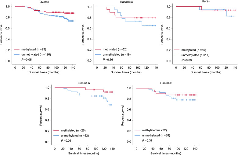
The Kaplan–Meier estimator of the survivorship function is generally used to evaluate the impact of aberrant gene methylation on the survival of breast cancer patients. In the present study, we found that high expression of TRPM7 trended to be associated with poor survival only in the Lumina A breast cancer patients by analyzing TCGA database (Fig. 2).This was also supported by our data showing a negative relationship betweenTRPM7methylation with survival times after surgery (median survival time: 133.3 vs 125.1 months; P = 0.05) in our cohort (Fig. 3). Further analysis showed that TRPM7 methylation was negatively associated with poor survival only in patients with Lumina A subtype, but not in other subtypes, further supporting the above conclusion. Interestingly, TRPM7 methylation also trended to predict longer local failure free survival time in this subtype (Fig. 3). Median time before disease recurrence was 129.2 vs 111.7 months in the whole cohort, while no disease recurrence was seen in the methylated TRPM7 group in in the Lumina Asubtype. These results, taken together, suggest that TRPM7 methylation may predict better prognosis in patients with luminal A breast cancers.
Fig. 3.
Methylation of TRPM7 predicted longer survival time in Lumina A breast cancer patients. The Kaplan–Meier estimator of the survivorship function was used to evaluate the impact of aberrant methylation of TRPM7 in the whole cohort breast cancer patients and the different molecular subtype cancers
Discussion
Cancer is still a leading cause of death worldwide and the incidence and mortality is growing rapidly due to the increased life expectancy and lifestyle issues [36].The complexity of breast cancer development and the recurrence of this disease spur the researchers for developing new targets for breast cancer diagnosis and treatment [37–39]. Previous studies have shown that TRPM7 might be considered as a potential target for breast cancer treatment [29].
Numerous studies have shown that the TRP ion channels family genes now consists of more than 30 candidates, most of which are permeable for Zn2+, Ca2+, and some also for Mg2+ [18–22, 40, 41]. TRPM7 and TRPM8 has proved act as oncogenes in breast cancer tissues compared with normal tissues, and is correlated with the Scarff-Bloom-Richardson (SBR) grade, Ki67 and tumor size [31]. TRPM7 can also participate in cancer cell adhesion and migration via myosin-IIA filament and the MAPK signaling pathways. Besides, it can also affect protein localization by phosphorylating the heavy chain [32, 42].
Although the molecular function of TRPM7 in breast cancer cells has been widely studied, promoter methylation of TRPM7 in breast cancers and its correlationship with clinicopathological characteristics of patients remains largely unclear. In the present study, we investigated promoter methylation of TRPM7 in a cohort of breast cancers. Firstly, we analyzed the expression of TRPM7 using TCGA database, and found that its expression was higher in breast cancers than that in control subjects. In addition, we also observed that increased expression of TRPM7 predict poor survival in Lumina A breast cancer patients. We then examined promoter methylation of TRPM7 using MSP approach in a cohort of breast cancers and control subjects, and found the methylation frequency of TRPM7 was 42.7% in the whole cohort. However, different molecular subtypes seem differs in the methylation rate. Next, we further examined the relationship of TRPM7 methylation with clinicopathological characteristics, and found that TRPM7methylation was found to be significantly associated with molecular subtypes, lymph node metastasis, disease recurrence and cancer related death. In addition, univariate and multivariate Cox regression showed that TRPM7 methylation served as a predictor of better survival in Lumina A breast cancer patients. The Kaplan–Meier estimator of the survivorship function also indicated that TRPM7 methylation was negatively associated with longer survival time of breast cancer patients either after surgery or before disease recurrence.
Conclusion
In conclusion, this is the first study which investigates the methylation status of TRPM7 gene in breast cancers, providing the evidences for a possible pathogenic role of TRPM7 in the development of breast cancer and suggesting that TRPM7 methylation predicts a better patient prognosis in patients with Lumina A breast cancers. However, the limitation of our study is that we do not determine the reason why methylation frequency of TRPM7 varies in different subtypes of breast cancer, and the relationship between TRPM7 methylation and gene expression status.
Supplementary Information
Additional file 1: Figure S1. Univariate analysis of TRPM7 methylation with clinicopathological characteristics in breast cancers. TRPM7 methylation negatively associated with lymph node metastasis and cancer related death.
Additional file 2: Figure S2. Univariate analysis of TRPM7 methylation with clinicopathological characteristics in Her2 positive breast cancers. TRPM7 methylation was negatively associated with lymph node metastasis and radiotherapy in patients with Her2 positive cancer patients.
Additional file 3: Figure S3. Univariate analysis of TRPM7 methylation with clinicopathological characteristics in Luminal A breast cancers. TRPM7 methylation was negatively associated with lymph node metastasis and radiotherapy in patients with Liminal A cancer patients.
Additional file 4: Figure S4. Univariate analysis of TRPM7 methylation with clinicopathological characteristics in Luminal B breast cancers. TRPM7 methylation was negatively associated with lymph node metastasis and radiotherapy, while positively associated with endocrine therapy in Luminal B breast cancers.
Additional file 5: Figure S5. Prognostic value of clinicopathological factors and TRPM7 methylation using univariate Cox regression analysis in breast cancers. TRPM7 methylation was a potential predictor of better survival for the whole cohort breast cancer patients.
Additional file 6: Figure S6. Prognostic value of clinicopathological factors and TRPM7 methylation using multivariate Cox regression analysis in Luminal A breast cancers. Cox multivariate regression showed that TRPM7 methylation and endocrine therapy is a predictor of better survival in Lumina A patients as an independently variable with respect to the age, LNM, radiotherapy and chemotherapy.
Acknowledgements
Not applicable.
Authors’ contributions
YQ and PH conceived and designed this study. YW and PC performed experimental work. RL and RC analyzed the data. YQ and PH produced the text and the figures. MJ and XZ collected the patient data. All authors read and approved the final manuscript.
Funding
This work was supported by the Nature Science Foundation of Shaanxi (No. 2020JQ-503) and Fundamental Research Funds for the Central Universities (No.xzy012019092).
Availability of data and materials
All data generated or analyzed during this study are included in this article. Further enquiries can be directed to the corresponding author.
Declarations
Ethics approval and consent to participate
This study was approved by the Ethics Committee of the First Affiliated Hospital of Xi'an Jiaotong University. All methods in the study were performed in accordance with relevant institutional/national/international guidelines. All individuals signed informed consent to participate in the study.
Consent for publication
Not applicable.
Competing interests
All authors contributed to and approved the final manuscript. The authors declare no conflict of interest.
Footnotes
Publisher’s Note
Springer Nature remains neutral with regard to jurisdictional claims in published maps and institutional affiliations.
Contributor Information
Peng Hou, Email: phou@xjtu.edu.cn.
Yiping Qu, Email: 15398036489@163.com.
References
- 1.Siegel RL, Miller KD, Fuchs HE, Jemal A. Cancer statistics, 2022. CA Cancer J Clin. 2022;72(1):7–33. doi: 10.3322/caac.21708. [DOI] [PubMed] [Google Scholar]
- 2.Tsang JYS, Tse GM. Molecular Classification of Breast Cancer. Adv Anat Pathol. 2020;27(1):27–35. doi: 10.1097/PAP.0000000000000232. [DOI] [PubMed] [Google Scholar]
- 3.Chen M, Liu D, Chen W, Chen W, Shen K, Wu J, Zhu L. Impact of Different Modules of 21-Gene Assay in Early Breast Cancer Patients. Front Endocrinol. 2021;12:759338. doi: 10.3389/fendo.2021.759338. [DOI] [PMC free article] [PubMed] [Google Scholar]
- 4.Masucci L, Torres S, Eisen A, Trudeau M, Tyono I, Saunders H, Chan KW, Isaranuwatchai W. Cost-utility analysis of 21-gene assay for node-positive early breast cancer. Current oncology (Toronto, Ont) 2019;26(5):307–318. doi: 10.3747/co.26.4769. [DOI] [PMC free article] [PubMed] [Google Scholar]
- 5.Jin L, Han B, Siegel E, Cui Y, Giuliano A, Cui X. Breast cancer lung metastasis: Molecular biology and therapeutic implications. Cancer Biol Ther. 2018;19(10):858–868. doi: 10.1080/15384047.2018.1456599. [DOI] [PMC free article] [PubMed] [Google Scholar]
- 6.Pedrosa R, Mustafa DA, Soffietti R, Kros JM. Breast cancer brain metastasis: molecular mechanisms and directions for treatment. Neuro Oncol. 2018;20(11):1439–1449. doi: 10.1093/neuonc/noy044. [DOI] [PMC free article] [PubMed] [Google Scholar]
- 7.O'Grady S, Morgan MP. Microcalcifications in breast cancer: From pathophysiology to diagnosis and prognosis. Biochim Biophys Acta. 2018;1869(2):310–320. doi: 10.1016/j.bbcan.2018.04.006. [DOI] [PubMed] [Google Scholar]
- 8.Clemenceau A, Michou L, Diorio C, Durocher F. Breast Cancer and Microcalcifications: An Osteoimmunological Disorder? Int J Mol Sci. 2020;21(22):8613. [DOI] [PMC free article] [PubMed]
- 9.O'Grady S, Morgan MP. Calcium transport and signalling in breast cancer: Functional and prognostic significance. Semin Cancer Biol. 2021;72:19–26. doi: 10.1016/j.semcancer.2019.12.006. [DOI] [PubMed] [Google Scholar]
- 10.Azimi I, Roberts-Thomson SJ, Monteith GR. Calcium influx pathways in breast cancer: opportunities for pharmacological intervention. Br J Pharmacol. 2014;171(4):945–960. doi: 10.1111/bph.12486. [DOI] [PMC free article] [PubMed] [Google Scholar]
- 11.Déliot N, Constantin B. Plasma membrane calcium channels in cancer: Alterations and consequences for cell proliferation and migration. Biochim Biophys Acta. 2015;1848(10):2512–2522. doi: 10.1016/j.bbamem.2015.06.009. [DOI] [PubMed] [Google Scholar]
- 12.Hantute-Ghesquier A, Haustrate A, Prevarskaya N, Lehen'kyi V. TRPM Family Channels in Cancer. Pharmaceuticals (Basel, Switzerland). 2018;11(2):58. [DOI] [PMC free article] [PubMed]
- 13.Schönherr R. Clinical relevance of ion channels for diagnosis and therapy of cancer. J Membr Biol. 2005;205(3):175–184. doi: 10.1007/s00232-005-0782-3. [DOI] [PubMed] [Google Scholar]
- 14.Zou ZG, Rios FJ, Montezano AC, Touyz RM. TRPM7, Magnesium, and Signaling. Int J Mol Sci. 2019;20(8):1877. [DOI] [PMC free article] [PubMed]
- 15.Meng S, Alanazi R, Ji D, Bandura J, Luo ZW, Fleig A, Feng ZP, Sun HS. Role of TRPM7 kinase in cancer. Cell Calcium. 2021;96:102400. doi: 10.1016/j.ceca.2021.102400. [DOI] [PubMed] [Google Scholar]
- 16.Huang Y, Fliegert R, Guse AH, Lü W, Du J. A structural overview of the ion channels of the TRPM family. Cell Calcium. 2020;85:102111. doi: 10.1016/j.ceca.2019.102111. [DOI] [PMC free article] [PubMed] [Google Scholar]
- 17.Visser D, Middelbeek J, van Leeuwen FN, Jalink K. Function and regulation of the channel-kinase TRPM7 in health and disease. Eur J Cell Biol. 2014;93(10–12):455–465. doi: 10.1016/j.ejcb.2014.07.001. [DOI] [PubMed] [Google Scholar]
- 18.Dhennin-Duthille I, Gautier M, Korichneva I, Ouadid-Ahidouch H. TRPM7 involvement in cancer: a potential prognostic factor. Magnes Res. 2014;27(3):103–112. doi: 10.1684/mrh.2014.0367. [DOI] [PubMed] [Google Scholar]
- 19.Kollewe A, Chubanov V, Tseung FT, Correia L, Schmidt E, Rössig A, Zierler S, Haupt A, Müller CS, Bildl W et al: The molecular appearance of native TRPM7 channel complexes identified by high-resolution proteomics. eLife 2021, 10. [DOI] [PMC free article] [PubMed]
- 20.Monteilh-Zoller MK, Hermosura MC, Nadler MJ, Scharenberg AM, Penner R, Fleig A. TRPM7 provides an ion channel mechanism for cellular entry of trace metal ions. J Gen Physiol. 2003;121(1):49–60. doi: 10.1085/jgp.20028740. [DOI] [PMC free article] [PubMed] [Google Scholar]
- 21.Schmidt E, Narangoda C, Nörenberg W, Egawa M, Rössig A, Leonhardt M, Schaefer M, Zierler S, Kurnikova MG, Gudermann T, et al. Structural mechanism of TRPM7 channel regulation by intracellular magnesium. Cellular and molecular life sciences : CMLS. 2022;79(5):225. doi: 10.1007/s00018-022-04192-7. [DOI] [PMC free article] [PubMed] [Google Scholar]
- 22.Mittermeier L, Demirkhanyan L, Stadlbauer B, Breit A, Recordati C, Hilgendorff A, Matsushita M, Braun A, Simmons DG, Zakharian E, et al. TRPM7 is the central gatekeeper of intestinal mineral absorption essential for postnatal survival. Proc Natl Acad Sci USA. 2019;116(10):4706–4715. doi: 10.1073/pnas.1810633116. [DOI] [PMC free article] [PubMed] [Google Scholar]
- 23.Yee NS. Role of TRPM7 in Cancer: Potential as Molecular Biomarker and Therapeutic Target. Pharmaceuticals (Basel, Switzerland). 2017;10(2):39. [DOI] [PMC free article] [PubMed]
- 24.Park HS, Hong C, Kim BJ, So I. The Pathophysiologic Roles of TRPM7 Channel. Korean J Physiol Pharmacol. 2014;18(1):15–23. doi: 10.4196/kjpp.2014.18.1.15. [DOI] [PMC free article] [PubMed] [Google Scholar]
- 25.Chen L, Cao R, Wang G, Yuan L, Qian G, Guo Z, Wu CL, Wang X, Xiao Y. Downregulation of TRPM7 suppressed migration and invasion by regulating epithelial-mesenchymal transition in prostate cancer cells. Med Oncol. 2017;34(7):127. doi: 10.1007/s12032-017-0987-1. [DOI] [PubMed] [Google Scholar]
- 26.Yang F, Cai J, Zhan H, Situ J, Li W, Mao Y, Luo Y. Suppression of TRPM7 Inhibited Hypoxia-Induced Migration and Invasion of Androgen-Independent Prostate Cancer Cells by Enhancing RACK1-Mediated Degradation of HIF-1α. Oxid Med Cell Longev. 2020;2020:6724810. doi: 10.1155/2020/6724810. [DOI] [PMC free article] [PubMed] [Google Scholar]
- 27.Yee NS, Kazi AA, Li Q, Yang Z, Berg A, Yee RK. Aberrant over-expression of TRPM7 ion channels in pancreatic cancer: required for cancer cell invasion and implicated in tumor growth and metastasis. Biology open. 2015;4(4):507–514. doi: 10.1242/bio.20137088. [DOI] [PMC free article] [PubMed] [Google Scholar]
- 28.Kim BJ, Hong C. Role of transient receptor potential melastatin type 7 channel in gastric cancer. Integrative medicine research. 2016;5(2):124–130. doi: 10.1016/j.imr.2016.04.004. [DOI] [PMC free article] [PubMed] [Google Scholar]
- 29.Kim BJ, Kim SY, Lee S, Jeon JH, Matsui H, Kwon YK, Kim SJ, So I. The role of transient receptor potential channel blockers in human gastric cancer cell viability. Can J Physiol Pharmacol. 2012;90(2):175–186. doi: 10.1139/y11-114. [DOI] [PubMed] [Google Scholar]
- 30.Calik M, Calik I, Artas G, Ozercan IH. Prognostic Value of TRPM7 Expression and Factor XIIIa-Expressing Tumor-Associated Macrophages in Gastric Cancer. Gastroenterology research and practice. 2021;2021:7249726. doi: 10.1155/2021/7249726. [DOI] [PMC free article] [PubMed] [Google Scholar]
- 31.Dhennin-Duthille I, Gautier M, Faouzi M, Guilbert A, Brevet M, Vaudry D, Ahidouch A, Sevestre H, Ouadid-Ahidouch H. High expression of transient receptor potential channels in human breast cancer epithelial cells and tissues: correlation with pathological parameters. Cell Physiol Biochem. 2011;28(5):813–822. doi: 10.1159/000335795. [DOI] [PubMed] [Google Scholar]
- 32.Meng X, Cai C, Wu J, Cai S, Ye C, Chen H, Yang Z, Zeng H, Shen Q, Zou F. TRPM7 mediates breast cancer cell migration and invasion through the MAPK pathway. Cancer Lett. 2013;333(1):96–102. doi: 10.1016/j.canlet.2013.01.031. [DOI] [PubMed] [Google Scholar]
- 33.Cordier C, Prevarskaya N, Lehen'kyi V. TRPM7 Ion Channel: Oncogenic Roles and Therapeutic Potential in Breast Cancer. Cancers. 2021;13(24):6322. [DOI] [PMC free article] [PubMed]
- 34.Song C, Choi S, Oh KB, Sim T. Suppression of TRPM7 enhances TRAIL-induced apoptosis in triple-negative breast cancer cells. J Cell Physiol. 2020;235(12):10037–10050. doi: 10.1002/jcp.29820. [DOI] [PubMed] [Google Scholar]
- 35.Cui R, Chen P, Wang Y, Lu R, Ji M, Hou P, Qu Y. Cohesin RAD21 Gene Promoter Methylation Correlated with Better Prognosis in Breast Cancer Patients. Cytogenetic Genome Res. 2022:1–10 (Online ahead of print). [DOI] [PubMed]
- 36.Xia C, Dong X, Li H, Cao M, Sun D, He S, Yang F, Yan X, Zhang S, Li N, et al. Cancer statistics in China and United States, 2022: profiles, trends, and determinants. Chin Med J. 2022;135(5):584–590. doi: 10.1097/CM9.0000000000002108. [DOI] [PMC free article] [PubMed] [Google Scholar]
- 37.Kirkham AA, Gelmon KA, Van Patten CL, Bland KA, Wollmann H, McKenzie DC, Landry T, Campbell KL. Impact of Exercise on Chemotherapy Tolerance and Survival in Early-Stage Breast Cancer: A Nonrandomized Controlled Trial. J Natl Compr Canc Netw. 2020;18(12):1670–1677. doi: 10.6004/jnccn.2020.7603. [DOI] [PubMed] [Google Scholar]
- 38.Delgado-Ramos GM, Nasir SS, Wang J, Schwartzberg LS. Real-world evaluation of effectiveness and tolerance of chemotherapy for early-stage breast cancer in older women. Breast Cancer Res Treat. 2020;182(2):247–258. doi: 10.1007/s10549-020-05684-5. [DOI] [PubMed] [Google Scholar]
- 39.Vrbic S, Pejcic I, Filipovic S, Kocic B, Vrbic M. Current and future anti-HER2 therapy in breast cancer. J Buon. 2013;18(1):4–16. [PubMed] [Google Scholar]
- 40.Li H. TRP Channel Classification. Adv Exp Med Biol. 2017;976:1–8. doi: 10.1007/978-94-024-1088-4_1. [DOI] [PubMed] [Google Scholar]
- 41.Moran MM. TRP Channels as Potential Drug Targets. Annu Rev Pharmacol Toxicol. 2018;58:309–330. doi: 10.1146/annurev-pharmtox-010617-052832. [DOI] [PubMed] [Google Scholar]
- 42.Guilbert A, Gautier M, Dhennin-Duthille I, Rybarczyk P, Sahni J, Sevestre H, Scharenberg AM, Ouadid-Ahidouch H. Transient receptor potential melastatin 7 is involved in oestrogen receptor-negative metastatic breast cancer cells migration through its kinase domain. Eur J Cancer. 2013;49(17):3694–3707. doi: 10.1016/j.ejca.2013.07.008. [DOI] [PubMed] [Google Scholar]
Associated Data
This section collects any data citations, data availability statements, or supplementary materials included in this article.
Supplementary Materials
Additional file 1: Figure S1. Univariate analysis of TRPM7 methylation with clinicopathological characteristics in breast cancers. TRPM7 methylation negatively associated with lymph node metastasis and cancer related death.
Additional file 2: Figure S2. Univariate analysis of TRPM7 methylation with clinicopathological characteristics in Her2 positive breast cancers. TRPM7 methylation was negatively associated with lymph node metastasis and radiotherapy in patients with Her2 positive cancer patients.
Additional file 3: Figure S3. Univariate analysis of TRPM7 methylation with clinicopathological characteristics in Luminal A breast cancers. TRPM7 methylation was negatively associated with lymph node metastasis and radiotherapy in patients with Liminal A cancer patients.
Additional file 4: Figure S4. Univariate analysis of TRPM7 methylation with clinicopathological characteristics in Luminal B breast cancers. TRPM7 methylation was negatively associated with lymph node metastasis and radiotherapy, while positively associated with endocrine therapy in Luminal B breast cancers.
Additional file 5: Figure S5. Prognostic value of clinicopathological factors and TRPM7 methylation using univariate Cox regression analysis in breast cancers. TRPM7 methylation was a potential predictor of better survival for the whole cohort breast cancer patients.
Additional file 6: Figure S6. Prognostic value of clinicopathological factors and TRPM7 methylation using multivariate Cox regression analysis in Luminal A breast cancers. Cox multivariate regression showed that TRPM7 methylation and endocrine therapy is a predictor of better survival in Lumina A patients as an independently variable with respect to the age, LNM, radiotherapy and chemotherapy.
Data Availability Statement
All data generated or analyzed during this study are included in this article. Further enquiries can be directed to the corresponding author.


