Abstract
Mitochondrial encephalomyopathy, lactic acidosis, and stroke-like episodes syndrome is a multisystem, progressive neurodegenerative condition, and the most common mitochondrial cytopathy. While not a primary characteristic, sensorineural hearing loss is a common additional symptom reported in up to 78% of cases. This article presents 2 cases of cochlear implantation in patients with mitochondrial encephalomyopathy, lactic acidosis, and stroke-like episodes syndrome. Both cases demonstrated significantly improved speech recognition, with results significantly better than previous case reports. Cochlear implants are an appropriate treatment for severe-profound hearing loss in mitochondrial encephalomyopathy, lactic acidosis, and stroke-like episodes syndrome. While anesthetic risks and cognitive skills need to be taken into consideration, routine programming and rehabilitation pathways may be appropriate for this cohort.
Keywords: Cochlear implants, genetic deafness, sensorineural hearing loss
Introduction
Mitochondrial encephalomyopathy, lactic acidosis, and stroke-like episodes (MELAS) syndrome is a multisystem, progressive neurodegenerative condition characterized by myelopathy, encephalopathy, lactic acidosis, and stroke-like episodes. Additional symptoms include sensorineural hearing loss (SNHL), short stature, cardiomyopathy, diabetes mellitus, headaches, fatigue, muscle weakness, and respiratory problems.1 Mitochondrial encephalomyopathy, lactic acidosis, and stroke-like episodes result from mitochondrial DNA variants with the m.3243A>G and m.3271T>C variants accounting for 80% and 10% of the patients, respectively.2,3 Sensorineural hearing loss in MELAS has a reported incidence between 27% and 78%, with approximately 20% severe-profound.4 The progressive nature of hearing loss is related to the severity of the mitochondrial disorder and the accumulation of mutated mtDNA within the cochlea. Hearing loss occurs as the result of deficient intracellular adenosine triphosphate energy release within the stria vascularis and the cochlear hair cells.4,5 Structural lesions have been reported in MELAS, with imaging studies reporting focal lesions affecting the auditory pathway, the occipital and parietal lobes, and the cerebellum.5 Hearing aids are the primary treatment for SNHL; however, where individuals no longer gain sufficient benefit from amplification, cochlear implantation may be suitable. Scarpelli et al6 suggested that “patients with mitochondrial disease are ideal recipients of a cochlear implant because the hearing loss develops well after speech development.”
Case Presentation
Case 1
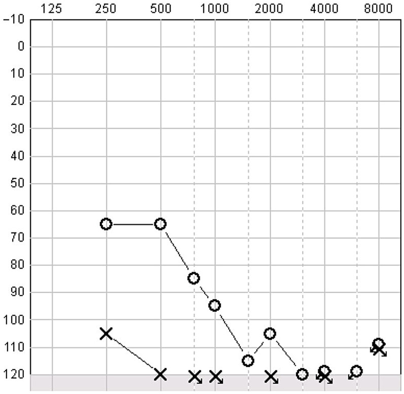
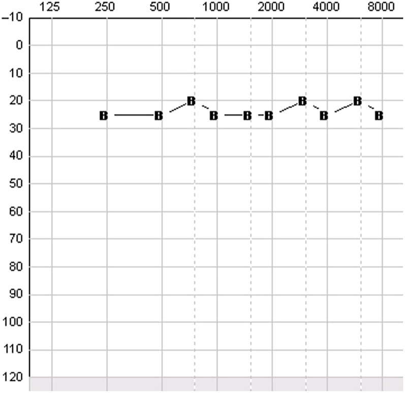
Case 1 is a 53-year-old male with progressive bilateral hearing loss, first noticed when aged 28. He had worn hearing aids bilaterally since age 33 but stopped using the left hearing aid due to poor benefit. Genetic testing confirmed MELAS with the m.3243 A>G variant. Pure tone audiometry showed a left profound loss and right severe-profound high-frequency sloping loss (Figure 1). Aided speech testing scored 2% & 32% for female and male voices, respectively, using Bamford–Kowal–Bench (BKB) sentences at 70 dB. The patient was implanted with a right CI522. Programming sessions followed routine protocols and manufacturer-recommended parameters (Cochlear N7: ACE, MP1+2, Rate 900, Maxima 8, Pulse Width 37) with behaviorally set threshold and comfort levels. After 1-year of implantation, he reported great satisfaction with his hearing and was able to listen to and enjoy music. Aided sound field-testing showed good thresholds (Figure 2). Auditory speech sound evaluation phoneme discrimination was excellent (20/20—100%). Speech recognition was assessed 4 months post-implantation and was scored 80% (female BKB auditory was only at 70 dB).
Figure 1.
Pure Tone Audiogram, Case 1.
Figure 2.
1 year post implant, Aided Sound Field Testing, Case 1.
Case 2
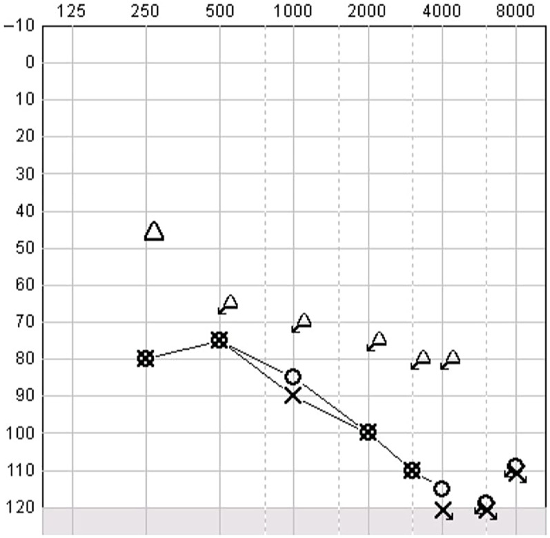
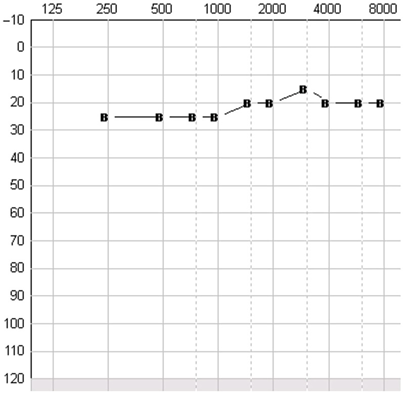
Case 2 is a 47-year-old male who first noticed progressive hearing loss in his mid-20s. He had consistently worn hearing aids since diagnosis. Mitochondrial encephalomyopathy, lactic acidosis, and stroke-like episodes syndrome was confirmed by genetic testing (variant unknown). Pure tone audiometry showed bilateral severe-profound high-frequency sloping loss (Figure 3). Aided auditory and aided speech testing scored 26% (right only), 26% (left only), and 42% (bilateral aids) (70 dB female BKB sentences in quiet). The patient was implanted with a left advanced bionics ultra mid scala electrode. Programming sessions followed routine protocols and manufacturer-recommended parameters (Naida Q90: HiRes Optima P, APW2 18us 3712pps) with behaviorally set threshold and most comfortable levels. After only 2 weeks, optimal settings were found, which the patient has successfully used for over 2 years. After 1-year of implantation, he reported being “very content,” was able to use the phone, and enjoyed both familiar and unfamiliar music. Aided sound field-testing showed good thresholds (Figure 4). Auditory speech sound evaluation phoneme discrimination was excellent (20/20—100%). Aided speech testing at 70 dB had significantly improved: BKB female in quiet 94%, male in quiet 98%, female in noise 100%, and male voice in noise 98%. These results were maintained for 2 years post-implantation.
Figure 3.
Pure Tone Audiogram, Case 2.
Figure 4.
1 year post implant, aided Sound Field Testing, Case 2.
Written informed consent was obtained from all participants who participated in this study.
Discussion
The number of cases of cochlear implantation in MELAS reported in the literature is low. The previous cases in the literature fail to provide adequate information for the effective comparison between cases. Three did not provide pre-implant speech outcomes.6-8 Two did not provide post-implant outcomes.8,9 Where post-implant outcomes are provided, 3 provide only qualitative statements of benefit.6,7,9 In both presented cases, results were acquired through routine programming protocols, and both patients rapidly settled on optimized mapping with excellent threshold and speech discrimination outcomes. The speech outcomes were better than those reported in the studies of Rosenthal et al10 and Yasumura et al.1 Post-implantation-aided sound field thresholds compare favorably to those reported by Rosenthal et al10 and Karkos et al.9 Despite significant central nervous system degeneration associated with MELAS Rosenthal et al10 reports that higher auditory pathways may be preserved. No specific tests for retro-cochlear pathology were performed in our case reports, but successful outcomes maintained over the follow-up period are more consistent with cochlear pathology. Cognitive impairment may limit auditory rehabilitation, and Chinnery et al5 suggest that “it may not be prudent to invest in a cochlear implant in a patient with very poor prognosis from the outset.” Our 2 cases followed normal rehabilitation plans and were able to complete all tasks allocated with ease.
Conclusion
Cochlear implantation is the appropriate treatment for severe hearing loss in MELAS. While anesthetic risks and cognitive skills need to be taken into consideration, routine programming and rehabilitation pathways may be appropriate for this cohort.
Funding Statement
The authors declared that this study has received no financial support.
Footnotes
Informed Consent: Written informed consent was obtained from all participants who participated in this study.
Peer-review: Externally peer-reviewed.
Author Contributions: Concept – G.C.; Design – G.C.; Supervision – G.C., M.B.; Resources – G.C.; Materials – G.C.; Data Collection and/or Processing – G.C.; Analysis and/or Interpretation – G.C., M.B., P.K.; Literature Search – G.C., P.K.; Writing Manuscript – G.C.; Critical Review – G.C., M.B., P.K.
Conflict of Interest: The authors have no conflicts of interest to declare.
References
- 1. Yasumura Y, Aso S, Fujisaka M, Watanabe Y. Cochlear implantation in a patient with mitochondrial encephalomyopathy, lactic acidosis and stroke-like episodes syndrome. Acta Otolaryngol. 2003;123:55–58.. [DOI] [PubMed] [Google Scholar]
- 2. Thompson VA, Wahr JA. Anesthetic considerations in patients presenting with mitochondrial myopathy, encephalopathy, lactic acidosis and stroke like episodes (MELAS) syndrome. Anesth Analg. 1997;85(6):1404–1406.. 10.1097/00000539-199712000-00041) [DOI] [PubMed] [Google Scholar]
- 3. Hougaard DD, Hestoy DH, Hojland AT, Gailhede M, Petersen MB. Audiological and vestibular findings in subjects with MELAS syndrome. J Int Adv Otol. 2019;15(2):296–303.. 10.5152/iao.2019.5913) [DOI] [PMC free article] [PubMed] [Google Scholar]
- 4. Handzel O, Ungar OJ, Lee DJ, Nadol JB. Temporal bone histopathology in MELAS syndrome. Laryngoscope Investig Otolaryngol. 2020;5(1):152–156.. 10.1002/lio2.344) [DOI] [PMC free article] [PubMed] [Google Scholar]
- 5. Chinnery PF, Elliott C, Green GR.et al. The spectrum of hearing loss due to mitochondrial DNA defects. Brain. 2000;123(1):82–92.. 10.1093/brain/123.1.82) [DOI] [PubMed] [Google Scholar]
- 6. Scarpelli M, Zappini F, Filosto M, Russignan A, Tonin P, Tomelleri G. Mitochondrial sensorineural hearing loss: a retrospective study and a description of cochlear implantation in a MELAS patient. Genet Res Int. 2012;2012:287432. 10.1155/2012/287432) [DOI] [PMC free article] [PubMed] [Google Scholar]
- 7. Sue CM, Lipsett LJ, Crimmins DS.et al. Cochlear origin of hearing loss in MELAS syndrome. Ann Neurol. 1998;43(3):350–359.. 10.1002/ana.410430313) [DOI] [PubMed] [Google Scholar]
- 8. Randhawa N, Wilson L, Mann S, Sirrs S, Benavente O. Clinical reasoning: a complicated case of MELAS. Neurology. 2016;87(16):e189–e195.. 10.1212/WNL.0000000000003222) [DOI] [PubMed] [Google Scholar]
- 9. Karkos, Anari, Johnson. Cochlear implantation in patients with MELAS syndrome. Eur Arch Oto-Rhino-Laryngol. 2004;262(4):322–324.. [DOI] [PubMed] [Google Scholar]
- 10. Rosenthal EL, Kileny PR, Boerst A, Telian SA. Successful cochlear implantation in a patient with MELAS syndrome. Am J Otol. 1999;20(2):187–90.. [PubMed] [Google Scholar]


 Content of this journal is licensed under a


