Abstract
Rickettsia prowazekii, the etiologic agent of epidemic typhus, is an obligate, intracytoplasmic, parasitic bacterium. Recently, the transformation of this bacterium via electroporation has been reported. However, in these studies identification of transformants was dependent upon either selection of an R. prowazekii rpoB chromosomal mutation imparting rifampin resistance or expression of the green fluorescent protein and flow cytometric analysis. In this paper we describe the expression in R. prowazekii of the Escherichia coli ereB gene. This gene codes for an erythromycin esterase that cleaves erythromycin. To the best of our knowledge, this is the first report of the expression of a nonrickettsial, antibiotic-selectable gene in R. prowazekii. The availability of a positive selection for rickettsial transformants is an important step in the characterization of genetic analysis systems in the rickettsiae.
Rickettsia prowazekii, the etiologic agent of epidemic typhus, is an obligate, intracellular, parasitic bacterium. Unlike the obligate, intracellular bacteria of the genera Chlamydia, Coxiella, and Ehrlichia, R. prowazekii grows only within the cytoplasm of the eucaryotic host cell rather than within an intracytoplasmic vesicle (9, 20, 22). Rickettsiae are capable of entering a wide range of eucaryotic cells by a process of induced phagocytosis and are well adapted to exploit the cytoplasmic environment (22). After reaching the cytoplasm they transport high-energy compounds, such as ATP, using specialized transport systems (21), but they also retain the ability to generate ATP via an intact tricarboxylic acid cycle and oxidative phosphorylation.
Recently, the genome sequence of R. prowazekii was published, providing a complete rickettsial genotype for analysis (1). In addition, progress in the development of techniques for the genetic manipulation of rickettsiae has been made. Rachek et al. (13) described the transformation of R. prowazekii to rifampin resistance via electroporation of a rickettsial rpoB gene containing a rifampin resistance mutation. Similarly, Troyer et al. (18) described the successful transformation of the closely related Rickettsia typhi, using green fluorescent protein to screen for transformants. Unfortunately, a method for the positive selection of R. prowazekii transformants, using an antibiotic resistance gene encoding a product that would inactivate or destroy the antibiotic, has not been described.
Erythromycin is a macrolide antibiotic that binds the procaryotic ribosome, inhibiting protein synthesis. R. prowazekii is sensitive to this drug in in vitro assays, and erythromycin can be used in the laboratory since it is not recommended for the treatment of R. prowazekii infections (14, 15). In clinical isolates ribosomal modification is the predominant mechanism of resistance to erythromycin, although active efflux of the drug and antibiotic inactivation mechanisms have been identified (10, 11, 19). One example of resistance resulting from the latter mechanism is the production of esterases that hydrolyze the lactone ring of the erythromycin molecule. Two genes (ereA and ereB) have been identified in Escherichia coli that code for such esterases (2, 3, 12). Interestingly, ereB exhibits a usage of A+T-rich codons, unusual for E. coli but similar to that of rickettsial genes (2). In this report we describe the use of ereB as a selectable marker in R. prowazekii transformation and demonstrate its insertion into a selected site of the R. prowazekii genome.
Bacterial strains, plasmids, and oligonucleotides used in this study are listed in Table 1. E. coli strains were grown on Luria-Bertani medium (4). When required for selection of E. coli transformants, the antibiotic ampicillin or erythromycin was added to a final concentration of 50 or 200 μg/ml, respectively. R. prowazekii Madrid E strain seed pool passage 282 was used for infecting mouse L929 fibroblasts. Rickettsia-infected L929 cells were grown in an atmosphere of 5% CO2 at 34°C in modified Eagle medium supplemented with 10% newborn calf serum (Sigma, St. Louis, Mo.) and 1 mM l-glutamine (Sigma). For selection, erythromycin (Fisher Scientific, Pittsburgh, Pa.) was added to supplemented modified Eagle medium at a final concentration of 200 ng/ml, and the erythromycin-containing medium was changed every 2 to 3 days. Rickettsial growth was monitored by microscopic examination of Gimenez-stained (8) infected cells growing on glass coverslips. All DNA manipulations were performed as described previously (13). PCR amplifications for detection of R. prowazekii and integration of the transforming plasmid into the rickettsial chromosome were performed with the oligonucleotide primers listed in Table 1. For DNA sequencing, the PCR products obtained were purified using a GeneClean II kit (Bio 101, La Jolla, Calif.) and sequenced directly using a ThermoSequenase cycle sequencing kit from Amersham Life Science, Inc. (Cleveland, Ohio). Probes used in Southern hybridizations (16) were 32P labeled using the Multiprime DNA labeling system (Amersham) and [α-32P]dATP (ICN, Irvine, Calif.).
TABLE 1.
Strains, plasmids, and oligonucleotides
| Strain, plasmid, or oligonucleotide | Description | Source and/or reference |
|---|---|---|
| Strains | ||
| R. prowazekii Madrid E | Ems | 7 |
| E. coli XL1-Blue | recA1 endA1 gvrA96 thi-1 hsdR17 supE44 relA1 lac [F′ proAB lacIqZΔM15 Tn10 (Tetr)] | Stratagene (5) |
| Plasmids | ||
| pMOB | Apr | Gold BioTechnology (17)a |
| pAT72 | Emr | 2 |
| pMW1041 | Aps Emr | This study |
| pMW264 | pBR322 with inserted 1,881-bp R. prowazekii EcoRV fragment containing the gltA gene | 23 |
| pMW1047 | pMW1041 with inserted 1,881-bp EcoRV fragment from pMW264 | This study |
| Deoxyoligonucleotidesb | ||
| DW318 | CATGAACCTTACACGTTGAGC (gltA) | DNAgencyc |
| DW331 | CACCTAATGCTACAACTCGGG (ereB) | DNAgency |
| DW337 | GGAGATACCCGAGTTGTAGC (ereB) | Life Technologiesd |
| DW338 | GGCTGCCTGTGATGTGGAG (ereB) | Life Technologies |
Gold Biotechnology, St. Louis, Mo.
All sequences are written 5′-3′.
DNAgency, Ashton, Pa.
Life Technologies, Rockville, Md.
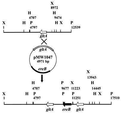
The transforming plasmid used in these studies was constructed with the plasmid pMOB (17) as the base replicon. A 735-bp portion of the ampicillin resistance gene of pMOB was removed by SspI and BsaI digestion and replaced with a 1,918-bp HincII-SmaI fragment containing the E. coli ereB gene from pAT72 (2). The resulting plasmid, pMW1041, conferred an erythromycin resistance phenotype when introduced into E. coli strains. To provide a target for homologous recombination into the R. prowazekii genome, a 1,881-bp R. prowazekii EcoRV fragment from plasmid pMW264 (23), containing the gltA gene with its constitutive promoter (6), was cloned into the EcoRV site of pMW1041. This generated the plasmid, designated pMW1047 (Fig. 1), used in the transformation experiments. The entire coding sequence of gltA was included in this construct to ensure that a single crossover event within this region would not destroy the gene but would instead produce a gltA duplication.
FIG. 1.
Restriction maps of the Madrid E gltA locus (top) and predicted pMW1047 insertion in RPMOB.001 (bottom). H, HindIII; P, PstI; X, XbaI. Numbers below the relevant restriction sites indicate the location of the restriction site on the map. Arrows identify the location and orientation of the gltA and ereB genes.
Following electroporation with pMW1047, the rickettsiae were allowed to infect L929 cells, which were then incubated for 24 h. At 24 h after infection, nearly 100% of the L929 cells were infected, with each host cell containing approximately 10 to 15 rickettsiae/cell. Erythromycin (200 ng/ml) was added, and incubation was continued. Rickettsiae were slowly cleared from the host cells following erythromycin addition. At 7 days postinfection, rickettsiae could not be visually detected on stained coverslips or were detected at very low levels. However, at 21 days rickettsiae could once again easily be detected visually, with approximately 1% of the host cells containing 200 to 300 rickettsiae per cell. The cell cultures were incubated until approximately 20% of the host cells were infected before the L929 cells were harvested. Rickettsiae were isolated for analysis of their chromosomal DNA. PCR assays using primers DW318 (located upstream from the gltA gene) and DW331 (located within the ereB gene), which would generate a PCR product only if pMW1047 was inserted into the chromosome at the gltA gene, yielded the predicted fragment when DNA from the erythromycin-resistant rickettsiae was used as a template (data not shown). Direct sequencing of this PCR fragment revealed that the fragment consisted of gltA and ereB sequences, confirming that ereB-containing rickettsial transformants were present in the Emr population.
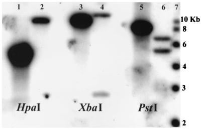
Since genetic analysis requires the isolation of clones derived from a single transformed bacterium, clonal isolates of rickettsial Emr transformants were obtained by limiting dilution. Rickettsia-positive microtiter dish wells were identified by PCR using primers targeted to a rickettsial chromosomal gene. Wells positive for rickettsiae were then analyzed for the ereB gene using ereB-specific primers DW337 and DW338 (Table 1). A single clone, designated RPMOB.001, was selected for additional characterization. The replication time of 10 h for this clone did not differ from that of the Madrid E parent strain. However, the MIC of erythromycin for RPMOB.001 was determined to be 2 μg/ml, in contrast to 0.2 μg/ml for the Madrid E strain. Southern hybridization analysis (16) revealed the changes in mobility of gltA-hybridizing sequences predicted from the genome sequence and restriction map of pMW1047 (Fig. 1). For Madrid E chromosomal DNA, the expected HpaI, XbaI, and PstI fragments of 4,767, 8,971, and 7,742 bp, respectively (Fig. 2, lanes 1, 3, and 5), were observed. For RPMOB.001 DNA, the predicted fragment of 9,738 bp for HpaI, two fragments of 2,720 and 11,222 bp for XbaI, and two fragments of 4,880 and 6,259 bp for PstI were observed (Fig. 2, lanes 2, 4, and 6), confirming the presence of a pMW1047 insertion at the gltA locus of R. prowazekii. DNA sequencing of PCR products spanning this region confirmed the insertion of the ereB gene into the gltA locus. All of the clones obtained from transformations using pMW1047 were found to have the gene inserted at the same site as in RPMOB.001. No Emr clones were obtained in which pMW1047 was inserted at another chromosomal site or in which the plasmid replicated autonomously.
FIG. 2.
Hybridization of the R. prowazekii gltA gene to a Southern blot of R. prowazekii chromosomal DNA digested with the indicated restriction enzymes and isolated from the Madrid E strain (lanes 1, 3, and 5) or RPMOB.001 (lanes 2, 4, and 6). Molecular size markers (lane 7) are indicated.
Initially, the efficiency of R. prowazekii erythromycin resistance transformation (successful entrance of DNA into the cell, successful recombination, and ereB expression) was low. We achieved only 1 successful experiment in 17 electroporations attempted. This is in contrast to the two of three successful rifampin resistance transformations obtained previously (13). This prompted us to evaluate our transformation conditions and generate a revised protocol, which yielded a success rate of two out of five electroporations. The revised protocol differs from that used in the rifampin selection transformations in several critical parameters. First, electroporation conditions were changed by increasing the field strength from 17 to 24 kV/cm. Rickettsial viability was reduced approximately 50% at the higher field strength. Second, five to seven times more L929 cells (1 × 10⁸ to 1.5 × 10⁸) were used to ensure that every electroporated rickettsia had the opportunity to infect a host cell. Finally, the number of rickettsiae per cell had to be evaluated to ensure selection. We discovered that erythromycin selection is dependent upon the number of rickettsiae per cell at the time of initial selection. If the number per cell is greater than 20 at the time of initial selection (24 h after selection), erythromycin at 200 ng/ml is unable to stop the growth of these rickettsiae at a rate sufficient to prevent them from destroying the host cell. Since reinfection by the rickettsiae is not efficient, lysis at this early stage, when transformants are few, results in the loss of potential transformants. At lower numbers per cell, the sensitive rickettsiae stop growing before the host cells lyse. Thus, the goal is to have one or two electroporated rickettsiae infect each host cell, followed by erythromycin selection at 24 h, when the rickettsial numbers will be less than 20 per host cell.
With the isolation of RPMOB.001, we have demonstrated that the E. coli ereB gene can be inserted at a selected target site into the R. prowazekii genome via homologous recombination. The identification of a selectable marker that can be targeted to any rickettsial gene of interest is a significant accomplishment in efforts to establish a genetic system in this organism and, to our knowledge, is the first demonstration of the expression of a nonrickettsial antibiotic resistance gene in R. prowazekii.
Acknowledgments
We thank Patrice Courvalin for providing pAT72, containing the ereB gene used in this study, and Priscilla Wyrick for helpful discussions on the use of erythromycin as a selective agent for rickettsiae.
This work was supported by NIH grant AI20384 to D.O.W.
REFERENCES
- 1.Andersson S G E, Zomorodipour A, Andersson J O, Sicheritz-Pontén T, Alsmark U C M, Podowdki R M, Näslund A K, Eriksson A-S, Winkler H H, Kurland C G. The genome sequence of Rickettsia prowazekii and the origin of mitochondria. Nature. 1998;396:133–143. doi: 10.1038/24094. [DOI] [PubMed] [Google Scholar]
- 2.Arthur M, Autissier D, Courvalin P. Analysis of the nucleotide sequence of the ereB gene encoding the erythromycin esterase type II. Nucleic Acids Res. 1986;14:4987–4999. doi: 10.1093/nar/14.12.4987. [DOI] [PMC free article] [PubMed] [Google Scholar]
- 3.Arthur M, Courvalin P. Contribution of two different mechanisms to erythromycin resistance in Escherichia coli. Antimicrob Agents Chemother. 1986;30:694–700. doi: 10.1128/aac.30.5.694. [DOI] [PMC free article] [PubMed] [Google Scholar]
- 4.Ausubel F, Brent R, Kingston R E, Moore D D, Seidman J G, Smith J A, Struhl K, editors. Current protocols in molecular biology. 1–3. New York, N.Y: John Wiley & Sons, Inc.; 1997. [Google Scholar]
- 5.Bullock W O, Fernandez J M, Short J M. A high efficiency plasmid transforming recA Escherichia coli strain with beta-galactosidase selection. BioTechniques. 1987;5:376–379. [Google Scholar]
- 6.Cai J, Pang H, Wood D O, Winkler H H. The citrate synthase-encoding gene of Rickettsia prowazekii is controlled by two promoters. Gene. 1995;163:115–119. doi: 10.1016/0378-1119(95)00365-d. [DOI] [PubMed] [Google Scholar]
- 7.Clavero G, Perez Gallardo F. Estudio experimental de una cepa apatogena e inmunizante de Rickettsia Prowazeki. Cepa E. Rev Sanid Hig Publica. 1943;17:1–27. [Google Scholar]
- 8.Gimenez D F. Staining rickettsiae in yolk-sac cultures. Stain Technol. 1964;39:135–140. doi: 10.3109/10520296409061219. [DOI] [PubMed] [Google Scholar]
- 9.Hackstadt T. The biology of rickettsiae. Infect Agents Dis. 1996;5:127–143. [PubMed] [Google Scholar]
- 10.LeClercq R, Courvalin P. Bacterial resistance to macrolide, lincosamide, and streptogramin antibiotics by target modification. Antimicrob Agents Chemother. 1991;35:1267–1272. doi: 10.1128/aac.35.7.1267. [DOI] [PMC free article] [PubMed] [Google Scholar]
- 11.LeClercq R, Courvalin P. Intrinsic and unusual resistance to macrolide, lincosamide, and streptogramin antibiotics in bacteria. Antimicrob Agents Chemother. 1991;35:1273–1276. doi: 10.1128/aac.35.7.1273. [DOI] [PMC free article] [PubMed] [Google Scholar]
- 12.Ounissi H, Courvalin P. Nucleotide sequence of the gene ereA encoding the erythromycin esterase in Escherichia coli. Gene. 1985;35:271–278. doi: 10.1016/0378-1119(85)90005-8. [DOI] [PubMed] [Google Scholar]
- 13.Rachek L I, Tucker A M, Winkler H H, Wood D O. Transformation of Rickettsia prowazekii to rifampin resistance. J Bacteriol. 1998;180:2118–2124. doi: 10.1128/jb.180.8.2118-2124.1998. [DOI] [PMC free article] [PubMed] [Google Scholar]
- 14.Rolain J M, Maurin M, Vestris G, Raoult D. In vitro susceptibilities of 27 rickettsiae to 13 antimicrobials. Antimicrob Agents Chemother. 1998;42:1537–1541. doi: 10.1128/aac.42.7.1537. [DOI] [PMC free article] [PubMed] [Google Scholar]
- 15.Saah A J. Rickettsia prowazekii (epidemic or louse-borne typhus) In: Mandell G L, Bennett J E, Dolin R, editors. Mandell, Douglas, and Bennett's principles and practice of infectious diseases. 5th ed. Vol. 2. Philadelphia, Pa: Churchill Livingstone; 2000. pp. 2050–2053. [Google Scholar]
- 16.Southern E M. Detection of specific sequences among DNA fragments separated by gel electrophoresis. J Mol Biol. 1975;98:503–517. doi: 10.1016/s0022-2836(75)80083-0. [DOI] [PubMed] [Google Scholar]
- 17.Strathmann M, Hamilton B A, Mayeda C A, Simon M I, Meyerowitz E M, Palazzolo M J. Transposon-facilitated DNA sequencing. Proc Natl Acad Sci USA. 1991;88:1247–1250. doi: 10.1073/pnas.88.4.1247. [DOI] [PMC free article] [PubMed] [Google Scholar]
- 18.Troyer J M, Radulovic S, Azad A F. Green fluorescent protein as a marker in Rickettsia typhi transformation. Infect Immun. 1999;67:3308–3311. doi: 10.1128/iai.67.7.3308-3311.1999. [DOI] [PMC free article] [PubMed] [Google Scholar]
- 19.Weisblum B. Erythromycin resistance by ribosome modification. Antimicrob Agents Chemother. 1995;39:577–585. doi: 10.1128/AAC.39.3.577. [DOI] [PMC free article] [PubMed] [Google Scholar]
- 20.Weiss E. The biology of rickettsiae. Annu Rev Microbiol. 1982;36:345–370. doi: 10.1146/annurev.mi.36.100182.002021. [DOI] [PubMed] [Google Scholar]
- 21.Winkler H H. Rickettsial permeability: an ADP-ATP transport system. J Biol Chem. 1976;251:389–396. [PubMed] [Google Scholar]
- 22.Winkler H H. Rickettsia species (as organisms) Annu Rev Microbiol. 1990;44:131–153. doi: 10.1146/annurev.mi.44.100190.001023. [DOI] [PubMed] [Google Scholar]
- 23.Wood D O, Williamson L R, Winkler H H, Krause D C. Nucleotide sequence of the Rickettsia prowazekii citrate synthase gene. J Bacteriol. 1987;169:3564–3572. doi: 10.1128/jb.169.8.3564-3572.1987. [DOI] [PMC free article] [PubMed] [Google Scholar]


