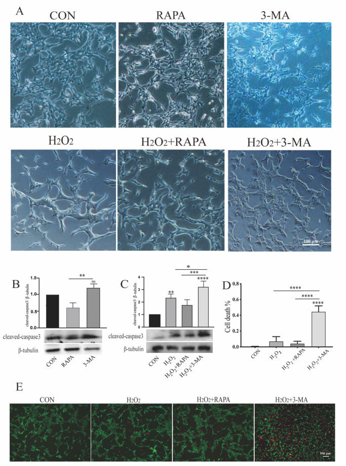
Figure 1.
Regulation of autophagy can modulate the morphologic alterations and apoptosis of MGCs under oxidative stress. r-MCs were treated with rapamycin or 3-MA under normal conditions or oxidative stress for 24 h. (A) Bright-field microscopy was used to detect the morphologic alterations of MGCs. r-MCs treated with H2O2 and 3-MA formed aggregates. Scale bar: 100 μm. (B,C) Apoptosis of MGCs was detected by changes in the expression of cleaved caspase 3 by western blotting. Appropriate downregulation of autophagy induced by 3-MA (1 mM) was not toxic to r-MCs under normoxia, while it could increase vulnerability to oxidative stress. Data are shown as means ± SEMs (n = 3 per group; * p < 0.05, ** p < 0.01, *** p < 0.001 and **** p < 0.0001). (D) Cell death in different groups was quantified. Data are shown as means ± SEMs (n = 3 per group; **** p < 0.0001). (E) Live/dead cell staining for oxidative stress and autophagic dysfunction showed obvious MGC apoptosis. Scale bar: 100 μm. RAPA, rapamycin; 3-MA, 3-methyladenine.

