

Abstract
Minocycline, a semisynthetic derivative of tetracycline that is used to treat various infectious and noninfectious conditions, can cause tissue hyperpigmentation. The skin, oral mucosa, sclera, and rarely the nails, can all be affected. The discoloration varies from blue, slate-gray, or brown, and it typically occurs in a dose-dependent fashion. The mechanism of hyperpigmentation, however, remains largely unknown. Herein, we present a case of gray-blue hyperpigmentation of the skin, sclera, and nails after long-term treatment with minocycline for acne.
Keywords: Hyperpigmentation, Skin, Nails, Sclera, Minocycline, Side effect, Acne
Introduction
Ingestion of minocycline, a highly lipid soluble semisynthetic derivative of tetracycline, used for a variety of conditions, including acne vulgaris, may cause hyperpigmentation of the skin, teeth, bone, sclera, mucous membranes, thyroid, trachea, and nails [1]. With the aim of alerting clinicians about the possibility of this adverse reaction, in this report, we present a case of minocycline-induced gray-blue pigmentation affecting multiple body tissues.
Case Presentation
An 83-year-old man presented with long-standing hyperpigmentation of the skin, oral mucosa, sclera, and nails. Past medical history included squamous cell carcinoma, atrial fibrillation, essential hypertension, and benign prostatic hyperplasia. Medications included apixaban, hydrochlorothiazide, and tamsulosin. He had taken minocycline 100 mg daily for more than 50 years for acne vulgaris (self-prescribed) prior to presentation. Physical examination was significant for gray-blue patches involving the shins and face (Fig. 1, 2, respectively), with diffuse gray-blue discoloration of the sclera (Fig. 3), gums, and all fingernails (Fig. 4). Visual function was intact. A clinical diagnosis of minocycline-induced pigmentation was favored. Other causes were excluded.
Fig. 1.

Diffuse gray to blue patches involving the left shin.
Fig. 2.

Gray to blue discoloration of the right temple.
Fig. 3.

Gray to blue discoloration of the sclera.
Fig. 4.
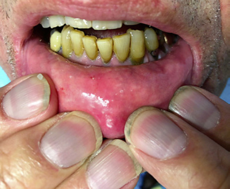
Gray to blue discoloration of the gums and fingernail beds.
Discussion
Tissue hyperpigmentation is a rare but possible adverse reaction associated with minocycline treatment for acne vulgaris. In a prospective study of 700 acne patients, 2.4% experienced hyperpigmentation, with an increased incidence with higher minocycline doses (p < 0.01); 4% in patients receiving 200 mg daily compared with 0.4% and 1.1% of those taking 100 mg and 100/200 mg daily and on alternate days, respectively [2]. The mechanism of hyperpigmentation is unclear; it may be due to chelation of a minocycline derivative with iron in macrophage lysosomes [3]. Three clinical pigmentation patterns exist [1]. Type I presents with gray-blue macules following inflammation or trauma (commonly involving the face and associated with acne) [1]. Type II, well circumscribed gray-blue pigmentation involving previously normal skin, most commonly involves the shins and forearms [1]. Type III presents as a diffuse muddy-brown discoloration of previously normal skin, accentuated in sun exposed areas [1]. Types II and III are associated with long-term minocycline treatment and high cumulative doses (70–100 g), while type I may occur only a few weeks after drug initiation [1]. On histopathology, types I and II show dermal pigment granules within macrophages concentrated around vessels and adnexa, respectively [3]. Type III shows increased melanin in basal keratinocytes and dermal melanophages, while iron is absent [3]. Minocycline-induced nail pigmentation is far less common than skin hyperpigmentation. It most frequently presents with a slate-gray discoloration of the proximal nail bed [1]. An alternative presentation is longitudinal melanonychia, but it is much less common [1]. Ocular pigment deposition is rare and can involve the sclera, conjunctiva, and retina [1]. Pigmentation of ocular structures does not seem to affect visual function [1, 4].
In the present case, the patient took minocycline for decades, easily exceeding the 100-g cumulative dose threshold associated with pigmentation. The clinical presentation is best categorized as type II. Differential diagnoses include stasis dermatitis of the lower extremities, erythema dyschromicum perstans, Addison's disease, and nevus of Ota. Differentials of gray-blue nail pigmentation include Wilson's disease, argyria (azure lunula), and other medications, including zidovudine, antimalarials, and amiodarone. Taking a thorough clinical history and recognizing classic pigmentation patterns may avoid unnecessary biopsies. While minocycline-associated pigmentation is uncommon, patients receiving treatment with minocycline should be informed of this possible side effect, especially those receiving long-term treatment with cumulative doses >100 g. Pigmentation patterns type I and II, as well as nail pigmentation, may persist for prolonged periods of time and resolve with drug cessation [1], while type III pigmentation is often permanent. There have been reports of successful treatment with Q-switched lasers [5].
Statement of Ethics
Written and informed consent was obtained from the patient for publication of this case report and accompanying images. Ethical approval was not required for this study in accordance with local or national guidelines.
Conflict of Interest Statement
The authors have no conflicts of interest to declare.
Funding Sources
This article has no funding source.
Author Contributions
Study conception and design and acquisition of data: Jose W. Ricardo and Shari R. Lipner. Drafting of the manuscript: Jose W. Ricardo, Shari R. Lipner, Kalee Shah, and Kira Minkis.
Data Availability Statement
All data generated or analyzed during this study are included in this article. Further inquiries can be directed to the corresponding author.
References
- 1.Eisen D, Hakim MD. Minocycline-induced pigmentation. Incidence, prevention and management. Drug Saf. 1998;18((6)):431–40. doi: 10.2165/00002018-199818060-00004. [DOI] [PubMed] [Google Scholar]
- 2.Goulden V, Glass D, Cunliffe WJ. Safety of long-term high-dose minocycline in the treatment of acne. Br J Dermatol. 1996;134((4)):693–5. doi: 10.1111/j.1365-2133.1996.tb06972.x. [DOI] [PubMed] [Google Scholar]
- 3.Bowen AR, McCalmont TH. The histopathology of subcutaneous minocycline pigmentation. J Am Acad Dermatol. 2007;57((5)):836–9. doi: 10.1016/j.jaad.2007.04.028. [DOI] [PubMed] [Google Scholar]
- 4.Maloney SM, Williams BK, Jr., Shields CL. Long-term minocycline therapy with scleral pigmentation simulating melanocytosis. JAMA Ophthalmol. 2018;136((11)):e183088. doi: 10.1001/jamaophthalmol.2018.3088. [DOI] [PubMed] [Google Scholar]
- 5.Collins P, Cotterill JA. Minocycline-induced pigmentation resolves after treatment with the Q-switched ruby laser. Br J Dermatol. 1996;135((2)):317–9. [PubMed] [Google Scholar]
Associated Data
This section collects any data citations, data availability statements, or supplementary materials included in this article.
Data Availability Statement
All data generated or analyzed during this study are included in this article. Further inquiries can be directed to the corresponding author.