Abstract
The current study analyzes the suitability and reliability of selected neurophysiological and vegetative nervous system markers as biomarkers for exercise and recovery in endurance sport. Sixty-two healthy men and women, endurance trained and moderately trained, performed two identical acute endurance tests (running trial 1 and running trial 2) followed by a washout period of four weeks. Exercise protocol consisted of an acute running trial lasting 60 minutes. An intensity corresponding to 95% of the heart rate at individual anaerobic threshold for 40 minutes was followed by 20 minutes at 110%. At pre-exercise, post-exercise, three hours post-exercise and 24 hours post-exercise, experimental diagnostics on Brain-derived neurotrophic factor (BDNF), heart rate variability (HRV), Stroop Color and Word Test (SCWT), and Short-Form McGill Pain Questionnaire (SF-MPQ) were performed. Significant changes over time were found for all parameters (p < .05). Furthermore, there was an approached statistical significance in the interaction between gender and training status in BDNF regulation (F(3) = 2.43; p = 0.06), while gender differences were found only for LF/HF-ratio (3hPoEx, F(3) = 3.40; p = 0.002). Regarding the reliability, poor ICC-values (< 0.5) were found for BDNF, Stroop sensitivity and pNN50, while all other parameters showed moderate ICC-values (0.5-0.75). Plasma-BDNF, SCWT performance, pain perception and all HRV parameters are suitable exercise-sensitive markers after an acute endurance exercise. Moreover, pain perception, SCWT reaction time and all HRV parameters show a moderate reliability, others rather poor. In summary, a selected neurophysiological and vegetative marker panel can be used to determine exercise load and recovery in endurance sports, but its repeatability is limited due to its vaguely reliability.
Key points.
Pain perception and the Stroop test have moderate reliability in the use of exercise and recovery markers after two identical exercise loads in endurance sports
Markers of heart rate variability also show moderate reliability as biomarkers after intense endurance exercise under identical conditions.
There are associations between neurophysiological markers and inflammatory blood markers after endurance exercise that are partially associated with gender and training status
Key words: NNervous system, monitoring training, biomarkers, reliability, executive function, heart rate variability
Introduction
The controlled monitoring of exercise and recovery cycles is fundamental in elite sports. A more precise diagnosis of how much exercise stress the athlete experiences and the recovery status using reliable parameters would help to better coordinate training loads. Moreover, individualized exercise loads, and the differentiated recovery management could be prescribed more precisely (Lee et al., 2017).
Currently, coaches, trainers, and athletes use many different diagnostic tools (Costache et al., 2021). Several blood-based markers, functional tests, and questionnaires are used despite having weaknesses and inaccuracies. Many of these tests are inaccurate, highly subjective, based on limited or non-validated markers. Some diagnostic tools are simply too complex or expensive. Therefore, there is a great need for subsidiary methods that have satisfactory reliability in addition to valid exercise sensitivity. Furthermore, diagnostics that can be combined and reflect a part of the whole physiological system quantitatively, are needed (Hacker et al., 2021; Reichel et al., 2020). While it is known that there are subjective and objective parameters that can be combined in response to an exercise stimulus, it is not known whether the parameters respond in a same way at different times under standardized conditions. For this purpose, reliability testing is of particular importance.
To find new tools or markers that allow a more accurate diagnosis of exercise and recovery cycles, it is certainly advisable to look at the physiological systems that show a sensitive response. Exercise is accompanied by changes in neurophysiological and vegetative function, such as brain health, improved cognition, and increased resistance to brain injury (Griesbach et al., 2004; Martínez-Díaz and Carrasco, 2021). An acute bout of exercise induces numerous neurophysiological processes depending on intensity and duration, local and central fatigue processes (Proschinger and Freese, 2019). A blood-based marker that has been studied repeatedly in the context of acute or chronic exercise tests is brain-derived neurotrophic factor (BDNF). BDNF is a neurotrophin, that is released into the blood by both the central nervous system (CNS) and musculature in response to exercise stress (Pedersen et al., 2009). Accordingly, there is an intensity-dependent increase during exercise, which is gradually regulated back to the baseline during the recovery phase (Reed et al., 2021; Winter et al., 2007).
Another method to analyse vegetative and recovery states represents the analysis of heart rate variability (HRV) (Du et al., 2005; Figueira et al., 2021). HRV is the fluctuation of beat-to-beat intervals, also known as R-R intervals in a defined period of time. During acute exercise and the subsequent recovery states, the duration of the R-R intervals changes due to an altered interplay between the sympathetic and parasympathetic nervous systems. Thereby, HRV initially decreases during physical exertion and gradually increases during the recovery period after an initial reduction (Gifford et al., 2018; Parekh and Lee, 2005). It is assumed that the vegetative state of exhaustion is reflected by HRV. A qualitative analysis of HRV is performed via the temporal representation of the R-R intervals in the form of tachograms. Thereby, numerous parameters of HRV can be derived. HRV is also a suitable physiological marker because measurement is non-invasive and available measurement devices are appropriate (Perini and Veicsteinas, 2003).
To include the executive functions into a test battery, the Stroop Color and Word Test (SCWT) is often used. Effects of acute exercise and training have already been shown for this test of selective attention (Alves et al., 2012). The Stroop effect can also be carried out relatively quickly and effortless, so that it is a potential tool for monitoring exercise and recovery cycles (Harveson et al., 2016). Finally, neurophysiological analyses often take place in connection with a subjective perception of pain. From a set of instruments, the Short-Form McGill Pain Questionnaire (SF-MPQ) is suitable for this purpose. Previous studies showed that the SF-MPQ reflects neurophysiological and muscular limitations during exercise, e.g. muscular pain and muscle damage (Krüger et al., 2015).
The present study used an innovative and explorative design to investigate the suitability and especially the reliability of selective parameters to analyze exercise and recovery cycles in endurance sports. Changes in neurophysiological, vegetative, or executive functions were examined, and differences between the sexes as well as the influence of training status was investigated. Briefly, the suitability of the markers are defined by exercise-sensitivity and reliability as a variable for an agreement of two repetitive measurements under standardized conditions (Pedlar et al., 2019; Reichel et al., 2020). For this purpose, moderately trained and trained participants of both sexes performed two almost identical endurance tests under highly controlled, almost identical conditions, four weeks apart. All markers were recorded at different times before and after the tests.
Methods
Participants
The study population is the same as published in a previous study and is therefore described briefly (Reichel et al., 2020). 62 participants (31 women and 31 men, age 19-43 years) were classified as either endurance trained (T) (n = 37) or moderately trained (MT) (n = 25) participants according to American College of Sports Medicine (ACSM) guidelines (Riebe et al., 2018). There were 25 trained and 6 moderately trained women and 12 trained and 19 moderately trained men. The T-group consisted mainly of runners, strength athletes and semi-professional team athletes such as soccer, handball, and volleyball players. All other subjects were defined as either recreationally active or rather athletically inactive. Anthropometric data were published previously (Reichel et al., 2020). Briefly, inclusion criteria were non-smoking, free of cardiovascular, metabolic or musculoskeletal diseases as well as free of acute infections, musculoskeletal injuries, and symptomatic respiratory deficits. The good health status was confirmed by a medical history questionnaire, a pulmonary function test (spirometry), an orthopedic and internal medical examination, an electrocardiogram (ECG), and blood pressure measurement. All participants signed a declaration of consent before participation. The research protocol complied with the principles of the Declaration of Helsinki, was approved by the local ethic committee of the Justus Liebig University Giessen (Germany) (Application number: 2017-0010), and fulfilled the international ethical standards (Harriss and Atkinson, 2015).
Study design
After recruitment, participants were tested for their endurance capacity via a continuous progressive exercise field test accompanied by lactate diagnostics and heart rate measurement.
The heart rate at the individual anaerobic threshold (IAT) was used to calculate the running speed at the two identical running trials (RTs). Thereby, the running pace was designed to implicate an individual physiological, vegetative and executive fatigue. Details of the test-retest design can be found at Reichel et al. (2020). Briefly, both RTs consisted of a 60-minute running field test: 40-minutes at an intensity corresponding to 95% of the heart rate at IAT, followed by a maximum of 20-minutes at 110% of the IAT. The RTs were interrupted by a four-week washout period.
The RTs took place one week after the lactate field test, specific requirement had to be met to guarantee a high controlled standardization that would ensure identical conditions:
Documentation of nutrition three days before the first RT. The protocol was replicated as a guideline for the food intake prior to the second RT.
It was not permitted to consume alcohol the day prior to the RT.
The participants were not allowed to carry out high-intensity exercise four days prior the RTs.
The participants were instructed not to change their lifestyle with regard to physical activity and dietary behavior during the study period. In order to detect deviations of their lifestyle between the testing days, participants had to fill out a questionnaire on lifestyle behaviour before each RT. Women completed a questionnaire on menstrual cycle.
Experimental diagnostics
Pre-exercise (PrEx), post-exercise (PoEx), three hours post-exercise (3hPoEx) and 24 hours post-exercise (24hPoEx) a test battery of experimental diagnostics was applied.
Blood analysis
For determination of BDNF, venous blood samples were collected from the arm vein with anticoagulated EDTA vacutainers. Subsequently samples were centrifuged at 2,500 x g for 10 min at 4°C to separate the blood into plasma and cellular fractions. The plasma samples were aliquoted and stored at -80°C. Finally, BDNF was analyzed by high-sensitivity enzyme-linked immunosorbent assay (ELISA) (Quantikine ELISA Kits: R&D Systems, analytical sensitivity: 20 pg/mL, detection range: 62.5-4,000 pg/mL; CV (%): 7,6; MVZ, Koblenz, Germany). The methodological analysis of other blood-based markers such as IL-1ra, IL-6 and IL-8, which were used for association purposes in this study, can be found in the study by Reichel et al. (2020).
Pain questionnaire
The McGill Pain Questionnaire (MPQ) is a widely used international questionnaire for assessing the quality and intensity of pain, originally developed by Melzack (1975) (Melzack, 1975). The German short form (SF) of this questionnaire, based on Radvila et al. (1987), focuses on the sensory (11 descriptors) and affective (4 descriptors) dimensions of pain perception (Radvila et al., 1987). A 3-value Likert-scale (0 = no pain is felt; 3 = strongly pain is felt) was used as a rating scheme. Furthermore, a rating of 0-100 millimeters on a visual analog scale (VAS) for momentary pain intensity, as well as a 5-value Likert scale (0 = no pain; 5 = excruciating) for overall pain experience, were used to quantify perceived pain. Achieving higher scores of all assessment parts represented a higher total score of pain.
Assessment of executive function
The SCWT is the most common neuropsychological procedure for assessing response inhibition (Chu et al., 2015; Scarpina and Tagini, 2017). The test involves the interference of words and colors. Participants had to determine the colors of words that appear on a computer screen whilst measuring their reaction time to a mouse clicks. It seems to be an automated task to determine the color of the word, but at the same time to prevent the judgement about right/wrong from changing by reading the word of the color seems to be more automated. The difficulty in inhibiting the more automated process is described by the Stroop effect (Scarpina and Tagini, 2017; Stroop, 1935). Here, the colored font is either congruent („matched correct“) or incongruent („nonmatched correct“) with the word. As proposed by Scarpina and Tagini (2017) the scoring method takes into consideration the speed (milliseconds) and accuracy (sensitivity index, dee prime (d´), according to Stanislaw and Todorov (1999)) of the participant’s response (Stanislaw and Todorov, 1999). Regarding the interpretation of d‘, a value of d‘ = 0 indicates that the participant was not able to inhibit cognitive interference (0%-hit-rate / 100%-false-alarm-rate). A value of d´=1 corresponds to perfect performance regarding the distinction of the stimuli between match and non-match (100%-hit-rate / 0%-false-alarm-rate) (Stanislaw and Todorov, 1999). When evaluating the speed of the subject’s response, the average of the reaction time for the conditions „matched correct“, and „nonmatched correct“ was considered.
Assessment of vegetative function
HRV was measured within a 12-minutes orthostatic stress test with spontaneous breathing in a quiet environment. To create standardized conditions and avoid influences of background sounds, each subject additionally got a pair of earplugs. Furthermore, participants should sit in an upright relaxed position and were asked to avoid any kind of movement. Each subject was provided with a wrist heart rate monitor (Polar RS800CX) and a compatible chest strap with a heart rate sensor (H3). The analysis was performed using the Polar Pro Trainer 5™ 5.40.170 software. R-R interval data were collected and converted to a computer for further analysis.
The selection of HRV parameters was based on scientific evidence-based exercise studies (Bellenger et al., 2016; da Silva et al., 2014). The outcomes were presented in a time domain plotting the R-R intervals in milliseconds (ms) against time (Borresen and Lambert, 2008). Time-domain parameters in this study were: Average R-R interval (ARR), square root of the mean of the sum of the squares of differences between adjacent normal R-R intervals (RMSSD), percentage of adjacent R-R intervals that differ from each other by more than 50 milliseconds (pNN50), standard deviation of instantaneous beat-to-beat R-R interval variability measured from Poincaré plots (SD1), and standard deviation of long-term beat-to-beat R-R interval variability measured from Poincaré plots (SD2) (Buchheit, 2014). The results of HRV measures were also presented in the frequency domain, namely the frequency of R-R interval changes. Frequencies in the range of 0.00 to 0.40 Hz were categorized into three groups: High frequency oscillations (HF) (0.15 to 0.40 Hz), low frequency oscillations (LF) (0.04-0.15), very low frequency oscillations (VLF) (0.003-0.04), and the ratio of HF and LF (HF/LF-ratio) (Buchheit, 2014).
Statistical analyses
Statistical analyses were conducted using SPSS version 25 (IBM® SPSS Statistics 25, IBM GmbH, Munich, Germany) and JASP (Version 0.14.1, JASP Team, Amsterdam, The Netherlands). Descriptive data represents mean ± standard deviation (SD) for the study population. First, the data was cleaned up and outliers were removed using z-transformation (z-score > 3 standard deviations) (Stocker and Steinke, 2016). Normal, or log-normal distributed data, determined by Kolmogorov-Smirnov-Test (KS-Test), were analyzed by a multi-factorial analysis of variance with repeated measures (ANOVA). Heterogeneous data were examined for effects concerning RT, time of measurement, training status, and the interactions of these variables. Taking into account sex, we separated the participants into same-sex subgroups and evaluated these with ANOVA analysis. For significant effects (p < .05), a Bonferroni post-hoc test was added. Test-retest reliability between the times of measurements of both RTs was estimated by intraclass correlation coefficient (ICC), based on a two-way mixed-effects model, single measurement, and absolute agreement. ICC values were classified as excellent (ICC > 0.9), good (ICC = 0.75-0.9), moderate (ICC = 0.5-0.75), or poor (ICC < 0.5) (Koo and Li, 2016). Finally, correlation analyses according to Pearson (parametric data) and Spearman rank (non-parametric data) were carried out for the heterogeneous total group as well as depending on training status and sex. For this purpose, difference values between the measurement time points (MTPs) were used. A p-value of ≤ .05 was accepted as statistically significant. Due to the explorative approach of the study, there was no multiple test adjustment of α-error. The Line Graphs with raw data were created using Prism 9 (GraphPad Software, San Diego, CA, USA).
Results
Brain-derived neurotrophic factor
Plasma BDNF concentrations show a significant difference between the multiple time points (MTPs) overall (F(3) =103.23; p < 0.001). As shown in Figure 1 (A), a significant increase in concentration at PrEx compared to PoEx was observed in post hoc analysis (p < 0.001). After the peak at PoEx, a significant decrease to 3hPoEx (p < .001) and 24hPoEx (p < 0.001) was determined. Also, a continuous reduction of the BDNF concentration was found from 3hPoEx to 24hPoEx (p = 0.009) at both RTs. The variance analysis revealed an influence of the variables gender and training status on the MTPs with (F(3) = 2.44; p = 0.067). Analysis of reliability shows a poor ICC of 0.33.
Figure 1.
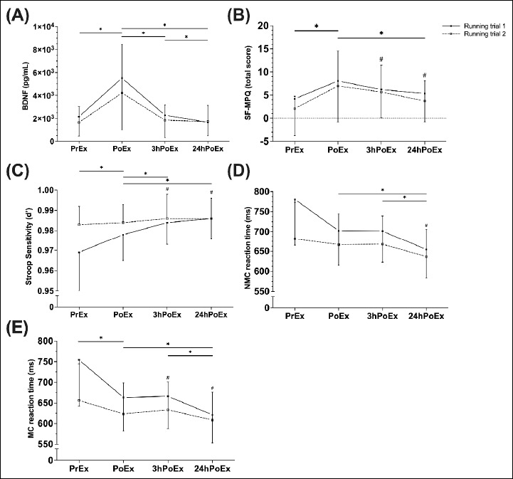
Plasma BDNF concentration (A), total score of SF-MPQ (B), and Stroop Test parameters (C – E) PrEx, PoEx, 3hPoEx and 24hPoEx for both RTs. Significant differences *(p < 0.05) to the previous MTP as well as significant differences #(p < 0.05) to the PrEx value. Values are means (±SD).
Short-form McGill Pain Questionnaire
Significant effects were found for the SF-MPQ total score between the MTPs (F(2,51) = 21.14; p < 0.001) (Figure 1B). The total score of the pain perception increased PoEx (p < 0.001) and was significantly higher compared to the baseline value at all MTPs except at 24hPoEx (PrEx - 3hPoEx, p < 0.001; PrEx - 24hPoEx, p < 0.001). After the peak PoEx, the total score significantly decreased back to baseline level up to 24hPoEx (p < 0.001). Moderate ICC-values of 0.54 were found for this parameter.
Stroop Color and Word Test
Differences for d’ across all MTPs in both RTs (F(2,56) = 23.92; p < 0.001) were found. The d’ increased significantly from MTP to MTP (PrEx – PoEx, p < 0.001; PoEx – 3hPoEx, p = 0.007; PoEx – 24hPoEx, p = 0.003), whereby no difference was detected between 3hPoEx and 24hPoEx. As shown in Figure 1 (C), all PoEx MTPs differed from the PrEx value (PrEx - PoEx/ 3hPoEx /24hPoEx, for all p < 0.001). A poor ICC of 0.30 was determined over the MTPs. For the reaction time of the Matched Correct (MC) values, differences could be found over all MTPs in both RTs (F(2,22) = 108.84; p < 0.001). Figure 1 (D) shows that the reaction time at PrEx is higher compared to PoEx MTPs (PrEx - PoEx/ 3hPoEx /24hPoEx, for all p < 0.001). The 24hPoEx reaction time represents the lowest value and differs significantly from the PoEx and 3hPoEx value (PoEx - 24hPoEx, p = 0.003; 3hPoEx - 24PoEx, p < 0.001). Moderate ICC-values were found for the participant’s reaction time (ICC = 0.62). Concerning the speed of the participant’s responds for the non-matched condition, statistical analysis yield a significant effect over all MTPs (F(1) = 5205.97; p < 0.001). Post hoc analysis revealed the lowest reaction time of the non-matched correct (NMC) values to the 24hPoEx MTP. Significant differences were found between the 24hPoEx value and all other MTPs (PrEx/ PoEx/ 3hPoEx - 24hPoEx, for all p < 0.001) (Figure 1E). The ICC value (0.58) was classified as moderate.
Heart rate variability
All results of the HRV time domain parameters showed a significant main effect between the MTPs on both RTs (ARR: F(3) = 104.65, p < 0.001; RMSSD: F(2,527) = 87.53, p < 0.001; pNN50: F(2,189) = 54.27, p < 0.001; SD1: F(2,43) = 89.79, p < 0.001; SD2: F(3) =58.09, p < 0.001). Post hoc analysis of all HRV time domain parameters demonstrated a decrease after the RTs (p < 0.001) (Figure 2A - E). Afterwards, the values increased continuously up to 3hPoEx and remained unchanged 24hPoEx, except for ARR (PoEx - 3hPoEx, p < 0.001; PoEx - 24hPoEx, p < 0.001). For ARR, another significant increase was found between 3hPoEx and 24hPoEx (p < 0.001). ICCs were classified as moderate for all parameters (ARR: 0.71, RMSSD: 0.58, pNN50: 0.49, SD1: 0.59, SD2: 0.61). For all HRV frequency domain parameters, significant differences were found over the MTPs (HF: F(3) = 89.65, p < 0.001; LF: F(2,556) = 30.33, p < 0.001; LF/HF: F(3) = 46.88, p < 0.001; VLF: F(3) = 81.68, p < 0.001) (Figure 3). A decrease was found at PoEx (p < 0.001), while at 3hPoEx, all parameters increased (p < 0.001) and still increased at 24hPoEx compared to PoEx (p < 0.001). Results of the variance analysis for LF/HF-ratio showed a significant main effect between the MTPs (F(3) = 46.88, p < 0.001) (Figure 3C). The values initially increased PoEx (p < 0.001) and continuously decreased to the baseline level until 24hPoEx (PoEx - 3hPoEx/ 24hPoEx, p < 0.001; 3hPoEx - 24hPoEx, p < 0.001). Regarding sex effects, a significant difference was found in the post hoc analysis of the 3hPoEx values (p = 0.002). The LF/HF ratio was almost twice as high for men as for women. The ICC values of the HRV frequency parameters over the MTPs were: HF = 0.52, LF = 0.64, for VLF = 0.57, and LF/HF-ratio = 0.63.
Figure 2.
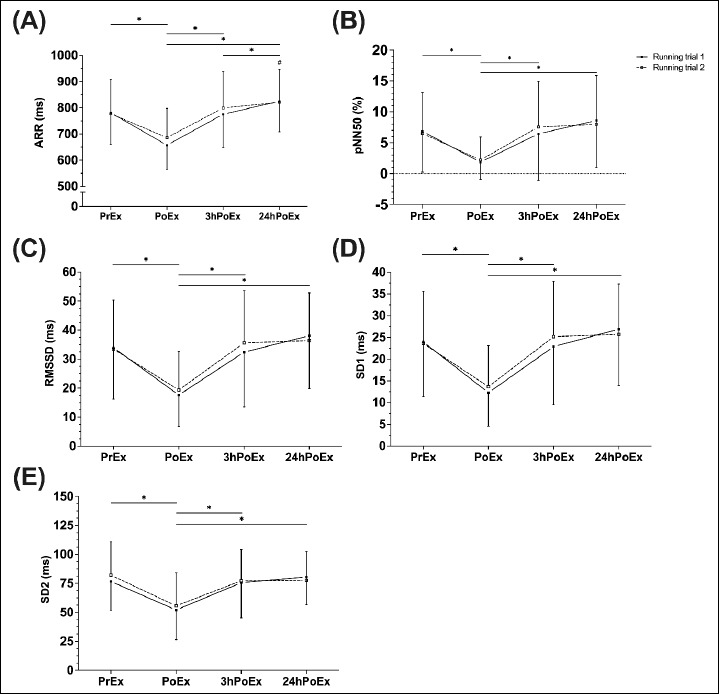
Changes in time domain HRV parameters PoEx, 3hPoEx, and 24hPoExat both RTs. *(p < 0.05) to the previous MTP as well as significant differences #(p < 0.05) to the PreExvalue. Values are means (±SD).
Figure 3.
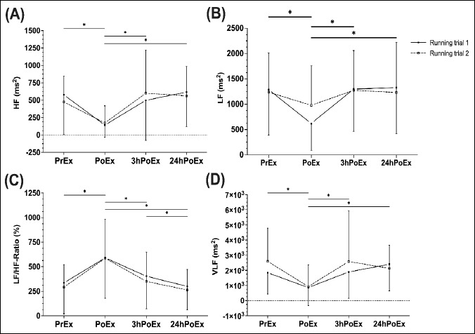
Changes in frequency domain HRV parameters PoEx, 3hPoEx, and 24hPoEx at both RTs. *(p < 0.05) to the previous MTP as well as significant differences #(p < 0.05) to the PrEx value. Values are means (±SD).
Associations between inflammatory markers and neurophysiological parameters
In order to identify associations between neurophysiological markers and other blood markers, analyses were carried out on markers that have already been published in the same study (Reichel et al., 2020). Only correlations that have shown a significance at both RTs or that correlated for one RT over time were considered. Correlation coefficients of r = -0.487, p = 0.018 (RT1) and r = -0.461, p = 0.027 (RT2) were found for IL-8 and VLF in men on both RTs in the recovery response after the RTs (Figure 4). We further found associations between IL-1ra and SF-MPQ total score (r = .622, p = .018/ r = .508, p = .031, RT1) (Figure 5 A1/2), IL-6 and SD2 (r = -0.542, p = .3/ r = -0.625, p = 0.006, RT2) (Figure 5 B1/2), and IL-8 and pNN50 (r = -0.586, p = 0.022/ r = -0.557, p = .016, RT2) (Figure 5 C1/2) in MT over time at one RT.
Figure 4.
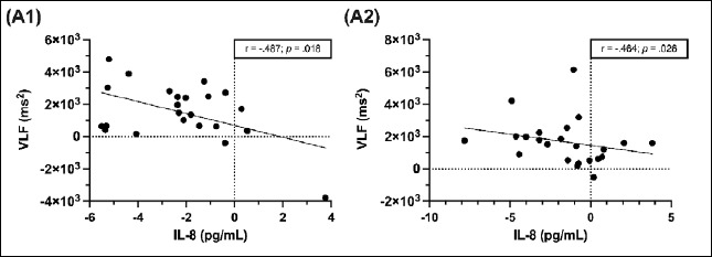
Correlations between IL-8 and VLF in men at both RTs in recovery phase after RTs. (A1) shows the recovery response of RT1, (A2) of RT2. Differences between the MTPs were used for the analyses. Significant correlations are presented with p < 0.05, n = 23.
Figure 5.
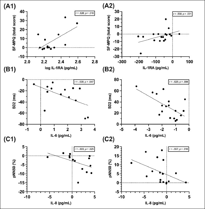
Correlations in moderately trained individuals (n = 14/ 18) (B1/2: IL-1ra and SF-MPQ total score, RT1; C1/2: IL-6 and SD2, RT2; D1/2: IL-8 and pNN50, RT2) at the exercise and recovery response on one RT. Number one of the graphs show the results of the exercise response, number two those of the recovery response. Differences between the MTPs were used for the analyses. Significant correlations are presented with p < 0.05.
Discussion
The current study found a significant immediate increase of BDNF, pain perception, and a significant decrease of most HRV parameters after both RTs, indicating their sensitivity to acute exercise. During 24h recovery, most parameters, except pain of perception, HR and mean of RR interval, returned to baseline levels. In contrast, for the Stroop sensitivity as well as the reaction time at the SCWT, a continuous increase over all MTPs was shown. Regarding their reliability, most parameters showed ICCs classified as moderate, while ICC of plasma BDNF, Stroop sensitivity, and pNN50 were classified as poor. Some neurophysiological markers showed a sex-specific regulation or a relation to training status. Only a few associations were found between neurophysiological parameters and blood-based inflammation markers.
At both RTs, plasma BDNF levels increased at PoEx (by 157% at the RT1 and 156% at the RT2). These results are consistent with the observation of Fonseca et al. (2021), who reported a significant increase in plasma BDNF following a single bout of progressive endurance exercise to fatigue. Accordingly, BDNF response to exercise depends on the volume of physical activity, identified by intensity and frequency as the determining predictors of biochemical upregulation (De Assis et al., 2018).
However, poor reliability was found for this neurophysiological parameter due to the large inter- and intrapersonal variation in plasma BDNF. Moreover, it has a complex genomic structure making it an ideal target for multiple and complex transcriptional regulations (Martinowich and Lu, 2008). Furthermore, the different occurrence of BDNF, as platelet-bound or free BDNF, can lead to variable and non-reproducible results (Walsh and Tschakovsky, 2018). Considering the effect of gender, it has been observed that both testosterone and oestrogen affect BDNF upregulation (Pluchino et al., 2013). However, no effect of the menstrual cycle on peripheral BDNF levels was found (Lommatzsch et al., 2005). The menstrual cycle was one of the reasons why we did the tests four weeks apart.
Despite all, BDNF appears to express a certain exercise sensitivity indicating that it could be worthwhile to keep searching for other reliable and robust parameters associated with BDNF upregulation to detect further innovative biomarkers for diagnostics in exercise and recovery.
For pain perception based on the SF-MPQ, similar results over MTPs were shown in a previous study (Krüger et al., 2015). Accordingly, the questionnaire can be classified as exercise sensitive. Recently, an association between pain perception and markers of inflammation was found (Krüger et al., 2015). Interestingly, we were able to show a comparable association with IL-1ra levels in MT. Such parallels are rooted in the nociceptive system and have already been established in medical studies (Slade et al., 2011). Mechanical or chemical stimuli are transferred into the central nervous system by afferent signals. This triggers inflammatory pathways with multiple signaling cascades resulting in the release of inflammatory cytokines (Ronchetti et al., 2017). Nevertheless, only moderate reliability can be shown for the SF-MPQ. Despite standardized test conditions, there are many factors affecting pain perception. Accordingly, consideration should be given to combining it with other tools in the context of exercise and training.
A closer look at the results of the SCWT revealed an exercise-sensitive increase. However, due to the steady increase across all MTPs, we suspect a learning effect in this test. After all, the increase of the subject’s performance on SCWT at RT2 did not enhance to the same extent as at RT1. This may indicate that participants’ improvement on SCWT weakens within the number of applications. Baseline average of Stroop sensitivity index or rather participant‘s reaction time varies widely comparing both RTs. Indeed, the baseline average of d’ at RT2 approximately corresponds with the average 3hPoEx at RT1. Correspondingly, poor reliability was found for d‘. Although reaction times are not identical on both RTs, the progression of reaction times within the MTPs between were largely consistent. Thus, participants’ reaction times do not differ to the same amount as d‘. Accordingly, moderate reliability was found for both conditions of reaction time.
Regarding the load sensitivity of the participant’s reaction times, an improvement at PoEx was initially found. Furthermore, no significant change was observed between PoEx and 3hPoEx. Since this consistency of stagnation on the subject’s performance can be observed at both RTs under both conditions (MC and NMC), it is speculated that there is a neurophysiological recovery status or a delayed mental impact due to the high level of physical exertion - inhibiting the participant’s enhancement on SCWT performance. This hypothesis should be analyzed in future investigations since there are previous findings that aerobic exercise has a positive impact on executive functions up to two hours after exercise (Alves et al., 2012; Chang et al., 2012).
The results of the various HRV parameters show relatively homogeneous exercise-sensitive characteristics that reflect the sympathetic and parasympathetic activity of the autonomic nervous system (ANS). A decrease can be recognized in the frequency- and time-domain parameters PoEx which is supported by previous studies (Saboul et al., 2016). Further, serveral studies proved reduced indices of parameters that reflect parasympathetic tonus (RMSSD, HF and pNN50) during exercise, because of the predominance of sympathetic tone (Makivić et al., 2013; Seiler et al., 2007). Due to the high intensity and its effect on the autonomic efferent activity, all values of the mentioned parameters present low values PoEx. Nevertheless, contradictory results can also be found in the literature for the frequency-based HRV parameters. For example, Vanderlei et al. (2008) found an increase in the parasympathetic HRV parameter (HF level) during 20 minutes of submaximal cycling at 60% of maximal heart rate (Vanderlei et al., 2008). It can be speculated that the type of exercise sensitivity may have different effects on HRV. Cottin et al., 2004 demonstrated higher values in LF and HF after moderate compared with high-intensity exercise in triathletes (Cottin et al., 2004).
As in previous studies, LF/HF ratio identified a sympathetic predominance after exercise at both RTs, which was indicated by an increased LF/HF ratio as well as a parasympathetic predominance 3hPoEx and 24hPoEx (Dong, 2016; Makivić et al., 2013). Especially in exercises requiring effort and stronger ANS activity, the LF/HF ratio indicates higher sympathetic activation (Shaffer et al., 2014). These results present a reduced HRV and therefore reduced adaptability to exogenous and endogenous physical stressors. In the sex-specific analysis, a difference in the frequency-based LF/HF-ratio 3hPoEx was found. The value was approximately twice as high for men as for women. These findings might reflect the increased cardiac sympathetic activity and increased sympathovagal balance after exercise in men compared to women (Boos et al., 2017; Koenig and Thayer, 2016).
The characteristics of parasympathetic parameters (HF, RMSSD, pNN50, SD1) as well as sympathetic parameters (LF, LF/HF, SD2) change similarly by a gradual increase back to baseline from PoEx to 3hPoEx and finally to 24hPoEx measurement at both RTs. This suggests that HRV parameters are valid markers representing the exercise-recovery cycle. Interestingly, the ARR showed an adaptation during recovery to 24hPoEx. The increase in R-R interval length between baseline and 24hPoEx may be due to short-term adaptive effects of exercise on the ANS. However, these may be only moderate related to other endogenous or exogenous factors, such as sleep quality (Busek et al., 2005), type of exercise (Kiviniemi et al., 2015), and outdoor temperature (Shaffer et al., 2014), which may compromise correct interpretation of R-R interval fluctuations.
Looking at the reliability analyses, ICCs of HRV parameters were classified as moderate. This initially shows that the vegetative parameters of HRV are relatively robust to standardized approaches and can be used as markers to monitor exercise and recovery. However, a misbehavior of the participants during the HRV measurement can be the reason for no higher ICC values. There are distinct guidelines for HRV measurement. However, restless behavior of the participants and irregular breathing, for example, cannot always be avoided(Saboul et al., 2016).
Interestingly, associations between HRV parameters and inflammatory markers were found on both RTs and in the exercise and recovery cycle. Previous studies have already demonstrated that the vagus nerve plays an important role in the regulation of inflammation (Tracey, 2007). For example, increased vagal activity led to a reduction in the production of pro-inflammatory cytokines such as TNF (Bernik et al., 2002). Furthermore, associations with IL-6 and CRP have been found specifically in reduced LF-HRV and HF-HRV (Cooper et al., 2015). This confirms not only a connection between the parasympathetic system but also the sympathetic system. However, it is controversially discussed that the sympathetic nervous system has both pro-inflammatory and anti-inflammatory properties (Koopman et al., 2011).
Finally, the study has limitations. Analysis of exercise capacity was performed by a lactate field test and not under laboratory conditions. This may be a reason for increased confounding variables on the results. However, this does not have to be a disadvantage, because we intend a quick transfer of our monitoring strategies into the application in sports practice.
Conclusion
In conclusion, plasma BDNF, SCWT performance, pain perception and HRV parameters are suitable exercise-sensitive markers after acute RTs. Some markers, such as pain perception, the reaction time of the SCWT, and all HRV parameters show moderate reliability, others rather poor. In addition, there were associations with other inflammatory markers, such as IL-1ra, IL-6, and IL-8, as well as classifications between sex and training status. However, these results are still very preliminary and need to be investigated in more detail in future studies. The extent to which the markers can then be used individually or as part of a test battery for practical monitoring of athletes should be shown in a consecutive step by individual time-series analyses. This involves repeated blood sampling at standardized times at rest and post-exercise – in combination with established monitoring tools – to record exercise load and the corresponding marker responses while establishing individualized reference values (Sperlich et al., 2016). The more individually a marker is regulated, the more important it is to perform serial measurements on an athlete over multiple load-recovery cycles to determine the individual ranges of its regulation (Becker et al., 2020). This is the task of scientists to evaluate such individual reference values of biomarker concentrations. It is recommended that athletes and coaches use a combination of blood markers, questionnaires, and cardiological parameters for monitoring exercise. The additional benefit of this combination of different diagnostics is that athletes and coaches can access subjective as well as objective parameters and thus draw conclusions about the current neurophysiological and vegetative state of the athletes from the perspective of several measurement parameters.
Acknowledgements
The experiments complied with the current laws of the country in which they were performed. The authors have no conflicts of interest to declare. The datasets generated and analyzed during the current study are not publicly available, but are available from the corresponding author who was an organizer of the study.
Biographies

Thomas REICHEL
Employment
Department of Exercise Physiology and Sports Therapy, Institute of Sports Science, Justus-Liebig-University Giessen, Germany
Degree
MSc
Research interests
Identification of moleclar biomarkers in sport; Evaluation of load-sensitive markers after acute exercise and adaptive responses to exercise training; Neurophysiological and psychometric parameters as markers for exercise and recovery
E-mail: Thomas.Reichel@sport.uni-giessen.de

Sebastian HACKER
Employment
Department of Exercise Physiology and Sports Therapy, Institute of Sports Science, Justus- Liebig-University Giessen, Germany
Degree
MSc
Research interests
Identification of moleclar biomarkers in sport; Individualised performance development of athletes usind blood-based biomarkers
E-mail: Sebastian.Hacker@sport.uni-giessen.de
Jana PALMOWSKI
Employment
Department of Exercise Physiology and Sports Therapy, Institute of Sports Science, Justus- Liebig-University Giessen, Germany
Degree
MSc
Research interests
Exercise physiology; Impact of exercise on Immunometabolism; low energy availability in female and male athletes
E-mail: Jana.Palmowski@sport.uni-giessen.de

Tim Konstantin BOßLAU
Employment
Department of Exercise Physiology and Sports Therapy, Institute of Sports Science, Justus- Liebig-University Giessen, Germany
Degree
MSc
Research interests
Exercise physiology; Effects of exercise combined with healthy diet on body composition and blood-based markers
E-mail: Tim.K.Bosslau@med.uni-giessen.de
Torsten FRECH
Employment
Department of Exercise Physiology and Sports Therapy, Institute of Sports Science, Justus- Liebig-University Giessen, Germany
Degree
PhD
Research interests
Exercise physiology; Endurance training status and molecular genetic adaptations; Exercise Intervention and Diabetes Mellitus
E-mail: Torsten.Frech@sport.uni-giessen.de

Paulos TIREKOGLOU
Employment
Department of Exercise Physiology and Sports Therapy, Institute of Sports Science, Justus- Liebig-University Giessen, Germany
Degree
MSc
Research interests
Exercise immunology; Heat shock protein in exercise physiology
E-mail: Paulos.Tirekoglou@sport.uni-giessen.de

Christopher WEYH
Employment
Department of Exercise Physiology and Sports Therapy, Institute of Sports Science, Justus- Liebig-University Giessen, Germany
Degree
PhD
Research interests
Investigation of the relationship between immune ageing, ageing of the vascular system and physical performance and the role of inflammation; Identification of molecular biomarkers in health and performance
E-mail: Christopher.Weyh@sport.uni-giessen.de
Evita BOTHUR
Employment
Medical Center for Laboratory Medicine and Microbiology, Koblenz-Mittelrhein, Germany
Degree
PhD
Research interests
Haemato-oncology - Cytomorphology - Quantification of shifts in cellular composition and disease-typical aberrations
E-mail: E.Bothur@labor-koblenz.com
Stefan SAMEL
Employment
Medical Center for Laboratory Medicine and Microbiology, Koblenz-Mittelrhein, Germany
Degree
PhD
Research interests
Analytics of clinical blood-based parameters; Therapeutic Drug Monitoring; Liquor protein diagnostics; Urine protein diagnostics
E-mail: S.Samel@labor-koblenz.com
Rüdiger WALSCHEID
Employment
Medical Center for Laboratory Medicine and Microbiology, Koblenz-Mittelrhein, Germany
Degree
PhD
Research interests
Laboratory medicine, microbiology, transfusion medicine, special diagnostics (molecular biology/human genetics, haemato-oncology)
E-mail: Walscheid@labor-koblenz.com

Karsten KRÜGER
Employment
Liebig-University Giessen, Germany
Degree
Prof. PhD. Research interests
Research interests
Applied human physiology with emphasis on the molecular and integrative mechanisms underlying acute exercise and adaptive responses to exercise training and their health-related implications; Exercise immunology focussed on the adaptation of the innate and adaptive immune system and the role of inflammation in adaptation processes; Mechanisms of anti- inflammatory effects of exercise training; Identification of molecular biomarkers in health and disease
E-mail: Karsten.Krueger@sport.uni-giessen.de
References
- Alves C.R., Gualano B., Takao P.P., Avakian P., Fernandes R.M., Morine D., Takito M.Y. (2012) Effects of acute physical exercise on executive functions: a comparison between aerobic and strength exercise. Journal of Sport and Exercise Psychology 34, 539-549. https://doi.org/10.1123/jsep.34.4.539 10.1123/jsep.34.4.539 [DOI] [PubMed] [Google Scholar]
- Becker M., Sperlich B., Zinner C., Achtzehn S. (2020) Intra-Individual and Seasonal Variation of Selected Biomarkers for Internal Load Monitoring in U-19 Soccer Players. Frontiers Physiology 11, 838. https://doi.org/10.3389/fphys.2020.00838 10.3389/fphys.2020.00838 [DOI] [PMC free article] [PubMed] [Google Scholar]
- Bellenger C.R., Fuller J.T., Thomson R.L., Davison K., Robertson E.Y., Buckley J.D. (2016) Monitoring Athletic Training Status Through Autonomic Heart Rate Regulation: A Systematic Review and Meta-Analysis. Sports Medicine 46, 1461-1486. https://doi.org/10.1007/s40279-016-0484-2 10.1007/s40279-016-0484-2 [DOI] [PubMed] [Google Scholar]
- Bernik T.R., Friedman S.G., Ochani M., DiRaimo R., Susarla S., Czura C.J., Tracey K.J. (2002) Cholinergic antiinflammatory pathway inhibition of tumor necrosis factor during ischemia reperfusion. Journal of Vascular Surgery 36, 1231-1236. https://doi.org/10.1067/mva.2002.129643 10.1067/mva.2002.129643 [DOI] [PubMed] [Google Scholar]
- Boos C.J., Vincent E., Mellor A., O'Hara J., Newman C., Cruttenden R., Scott P., Cooke M., Matu J., Woods D.R. (2017) The Effect of Sex on Heart Rate Variability at High Altitude. Medicine & Science in Sports & Exercise 49, 2562-2569. https://doi.org/10.1249/MSS.0000000000001384 10.1249/MSS.0000000000001384 [DOI] [PubMed] [Google Scholar]
- Borresen J., Lambert M.I. (2008) Autonomic control of heart rate during and after exercise : measurements and implications for monitoring training status. Sports Medicine 38, 633-646. https://doi.org/10.2165/00007256-200838080-00002 10.2165/00007256-200838080-00002 [DOI] [PubMed] [Google Scholar]
- Buchheit M. (2014) Monitoring training status with HR measures: do all roads lead to Rome? Frontiers Physiology 5, 73. https://doi.org/10.3389/fphys.2014.00073 10.3389/fphys.2014.00073 [DOI] [PMC free article] [PubMed] [Google Scholar]
- Busek P., Vanková J., Opavský J., Salinger J., Nevsímalová S. (2005) Spectral analysis of the heart rate variability in sleep. Physiological Research 54, 369-376. https://doi.org/10.33549/physiolres.930645 10.33549/physiolres.930645 [DOI] [PubMed] [Google Scholar]
- Chang Y.K., Labban J.D., Gapin J.I., Etnier J.L. (2012) The effects of acute exercise on cognitive performance: a meta-analysis. Brain Research 1453, 87-101. https://doi.org/10.1016/j.brainres.2012.02.068 10.1016/j.brainres.2012.02.068 [DOI] [PubMed] [Google Scholar]
- Chu C.H., Chen A.G., Hung T.M., Wang C.C., Chang Y.K. (2015) Exercise and fitness modulate cognitive function in older adults. Psychology and Aging 30, 842-848. https://doi.org/10.1037/pag0000047 10.1037/pag0000047 [DOI] [PubMed] [Google Scholar]
- Cooper T.M., McKinley P.S., Seeman T.E., Choo T.H., Lee S., Sloan R.P. (2015) Heart rate variability predicts levels of inflammatory markers: Evidence for the vagal anti-inflammatory pathway. Brain, Behavior, and Immunity 49, 94-100. https://doi.org/10.1016/j.bbi.2014.12.017 10.1016/j.bbi.2014.12.017 [DOI] [PMC free article] [PubMed] [Google Scholar]
- Costache A.D., Costache II, Miftode R., Stafie C.S., Leon-Constantin M.M., Roca M., Drugescu A., Popa D.M., Mitu O., Mitu I., Miftode L.I., Iliescu D., Honceriu C., Mitu F. (2021) Beyond the Finish Line: The Impact and Dynamics of Biomarkers in Physical Exercise-A Narrative Review. Journal of Clinical Medicine 10, 4478. https://doi.org/10.3390/jcm10214978 10.3390/jcm10214978 [DOI] [PMC free article] [PubMed] [Google Scholar]
- Cottin F., Médigue C., Leprêtre P.M., Papelier Y., Koralsztein J.P., Billat V. (2004) Heart rate variability during exercise performed below and above ventilatory threshold. Medicine & Science in Sports & Exercise 36, 594-600. https://doi.org/10.1249/01.MSS.0000121982.14718.2A 10.1249/01.MSS.0000121982.14718.2A [DOI] [PubMed] [Google Scholar]
- da Silva C.C., Pereira L.M., Cardoso J.R., Moore J.P., Nakamura F.Y. (2014) The effect of physical training on heart rate variability in healthy children: a systematic review with meta-analysis. Pediatric Exercise Science 26, 147-158. https://doi.org/10.1123/pes.2013-0063 10.1123/pes.2013-0063 [DOI] [PubMed] [Google Scholar]
- De Assis G.G., Gasanov E.V., de Sousa M.B.C., Kozacz A., Murawska-Cialowicz E. (2018) Brain derived neutrophic factor, a link of aerobic metabolism to neuroplasticity. Journal of Physiology and Pharmacology 69, 351-358. https://doi.org/10.26402/jpp.2018.3.12 10.26402/jpp.2018.3.12 [DOI] [PubMed] [Google Scholar]
- Dong J.G. (2016) The role of heart rate variability in sports physiology. Experimental and Therapeutic Medicine 11, 1531-1536. https://doi.org/10.3892/etm.2016.3104 10.3892/etm.2016.3104 [DOI] [PMC free article] [PubMed] [Google Scholar]
- Du N., Bai S., Oguri K., Kato Y., Matsumoto I., Kawase H., Matsuoka T. (2005) Heart rate recovery after exercise and neural regulation of heart rate variability in 30-40 year old female marathon runners. Journal of Sports Science and Medicine 4, 9-17. https://pubmed.ncbi.nlm.nih.gov/24431956/ [PMC free article] [PubMed] [Google Scholar]
- Figueira B., Gonçalves B., Abade E., Paulauskas R., Masiulis N., Kamarauskas P., Sampaio J. (2021) Repeated Sprint Ability in Elite Basketball Players: The Effects of 10 × 30 m Vs. 20 × 15 m Exercise Protocols on Physiological Variables and Sprint Performance. Journal of Human Kinetics 77, 181-189. https://doi.org/10.2478/hukin-2020-0048 10.2478/hukin-2020-0048 [DOI] [PMC free article] [PubMed] [Google Scholar]
- Fonseca T.R., Mendes T.T., Ramos G.P., Cabido C.E.T., Morandi R.F., Ferraz F.O., Miranda A.S., Mendonca V.A., Teixeira A.L., Silami-Garcia E., Nunes-Silva A., Teixeira M.M. (2021) Aerobic training modulates the increase in plasma concentrations of cytokines in response to a session of exercise. Journal of Environmental and Public Health 6, 1-13. https://doi.org/10.1155/2021/1304139 10.1155/2021/1304139 [DOI] [PMC free article] [PubMed] [Google Scholar]
- Gifford R.M., Boos C.J., Reynolds R.M., Woods D.R. (2018) Recovery time and heart rate variability following extreme endurance exercise in healthy women. Physiological Reports 6, e13905. https://doi.org/10.14814/phy2.13905 10.14814/phy2.13905 [DOI] [PMC free article] [PubMed] [Google Scholar]
- Griesbach G.S., Hovda D.A., Molteni R., Wu A., Gomez-Pinilla F. (2004) Voluntary exercise following traumatic brain injury: brain-derived neurotrophic factor upregulation and recovery of function. Neuroscience 125, 129-139. https://doi.org/10.1016/j.neuroscience.2004.01.030 10.1016/j.neuroscience.2004.01.030 [DOI] [PubMed] [Google Scholar]
- Hacker S., Reichel T., Hecksteden A., Weyh C., Gebhardt K., Pfeiffer M., Ferrauti A., Kellmann M., Meyer T., Krüger K. (2021) Recovery-Stress Response of Blood-Based Biomarkers. International Journal of Environmental Research and Public Health 18, 5776. https://doi.org/10.3390/ijerph18115776 10.3390/ijerph18115776 [DOI] [PMC free article] [PubMed] [Google Scholar]
- Harriss D.J., Atkinson G. (2015) Ethical Standards in Sport and Exercise Science Research: 2016 Update. International Journal of Sports Medicine 36, 1121-1124. https://doi.org/10.1055/s-0035-1565186 10.1055/s-0035-1565186 [DOI] [PubMed] [Google Scholar]
- Harveson A.T., Hannon J.C., Brusseau T.A., Podlog L., Papadopoulos C., Durrant L.H., Hall M.S., Kang K.D. (2016) Acute Effects of 30 Minutes Resistance and Aerobic Exercise on Cognition in a High School Sample. Research Quarterly for Exercise and Sport 87, 214-220. https://doi.org/10.1080/02701367.2016.1146943 10.1080/02701367.2016.1146943 [DOI] [PubMed] [Google Scholar]
- Kiviniemi A.M., Tulppo M.P., Eskelinen J.J., Savolainen A.M., Kapanen J., Heinonen I.H., Hautala A.J., Hannukainen J.C., Kalliokoski K.K. (2015) Autonomic Function Predicts Fitness Response to Short-Term High-Intensity Interval Training. International Journal of Sports Medicine 36, 915-921. https://doi.org/10.1055/s-0035-1549854 10.1055/s-0035-1549854 [DOI] [PubMed] [Google Scholar]
- Koenig J., Thayer J.F. (2016) Sex differences in healthy human heart rate variability: A meta-analysis. Neuroscience & Biobehavioral Reviews 64, 288-310. https://doi.org/10.1016/j.neubiorev.2016.03.007 10.1016/j.neubiorev.2016.03.007 [DOI] [PubMed] [Google Scholar]
- Koo T.K., Li M.Y. (2016) A Guideline of Selecting and Reporting Intraclass Correlation Coefficients for Reliability Research. Journal of Chiropractic Medicine 15, 155-163. https://doi.org/10.1016/j.jcm.2016.02.012 10.1016/j.jcm.2016.02.012 [DOI] [PMC free article] [PubMed] [Google Scholar]
- Koopman F.A., Stoof S.P., Straub R.H., Van Maanen M.A., Vervoordeldonk M.J., Tak P.P. (2011) Restoring the balance of the autonomic nervous system as an innovative approach to the treatment of rheumatoid arthritis. Molecular Medicine 17, 937-948. https://doi.org/10.2119/molmed.2011.00065 10.2119/molmed.2011.00065 [DOI] [PMC free article] [PubMed] [Google Scholar]
- Krüger K., Pilat C., Schild M., Lindner N., Frech T., Muders K., Mooren F.C. (2015) Progenitor cell mobilization after exercise is related to systemic levels of G-CSF and muscle damage. Scandinavian Journal of Medicine & Science in Sports 25, 283-291. https://doi.org/10.1111/sms.12320 10.1111/sms.12320 [DOI] [PubMed] [Google Scholar]
- Lee E.C., Fragala M.S., Kavouras S.A., Queen R.M., Pryor J.L., Casa D.J. (2017) Biomarkers in Sports and Exercise: Tracking Health, Performance, and Recovery in Athletes. Journal of Strength & Conditioning Research 31, 2920-2937. https://doi.org/10.1519/JSC.0000000000002122 10.1519/JSC.0000000000002122 [DOI] [PMC free article] [PubMed] [Google Scholar]
- Lommatzsch M., Zingler D., Schuhbaeck K., Schloetcke K., Zingler C., Schuff-Werner P., Virchow J.C. (2005) The impact of age, weight and gender on BDNF levels in human platelets and plasma. Neurobiology of Aging 26, 115-123. https://doi.org/10.1016/j.neurobiolaging.2004.03.002 10.1016/j.neurobiolaging.2004.03.002 [DOI] [PubMed] [Google Scholar]
- Makivić B., Nikić Djordjević M., Willis M.S. (2013) Heart Rate Variability (HRV) as a tool for diagnostic and monitoring performance in sport and physical activities. Journal of Exercise Physiology Online 16, 103-131. [Google Scholar]
- Martínez-Díaz I.C., Carrasco L. (2021) Neurophysiological Stress Response and Mood Changes Induced by High-Intensity Interval Training: A Pilot Study. International Journal of Environmental Research and Public Health 18, 7320. https://doi.org/10.3390/ijerph18147320 10.3390/ijerph18147320 [DOI] [PMC free article] [PubMed] [Google Scholar]
- Martinowich K., Lu B. (2008) Interaction between BDNF and serotonin: role in mood disorders. Neuropsychopharmacology 33, 73-83. https://doi.org/10.1038/sj.npp.1301571 10.1038/sj.npp.1301571 [DOI] [PubMed] [Google Scholar]
- Melzack R. (1975) The McGill Pain Questionnaire: major properties and scoring methods. Pain 1, 277-299. https://doi.org/10.1016/0304-3959(75)90044-5 10.1016/0304-3959(75)90044-5 [DOI] [PubMed] [Google Scholar]
- Parekh A., Lee C.M. (2005) Heart rate variability after isocaloric exercise bouts of different intensities. Medicine & Science in Sports & Exercise 37, 599-605. https://doi.org/10.1249/01.MSS.0000159139.29220.9A 10.1249/01.MSS.0000159139.29220.9A [DOI] [PubMed] [Google Scholar]
- Pedersen B.K., Pedersen M., Krabbe K.S., Bruunsgaard H., Matthews V.B., Febbraio M.A. (2009) Role of exercise-induced brain-derived neurotrophic factor production in the regulation of energy homeostasis in mammals. Experimental Physiology 94, 1153-1160. https://doi.org/10.1113/expphysiol.2009.048561 10.1113/expphysiol.2009.048561 [DOI] [PubMed] [Google Scholar]
- Pedlar C.R., Newell J., Lewis N.A. (2019) Blood Biomarker Profiling and Monitoring for High-Performance Physiology and Nutrition: Current Perspectives, Limitations and Recommendations. Sports Medicine 49, 185-198. https://doi.org/10.1007/s40279-019-01158-x 10.1007/s40279-019-01158-x [DOI] [PMC free article] [PubMed] [Google Scholar]
- Perini R., Veicsteinas A. (2003) Heart rate variability and autonomic activity at rest and during exercise in various physiological conditions. European Journal of Applied Physiology 90, 317-325. https://doi.org/10.1007/s00421-003-0953-9 10.1007/s00421-003-0953-9 [DOI] [PubMed] [Google Scholar]
- Pluchino N., Russo M., Santoro A.N., Litta P., Cela V., Genazzani A.R. (2013) Steroid hormones and BDNF. Neuroscience 239, 271-279. https://doi.org/10.1016/j.neuroscience.2013.01.025 10.1016/j.neuroscience.2013.01.025 [DOI] [PubMed] [Google Scholar]
- Proschinger S., Freese J. (2019) Neuroimmunological and neuroenergetic aspects in exercise-induced fatigue. Exercise Immunology Review 25, 8-19. [PubMed] [Google Scholar]
- Radvila A., Adler R.H., Galeazzi R.L., Vorkauf H. (1987) The development of a German language (Berne) pain questionnaire and its application in a situation causing acute pain. Pain 28, 185-195. https://doi.org/10.1016/0304-3959(87)90115-1 10.1016/0304-3959(87)90115-1 [DOI] [PubMed] [Google Scholar]
- Reed J.L., Terada T., Cotie L.M., Tulloch H.E., Leenen F.H., Mistura M., Hans H., Wang H.W., Vidal-Almela S., Reid R.D., Pipe A.L. (2021) The effects of high-intensity interval training, Nordic walking and moderate-to-vigorous intensity continuous training on functional capacity, depression and quality of life in patients with coronary artery disease enrolled in cardiac rehabilitation: A randomized controlled trial (CRX study). Progress in Cardiovascular Diseases 70, 73-78. https://doi.org/10.1016/j.pcad.2021.07.002 10.1016/j.pcad.2021.07.002 [DOI] [PubMed] [Google Scholar]
- Reichel T., Boßlau T.K., Palmowski J., Eder K., Ringseis R., Mooren F.C., Walscheid R., Bothur E., Samel S., Frech T., Philippe M., Krüger K. (2020) Reliability and suitability of physiological exercise response and recovery markers. Scientific Reports 10, 11924. https://doi.org/10.1038/s41598-020-69280-9 10.1038/s41598-020-69280-9 [DOI] [PMC free article] [PubMed] [Google Scholar]
- Riebe D., Ehrman J.K., Liguori G., Magal M., Medicine A.C.o.S. (2018) ACSM's guidelines for exercise testing and prescription. Wolters Kluwer. [Google Scholar]
- Ronchetti S., Migliorati G., Delfino D.V. (2017) Association of inflammatory mediators with pain perception. Biomedicine & Pharmacotherapy 96, 1445-1452. https://doi.org/10.1016/j.biopha.2017.12.001 10.1016/j.biopha.2017.12.001 [DOI] [PubMed] [Google Scholar]
- Saboul D., Balducci P., Millet G., Pialoux V., Hautier C. (2016) A pilot study on quantification of training load: The use of HRV in training practice. European Journal of Sport Science 16, 172-181. https://doi.org/10.1080/17461391.2015.1004373 10.1080/17461391.2015.1004373 [DOI] [PubMed] [Google Scholar]
- Scarpina F., Tagini S. (2017) The Stroop Color and Word Test. Frontiers Physiology 8, 557. https://doi.org/10.3389/fpsyg.2017.00557 10.3389/fpsyg.2017.00557 [DOI] [PMC free article] [PubMed] [Google Scholar]
- Seiler S., Haugen O., Kuffel E. (2007) Autonomic recovery after exercise in trained athletes: intensity and duration effects. Medicine & Science in Sports & Exercise 39, 1366-1373. https://doi.org/10.1249/mss.0b013e318060f17d 10.1249/mss.0b013e318060f17d [DOI] [PubMed] [Google Scholar]
- Shaffer F., McCraty R., Zerr C.L. (2014) A healthy heart is not a metronome: an integrative review of the heart's anatomy and heart rate variability. Frontiers Physiology 5, 1040. https://doi.org/10.3389/fpsyg.2014.01040 10.3389/fpsyg.2014.01040 [DOI] [PMC free article] [PubMed] [Google Scholar]
- Slade G.D., Conrad M.S., Diatchenko L., Rashid N.U., Zhong S., Smith S., Rhodes J., Medvedev A., Makarov S., Maixner W., Nackley A.G. (2011) Cytokine biomarkers and chronic pain: association of genes, transcription, and circulating proteins with temporomandibular disorders and widespread palpation tenderness. Pain 152, 2802-2812. https://doi.org/10.1016/j.pain.2011.09.005 10.1016/j.pain.2011.09.005 [DOI] [PMC free article] [PubMed] [Google Scholar]
- Sperlich B., Achtzehn S., de Marées M., von Papen H., Mester J. (2016) Load management in elite German distance runners during 3-weeks of high-altitude training. Physiological Reports 4, pp?. https://doi.org/10.14814/phy2.12845 10.14814/phy2.12845 [DOI] [PMC free article] [PubMed] [Google Scholar]
- Stanislaw H., Todorov N. (1999) Calculation of signal detection theory measures. Behavior Research Methods, Instruments, & Computers 31, 137-149. https://doi.org/10.3758/BF03207704 10.3758/BF03207704 [DOI] [PubMed] [Google Scholar]
- Stocker T.C., Steinke I. (2016) Statistik: Grundlagen und Methodik. De Gruyter. https://doi.org/10.1515/9783110353891 10.1515/9783110353891 [DOI] [Google Scholar]
- Stroop J.R. (1935) Studies of interference in serial verbal reactions. Journal of Experimental Psychology 18, 643. https://doi.org/10.1037/h0054651 10.1037/h0054651 [DOI] [Google Scholar]
- Tracey K.J. (2007) Physiology and immunology of the cholinergic antiinflammatory pathway. Journal of Clinical Investigation 117, 289-296. https://doi.org/10.1172/JCI30555 10.1172/JCI30555 [DOI] [PMC free article] [PubMed] [Google Scholar]
- Vanderlei L.C., Silva R.A., Pastre C.M., Azevedo F.M., Godoy M.F. (2008) Comparison of the Polar S810i monitor and the ECG for the analysis of heart rate variability in the time and frequency domains. Brazilian Journal of Medical and Biological Research 41, 854-859. https://doi.org/10.1590/S0100-879X2008005000039 10.1590/S0100-879X2008005000039 [DOI] [PubMed] [Google Scholar]
- Walsh J.J., Tschakovsky M.E. (2018) Exercise and circulating BDNF: Mechanisms of release and implications for the design of exercise interventions. Applied Physiology, Nutrition, and Metabolism 43, 1095-1104. https://doi.org/10.1139/apnm-2018-0192 10.1139/apnm-2018-0192 [DOI] [PubMed] [Google Scholar]
- Winter B., Breitenstein C., Mooren F.C., Voelker K., Fobker M., Lechtermann A., Krueger K., Fromme A., Korsukewitz C., Floel A., Knecht S. (2007) High impact running improves learning. Neurobiology of Learning and Memory 87, 597-609. https://doi.org/10.1016/j.nlm.2006.11.003 10.1016/j.nlm.2006.11.003 [DOI] [PubMed] [Google Scholar]


