Abstract
Patients with chronic postsurgical pain (CPSP) frequently exhibit comorbid cognitive deficits. Recent observations have emphasized the critical effects of gut microbial metabolites, like short-chain fatty acids (SCFAs), in regulating cognitive function. However, the underlying mechanisms and effective interventions remain unclear. According to hierarchical clustering and 16S rRNA analysis, over two-thirds of the CPSP rats had cognitive impairment, and the CPSP rats with cognitive impairment had an aberrant composition of gut SCFA-producing bacteria. Then, using feces microbiota transplantation, researchers identified a causal relationship between cognitive-behavioral and microbic changes. Similarly, the number of genera that generated SCFAs was decreased in the feces from recipients of cognitive impairment microbiota. Moreover, treatment with the SCFAs alleviated the cognitive-behavioral deficits in the cognitively compromised pain rats. Finally, we observed that SCFA supplementation improved histone acetylation and abnormal synaptic transmission in the medial prefrontal cortex (mPFC), hippocampal CA1, and central amygdala (CeA) area via the ACSS2 (acetyl-CoA synthetase2)-HDAC2 (histone deacetylase 2) axis. These findings link pain-related cognition dysfunction, gut microbiota, and short-chain fatty acids, shedding fresh insight into the pathogenesis and therapy of pain-associated cognition dysfunction.
Keywords: Chronic postsurgical pain, Cognitive function, Short-chain fatty acids, Gut microbiota, Histone acetylation, Synaptic transmission
Introduction
Chronic postsurgical pain (CPSP), frequently comorbid with cognition impairments such as learning and memory [1–3], has long piqued the interest of patients and medics due to its high incidence (approximately 10–50%) and deleterious effect on patients [4]. Conversely, cognitive functions can influence pain [5–7]. The mechanisms of chronic postsurgical pain-induced cognitive dysfunction are extremely complicated, and effective interventions remain unclear. The attentional cost hypothesis, brain plasticity, and structural changes have been proposed thus far [8].
Evidence [9, 10] has highlighted the importance of gut microbial metabolites in regulating cognitive function via regulation of the blood–brain barrier permeability [11], brain energy homeostasis [12], and synaptic transmission [13]. During anaerobic fermentation, the microbiota’s major products are host-absorbable short-chain fatty acids (SCFAs), including acetate and butyrate, which are the most vital and pleiotropic functional components of host-microbe crosstalk [14]. SCFAs have been shown to have promising therapeutic benefits in various neurological disorders, including autism [15], diabetes-induced cognitive impairment [10], and maternal obesity–induced cognitive dysfunctions [9]. However, whether SCFAs restore cognitive behavioral deficits in chronic postsurgical pain rats is uncertain.
Histone acetylation epigenetic modifications have been implicated in synaptic plasticity, learning, and memory. Specifically, histone deacetylase 2 (HDAC2) has been shown to regulate cognition formation negatively and is likely to be the most relevant isoform responsible for HDAC inhibitor–induced memory enhancement [16]. Upregulation of HDAC2 activity has been associated with reduced dendritic spine density, synapse number, and expression of several genes involved in learning and memory [17, 18]. Notably, sodium butyrate, an HDAC inhibitor, improved learning capacity even after massive neuronal loss [18] and recovered memory deficits by enhancing histone acetylation [19]. Using 13C-carbon tracing in conjunction with acetyl-proteomics, researchers discovered that even in the presence of excess glucose, lipid-derived acetyl-CoA was a major (up to 90%) source of carbon for histone acetylation and may affect the epigenome [20]. Acetyl-CoA synthetase2 (ACSS2), which employs acetate as a source of acetyl-CoA for nuclear synthesis [21], is a direct controller of histone acetylation and spatial memory in mammals. Attenuation of ACSS2 expression in adult mice affects spatial memory while simultaneously increasing the expression of memory-related neuronal faulty genes pre-bound by ACSS2 [22].
Given these findings linking gut microbiota and metabolism to histone acetylation and cognitive function, we conducted a comprehensive study to investigate the impact of SCFAs (acetate and butyrate) on chronic pain-related cognitive dysfunction and underlying mechanisms in skin/muscle incision and retraction (SMIR) rats. Here, we report that (1) chronic postsurgical pain causes cognitive behavioral deficits in rats, (2) the gut SCFA-producing microbiome mediates pain-related cognitive impairments, and (3) SCFA consumption alleviates chronic postsurgical pain–induced cognitive deficits by improving histone acetylation and synaptic transmission via the ACSS2-HDAC2 axis. Our results may offer a more detailed understanding of how SCFAs alleviate pain-related cognitive deficits, providing more therapeutic strategies for pain-related cognitive dysfunction.
Materials and Methods
Experiment Animals
Male Sprague–Dawley (SD) rats (age: 6 weeks) were obtained from the Animal Centre of Tongji Hospital and housed under standard conditions (22 ± 2 °C, 12-h light/dark cycle), with free access to food and water ad libitum. The rats were randomly assigned to different groups. The protocol of all experiments was followed as per the Guide for the Animal Care and Use Committee, Tongji Hospital, and complied with the Guide for the Care and Use of Laboratory Animals (the National Institutes of Health guidelines).
Behavioral Test
All behavioral tests were completed in awake, unrestrained, and habituated rats. The extent of mechanical sensitivity was determined to evaluate whether SMIR surgery evoked mechanical hypersensitivity. The open-field test was applied to assess locomotor activity. The Y-maze test was used to determine the spatial orientation learning memory ability, and the novel object preference test was performed to evaluate the non-spatial visual learning memory [23–25]. The test devices were cleaned with 75% ethanol after every trial to avoid the interference of any odor.
Assessment of Mechanical Sensitivity
As described previously, mechanical sensitivity was measured based on the paw withdrawal threshold (PWT) [26, 27]. Before conducting the test, the rats were allowed to acclimate for > 30 min in separate plastic chambers over a mesh platform. Then, according to the up-down paradigm, the filaments of sequentially increasing stiffness (1.0–15 g) were vertically applied to the suitable plantar surface until the filament bent. Rapid withdrawal or licking of the paw was defined as a positive response.
Open Field Test
After habituation, each rat was placed into a black open field chamber (100 × 100 × 40 cm3) to move spontaneously for 5 min. The locomotor activity was measured by the total distance travelled [23].
NOP
The test was completed in a black open field in two steps. After accommodation without objects, the rat had free access to the two identical objects pasted on the symmetrical positions of the open field for 5 min at the training stage. After 2 h, the rats were placed again in the same apparatus and allowed to freely explore the same for 5 min with one of the identical objects being replaced with a novel object (test session). The time spent exploring the familiar object (F), and the novel object (N), was recorded. The recognition index was applied to assess the memory function [24, 28].
Y-Maze Test
The Y-maze was performed with two stages in a Y-shaped maze with three identical arms, angled at 120°. The randomly named start arm (animal entry) and the common arm were always kept open. The new arm was blocked on the first trial, on which the rat explored the two arms for 5 min. After 2 h, all the arms were opened, and the rat had free access to the three arms for 5 min (test trial). Preference for a new arm revealed the short-term spatial reference memory [23, 29].
SMIR Model
After anesthesia with pentobarbital sodium (50 mg/kg, intraperitoneal), the rats were laid on the back, and their right medial thigh was shaved [30]. After sterilizing the shaved skin, a 1.5–2.0-cm incision, approximately 4–5 mm medial to the saphenous vein, was made in the medial thigh to reveal the muscle and vessels of the thigh. Then, a 1.0-cm incision, approximately 3.0 mm proximal to the saphenous nerve, was made in the superficial muscle layer. Finally, the superficial skin and the muscle were parted to mimic the clinical scenario to allow a retractor to insert into the incision site for retracting (2.0 cm) for 1 h.
16S rRNA Microbiome Sequencing
After the behavioral tests, the fecal samples were collected and stored at − 80 °C until DNA extraction [9]. The 16 S rRNA sequencing of the microbiota was performed at the Majorbio Bio-pharm Technology Co., Ltd. (Shanghai, China). After extraction, the total genomic DNA was quantified on 1% agarose gel. The V3–V4 region of the bacterial 16S rRNA was amplified by the universal primers: 338 F and 806R with the GeneAmp® 9700 PCR thermocycler (ABI, USA). After 2% agarose gel extraction, the PCR product was purified and quantified by the AxyPrep DNA Gel Extraction Kit and the Quantus™ Fluorometer, respectively. The adapter sequences were added to the purified amplicons, and paired-end sequencing was performed on the Illumina MiSeq instrument (Illumina, USA). The free Majorbio Cloud Platform (http://www.isanger.com/) was used to perform statistical analyses. Sequences with a similarity cut-off of 97% were grouped into operational taxonomic units (OTUs) (Uparse v7.0.1090).
Serum SCFA Assay
The concentrations of SCFAs were detected by gas chromatography [9]. For this, 25% metaphosphoric acid (100 mL) was mixed with a serum sample (200 mL) to remove the solid particles. After centrifugation, 4 mL of crotonic acid was added to the supernatants (200 mL), and the mixture was vortexed and then filtered. The SCFAs was separated and detected by gas chromatography spectrometry (Agilent Technologies 7820 GC system).
Microbiota Depletion and FMT
To generate the pseudo-germ-free animal model, we provided the experimental rats with vancomycin hydrochloride (500 mg/L), ampicillin sulfate (1 g/L), neomycin sulfate (1 g/L), and metronidazole (1 g/L) in their sterile drinking water for 2 weeks [9, 31]. Each mixed fecal sample (1 g) from different unsusceptible or susceptible rats was suspended in 10 mL of sterile phosphate-buffered saline and vigorously vortexed for 5 min. The suspensions were obtained after homogenization and centrifugation to colonize pseudo-germ-free rats [9, 31]. Regarding the European Consensus Conference for fecal microbiota transplantation in clinical practice [32, 33], 0.43 g of feces/kg body weight is recommended, and, accordingly, a 200 g rat should receive 0.86 g feces diluted in 0.86 mL PBS via gavage once a day for 2 consecutive weeks.
SCFA Supplementation
For the S-AA group, acetate (135 mM) [9] was dissolved in the drinking water supplied to the rats soon after the SMIR surgery until the last day for behavioral tests. For the S-BTA group, butyrate was dissolved in saline and intraperitoneally injected at a dose of 1.2 g/kg 24 h before behavioral tests [19]. The acetate and the butyrate were supplemented in sequence for the S-AA-BTA group.
Electrophysiology Recording in Brain Slices
As previously described [26, 27, 34], the whole-cell recordings in vitro from the medial prefrontal cortex (mPFC), hippocampal CA1, and central amygdala (CeA) neurons were used to examine the alterations of the synaptic transmission after SCFA treatment. To prepare the slices, we immediately immersed the brain into the cold oxygenated cutting solution after anesthesia and sliced the brain into a thin Sect. (300 μm thick) with a vibratome (VT1200S; Leica, Germany). After incubating for at least 1 h at 30 °C in oxygenated artificial cerebrospinal fluid (ACSF), an individual slice was placed in the submersion-recording chamber and continuously perfused with ACSF (2 mL/min). The ACSF consisted of (in mM): 125.0 NaCl, 1.2 NaH2PO4, 26.0 NaHCO3, 5.0 KCl, 2.6 CaCl2, 1.3 MgCl2, and 10.0 glucose. Only one neuron was recorded in each slice.
Data acquisition and analysis of the electrical signals were performed with the Axonpatch 700B Amplifier (Molecular Devices, USA), the Digidata 1500B Digital Converter (Molecular Devices, USA), and the pClamp 10 software (Molecular Devices, USA). Excitatory pyramidal neurons and inhibitory interneurons in different regions were identified by accommodating responses to a sustained depolarizing intracellular current stimulation. Regular spiking neurons with a firing rate < 5 Hz were believed to correspond to the excitatory pyramidal neurons versus fast-spiking neurons (firing rate > 10 Hz) with shorter duration waveforms believed to correspond to the inhibitory interneurons [35, 36]. The WPI recording pipettes (4–7 MΩ, USA) were filled with the pipette solution containing (in mM) 122.0 potassium-gluconate, 2.0 MgCl2, 0.3 CaCl2, 5.0 NaCl, 5.0 EGTA, 10.0 HEPES, 4.0 Mg-ATP, and 0.3 Na3-GTP (pH 7.30 and 300 mOsm/kg). The miniature excitatory postsynaptic currents (mEPSCs) were recorded at − 70 mV with picrotoxin (100 μM) and tetrodotoxin (TTX) (1 μM). A fixed 5-min length of traces was then analyzed.
Immunostaining
The experimental animals were transcardially perfused with PBS and paraformaldehyde, and their brain tissues were post-fixed in 4% paraformaldehyde/phosphate-buffered saline at 4 °C. Then, the tissues were cut into Sects. (20 mm) and stored at − 20 °C until immunostaining. After washing with PBS, the sections were fixed and permeabilized with 0.3% Triton and blocked with 5% goat serum in 1 × PBS buffer. These sections were then incubated with the rabbit anti-ACSS2 (1:100) or the rabbit anti-HDAC2 (1:100) at 4 °C overnight. After washing with the PBS buffer, the sections were treated with CY3 goat anti-rabbit (1:200; Jackson) secondary antibodies at room temperature for 2 h. Subsequently, the sections were treated with 4′,6-diamidino-2-phenylindole (DAPI) (nuclear staining, 1:8000) for 8 min. We then imaged the tissue sections with the OLYMPUS Slice Scanning System (6C08534, TOKYO, Japan).
Western Blotting
Following brain removal after deep anesthetization with sodium pentobarbital, the mPFC, hippocampus, and CeA were homogenized in lysis buffer. After centrifugation, the protein concentration of the supernatant was detected by using the bicinchoninic acid kit (Boster). Then, 20 μg of each nuclear protein sample was separated on 10% SDS-PAGE gels and transferred onto a polyvinylidene fluoride membrane, which was then blocked in 5% skim milk for 2 h at room temperature, and the membrane was incubated overnight at 4 °C with different primary antibodies. Then, the blots were washed thrice and incubated with the secondary antibody for 1 h at room temperature. Immunoreactivity was quantified by densitometry with enhanced chemiluminescence.
The following primary antibodies were used: anti-GAPDH (1:1000, A19056, ABclonal); anti-ACSS2 (1:1000, T55995, Abmart); anti-HDAC2 (1:1000, 57156S, CST); anti-Ace-H4K12 (1:1000, 13,944, CST); anti-Ace-Lysine (1:1000, 9441, CST).
Statistical Analysis
All data were reported as the mean ± SEM. The normality distribution assessment was evaluated by the Kolmogorov–Smirnov test, and differences among the groups were assessed by one-way analysis of variance (ANOVA), followed by Tukey’s test. The significance testing was two-tailed, and p < 0.05 was statistically significant. The rats that underwent the SMIR surgery were divided into the susceptible group (cognitively compromised pain rats) and the unsusceptible group (cognitively normal pain rats) based on the Ward method and Euclidean distance square as distance measurement using the hierarchical cluster analysis of the data from the novel object recognition test and the Y-maze test [31]. Statistical analyses and graphs (except the 16S rRNA microbiome sequencing analyses) were performed with the Adobe Illustrator Artwork 14.0 and GraphPad Prism 8.0.
Results
Chronic Postsurgical Pain Rats Displayed Cognitive Behavioral Deficits
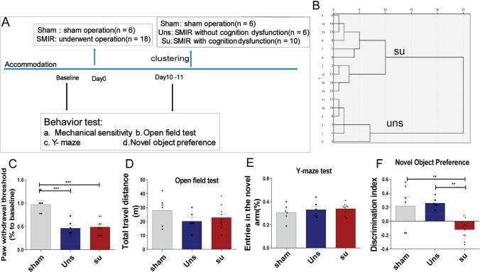
Using a hierarchical clustering analysis on cognitive function, the CPSP rats were divided into two groups: unsusceptible (no cognitive damage) (n = 10) and susceptible (cognitive impairment) (n = 6) 10 days after SMIR surgery (Fig. 1A and B). PWL (paw withdrawal threshold) in rats was dramatically reduced by SMIR surgery (Fig. 1C), although no motor impairment was seen (Fig. 1D). There was no significant difference in the Y-maze test (Fig. 1E). The susceptible rats performed poorly on the cognitive behavior test due to impaired non-spatial visual learning memory (Fig. 1F). According to these findings, SMIR surgery caused severe mechanical hypersensitivity and non-spatial memory problems in rats.
Fig. 1.
Chronic postsurgical pain–induced significant mechanical hypersensitivity and cognitive impairment. A Schematic illustration of the experimental design. B Dendrogram of hierarchical clustering analysis after SMIR surgery. Rats who underwent SMIR surgery were divided into su group (n = 10, cognitively compromised pain rats) and uns group (n = 6, cognitively normal pain rats) by hierarchical clustering analysis on the Y-maze test and the novel object preference test. Behavioral tests after SMIR surgery, including C mechanical sensitivity, D the open field test, E the Y-maze test, and F the novel object preference test. Tukey’s post hoc tests; **p < 0.01, ***p < 0.001, ****p < 0.0001
CPSP Rats with Cognitive Impairments Showed Abnormal Gut Microbiota Composition
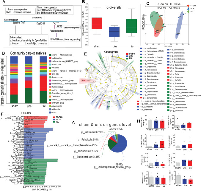
Metabolites produced by gut bacteria have influenced cognitive function [14]. As a result, 16S rRNA gene sequencing was used to analyze the changes in gut microbiota composition following SMIR (Fig. 2A). While bacterial α-diversity did not differ between the three groups (Fig. 2B), the β-diversity (PCoA, R2 = 0.3181, p = 0.003; Fig. 2C) showed notable differences between the three groups, which revealed that pain induced by SMIR or lower cognition induced by pain altered the microbiota composition. In addition, the microbiome structure was considerably altered after SMIR in rats (Fig. 2D and G), and the quantity of SCFAs-producing bacteria [37, 38], particularly Lachnospiraceae and Elusimicrobium, was decreased in the susceptible group (Fig. 2E−G).
Fig. 2.
The CPSP rats with cognitive impairment showed abnormal gut microbiota composition. A Schematic illustration of the experimental design. B Simpson index. C Principal coordinate analysis (PCoA). D The relative abundance of bacteria at the genus level. All genera with an average relative abundance below 1% were grouped to “others.” E–F Linear discriminant analysis effect size (LefSe) analysis: E cladogram and F linear discriminant analysis (LDA); red indicates clades enriched in the sham group, blue indicates clades enriched in the uns group (cognitively normal pain rats), whereas green indicates clades enriched in the su group (cognitively compromised pain rats). G Common species in the sham and uns group at the genus level. H SCFA concentrations in serum
Given the scarcity of SCFA-producing genera in the susceptible group, we looked at SCFAs in the serum of the three groups. Surprisingly, the sensitive rats had greater butyrate concentrations than the sham rats, despite no significant change in other SCFAs across groups (Fig. 2H). These findings suggested that the number of gut bacteria was strongly connected to cognitive impairments.
Microbiota Transplantation Induced Cognitive Impairment Behaviors and Reshaped the Gut Microbiome
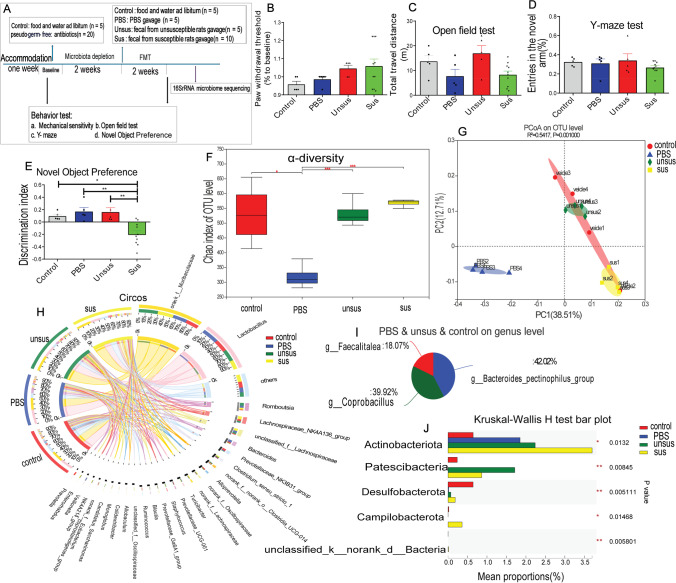
Since the CPSP rats with cognitive impairment had an abnormal gut microbiota composition, the fecal microbiota of unsusceptible rats (CPSP rats without cognitive impairment) or susceptible rats (CPSP rats with cognitive impairment) was transferred to germ-free rats (Fig. 3A). After FMT, recipients of susceptible microbiota demonstrated cognitive deficit behavior in the novel object preference test (Fig. 3E), but not in mechanical sensitivity (Fig. 3B), motor ability (Fig. 3C), and Y-maze test (Fig. 3D). These findings show that the fecal microbiota transplantation from cognitively compromised pain rats can induce the hallmarks of cognitive deficit behavior to germ-free recipient rats.
Fig. 3.
Microbiota from CPSP rats with poor cognition induced cognitive impairment behavior and induced abnormal gut microbiota composition. A Schematic illustration of the experimental design. Effect of microbiota transplantation on the B mechanical sensitivity, C open field test, D Y-maze test, and E novel object preference test. N = 5 for the control, PBS, and unsus group, n = 10 for the sus group. Tukey’s post hoc tests; *p < 0.05, **p < 0.01. F Chaos index, α diversity. G Principal coordinates analysis (PCoA), β diversity. H The proportion of dominant species at the genus level (Circos). I Common species in the control, PBS-treat, susceptible donors-transferred, and susceptible donor–transferred samples at the genus level. J Species with different abundance at the phylum level
As demonstrated in Fig. 3F, the α-diversity of chaos index decreased significantly in PBS-treat pseudo-germ-free rats because of microbiota depletion. While bacterial α-diversity did not differ between unsusceptible donor–transferred and susceptible donors-transferred pseudo-germ-free rats, the β-diversity (PCoA, R2 = 0.5417, p = 0.001; Fig. 3G) showed notable differences between these groups, which revealed that the transplantation of gut microbiota from cognitive impairment rats induced by pain altered the microbiota composition. The structure of the microbiome was significantly altered after fecal transplantation in rats (Fig. 3H), and SCFA-producing bacteria [37, 38], including Bacteroidaceae, Coprobacillus, and Fecalitalea, were prevalent in control, PBS-treat, and the unsusceptible donor–transferred samples, but absent in the majority of susceptible donor–transferred samples (Fig. 3I and J). These findings show a link between gut microbiome disruptions and cognitive abnormalities in chronic postoperative pain rats.
SCFA Intake Ameliorated Chronic Postsurgical Pain-Related Cognitive Deficits
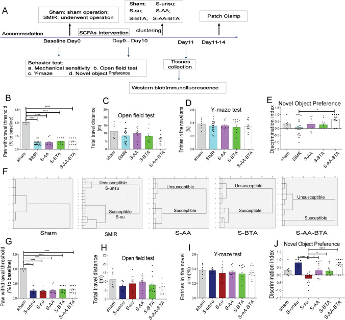
Previous research has revealed that the gut microbiome mediates pain-related cognitive deficits and that the genera that generate SCFAs (acetate and butyrate) were dramatically reduced not only in the susceptible group (Fig. 2) but also in the susceptible-transferred group (Fig. 3). As a result, we postulated that the drop in acetate and butyrate levels in susceptible rats was connected to their cognitive deficiencies. To put this theory to the test, CPSP rats were given acetate and butyrate supplements (Fig. 4A). SCFA supplementation did not influence mechanical sensitivity (Fig. 4B), motor ability (Fig. 4C), or spatial orientation learning memory capacity (Fig. 4D). As can be shown, only acetate and butyrate therapy corrected cognitive deficit behavior in a novel object preference test (Fig. 4E). Surprisingly, the hierarchical clustering analysis (Fig. 4F) revealed that SCFA administration considerably decreased the occurrence of cognitive impairment in the CPSP rats (SMIR group: 72.22%, S-AA group: 54.55%, S-BTA group: 45.46%, S-AA-BTA group: 20%).
Fig. 4.
SCFA treatment alleviated pain-induced cognitive deficits in rats. A Schematic of the SCFA intervention on the pain-induced cognitive deficits. Behavioral phenotypes after SCFA supplementation (acetate and butyrate), including B mechanical sensitivity, C the open field test, D Y-maze test, and E the novel object preference test in the sham group, SMIR group, S-AA group (SMIR rats with acetate), S-BTA group (SMIR rats with butyrate), S-AA-BTA group (SMIR rats with acetate and butyrate). F Dendrogram of hierarchical clustering analysis on the Y-maze test and the novel object preference test. sham group, n = 7; SMIR group, n = 18, rats in SMIR group were divided into S-su group (n = 13, SMIR rats with cognitive dysfunction) and S-unsugroup (n = 5, SMIR rats without cognitive dysfunction) by hierarchical clustering analysis; S-AA group, n = 11; S-BTA group, n = 11; S-AA-BTA group, n = 10. G–J Behavioral phenotypes in CPSP rats after hierarchical clustering analysis, including G mechanical sensitivity, H the open field test, I Y-maze, and J the novel object preference test. Results are expressed as mean ± SEM; Tukey’s post hoc tests; *p < 0.05, ***p < 0.001
Then, by hierarchical clustering, the rats that underwent SMIR surgery were divided into the cognitively normal pain rats (s-unsu group) and cognitively compromised pain rats (s-su group). We next evaluated the difference among these groups. As a result, SCFA therapy considerably enhanced the discrimination index compared to the s-su group (Fig. 4J). In comparison, there was no difference in mechanical sensitivity (Fig. 4G), motor ability (Fig. 4H), or spatial orientation learning memory capacity (Fig. 4I). These findings show that SCFA supplementation can reverse CPSP-induced cognitive impairment.
SCFA Treatment Regulated Histone Acetylation Through the ACCS2-HDAC2 Axis
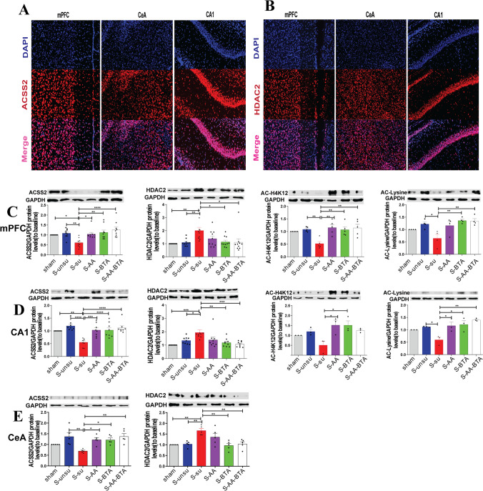
Sodium butyrate, an HDAC inhibitor, has improved memory via increasing histone acetylation [19]. Acetyl-CoA synthetase 2 (ACSS2) is a direct controller of histone acetylation and spatial memory that employs acetate as a source for nuclear acetyl-CoA synthesis [21]. Anatomically, the hippocampus, central amygdala, and medial prefrontal cortex—all involved in cognitive modulation—are also involved in pain control [8, 39, 40]. Given this, we investigated whether applying direct SCFAs to CPSP rats may ameliorate pain-induced cognitive impairments by increasing histone acetylation via the ACCS2-HDAC2 axis in these regions (Fig. 5). ACSS2 and HDAC2 immunostaining demonstrated that they were located in the nucleus (Fig. 6 A and B), consistent with prior results indicating ACSS2 and HDAC2 are abundantly localized to the nucleus of neurons [18, 22]. In the western blot test, we discovered that HDAC2 expression was dramatically raised, while ACSS2 expression was reduced in the mPFC (Fig. 6C), hippocampal CA1 (Fig. 6D), and CeA areas (Fig. 6E) of the susceptible group, but SCFA therapy restored them to normal levels. In keeping with these findings, we noticed reduced acetylation of H4K12 and lysine in the susceptible group’s mPFC and hippocampal CA1 region. Similarly, supplementing with SCFAs restored H4K12 and lysine acetylation. These data suggest that SCFA administration to CPSP rats alleviates pain-induced cognitive impairments by increasing histone acetylation via the ACCS2-HDAC2 axis.
Fig. 5.
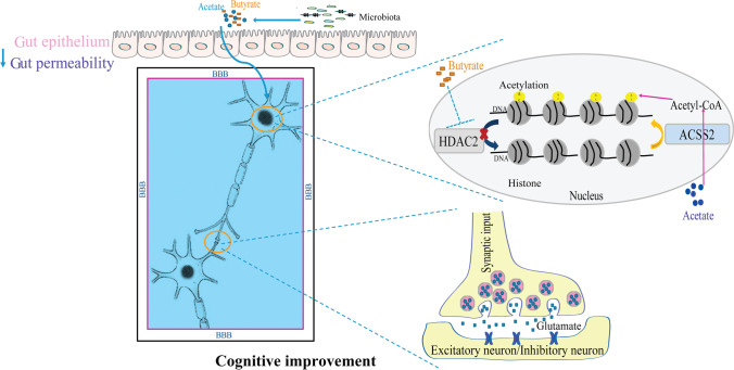
A hypothetical mechanism for SCFAs alleviates pain-related cognitive deficits by improving histone acetylation and synaptic transmission. The gut microbiota metabolites (acetate and butyrate) approach brain tissue by blood flow and reduce permeability to the gut and BBB (blood–brain barrier). In the nucleus, this acetate was converted into acetyl-CoA by ACSS2 (acetyl-CoA synthetase2) to supply carbon for histone acetylation. Butyrate, an HDAC inhibitor, inhibits histone deacetylase activity to increase histone acetylation. Improved histone acetylation restores the excitatory synaptic input to the excitatory and inhibitory neurons in the mPFC, CeA, and hippocampal CA1 areas in the synapses. Then, improved histone acetylation and synaptic transmission alleviate pain-related cognitive deficits
Fig. 6.
SCFA treatment regulated histone acetylation through the ACCS2-HDAC2 axis. A–B Representative immunofluorescent images of ACSS2 (red) and HDAC2 (red) in mPFC, CeA, and hippocampal CA1. Nuclei are stained blue. C Western blot analysis of ACSS2, HDAC2, ace-H4K12, and ace-lysine in mPFC in rats after SCFA treatment. D The expression level of ACSS2, HDAC2, ace-H4K12, and ace-lysine in hippocampal CA1 area after SCFAs treatment. E Representative western blots showing ACSS2 and HDAC2 expression in CeA area after SCFA treatment. Results are expressed as mean ± SEM; Tukey’s post hoc tests; *p < 0.05, **p < 0.01, ***p < 0.001, ****p < 0.0001
SCFA Treatment Restored Synaptic Deficits in Cognitively Compromised Pain Rats
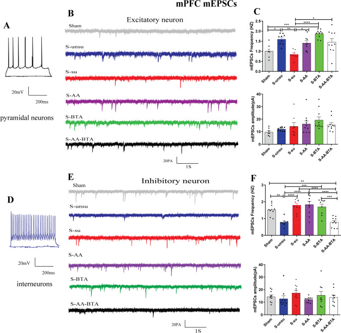
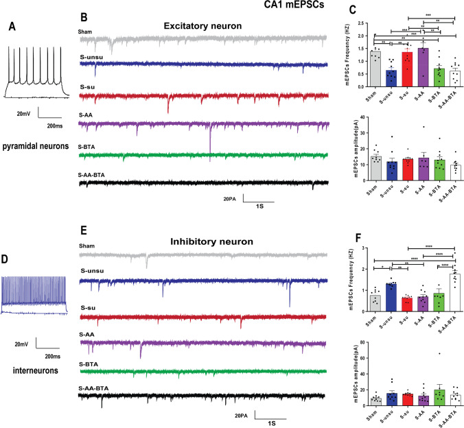
Histone acetylation epigenetic alterations have been connected to dendritic spine density and synapse number [17, 18]. SCFA therapy has been shown to improve synaptic function in the hippocampus and PFC, hence restoring maternal obesity-induced cognitive-behavioral impairments in children [9]. The next step was to see if SCFA therapy might reverse synaptic impairments in pain rats with cognitive impairment. Excitatory pyramidal neurons and inhibitory interneurons in mPFC (Fig. 7A and D), CeA (Fig. 8A and D), and hippocampal CA1 (Fig. 9A and D) were identified by accommodating responses to a sustained depolarizing intracellular current stimulation. An examination of miniature excitatory postsynaptic currents (mEPSCs) on mPFC excitatory neurons indicated that the frequency of mEPSCs in the susceptible group was considerably lower than in the unsusceptible group, despite no discernible change in amplitude (Fig. 7B and C). Significantly, acetate and butyrate supplementation increased the frequency of mEPSCs from the mPFC excitatory neurons in the S-AA, S-BTA, and S-AA-BTA groups (Fig. 7E and F).
Fig. 7.
SCFA treatment rescued synaptic deficits in layer II/III of the mPFC neurons in cognitively compromised pain rats. A–C Effect of the SCFAs intervention on the spontaneous synaptic transmission of excitatory neurons in pain-induced cognitive deficits rats. A Current-clamp recordings to identify excitatory neurons in the mPFC slices. B Original traces of mEPSCs of excitatory neurons recorded from the six groups. C Bar graphs showing the frequency and the amplitude of mEPSCs in mPFC excitatory neurons. N = 5 rats for each group. D–F Spontaneous synaptic transmission of the mPFC inhibitory neurons in slices. D Current-clamp recordings to identify inhibitory neurons in the mPFC slices. E Original traces of mEPSCs in slices from the six groups. F Bar graphs showing the frequency and the amplitude of mEPSCs in mPFC inhibitory neurons. Results are expressed as mean ± SEM; n = 5 rats for each group. Tukey’s post hoc tests; *p < 0.05, **p < 0.01, ***p < 0.001, ****p < 0.0001
Fig. 8.
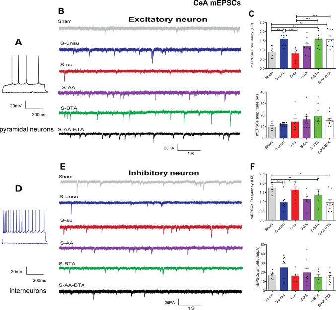
SCFA treatment improved synaptic deficits in CeA neurons in cognitively compromised pain rats. A–C Effect of the SCFAs intervention on the spontaneous synaptic transmission of excitatory neurons in pain-induced cognitive deficits rats. A Current-clamp recordings to identify excitatory neurons in the CeA. B Examples of mEPSCs of excitatory neurons recorded from the six groups. C Bar graphs showing the frequency and the amplitude of mEPSCs in CeA excitatory neurons. N = 5 rats for each group. D–F Effect of the SCFA intervention on the spontaneous synaptic transmission of CeA inhibitory neurons in slices. D Current-clamp recordings to identify inhibitory neurons in the CeA. E Examples of mEPSCs of inhibitory neurons recorded from the six groups. F Bar graphs showing the frequency and the amplitude of mEPSCs in the inhibitory neurons. Results are expressed as mean ± SEM; n = 5 rats for each group. Tukey’s post hoc tests; *p < 0.05, **p < 0.01, ***p < 0.001, ****p < 0.0001
Fig. 9.
SCFA treatment restored synaptic deficits in hippocampal CA1 neurons in cognitively compromised pain rats. A–C Effect of the SCFA intervention on the spontaneous synaptic transmission of excitatory neurons in hippocampal CA1 slices neurons. A Current-clamp recordings to identify excitatory neurons in the hippocampal CA1. B Representative mEPSC recording traces from the six groups. C Bar graphs showing the frequency and the amplitude of mEPSCs in hippocampal CA1 excitatory neurons. N = 5 rats for each group. D–F Effect of the SCFA intervention on the spontaneous synaptic transmission of hippocampal CA1 inhibitory neurons. D Current-clamp recordings to identify inhibitory neurons in the hippocampal CA1. E Representative mEPSCs of inhibitory neurons recording traces from the six groups. F Bar graphs showing the frequency and the amplitude of mEPSCs in hippocampal CA1 inhibitory neurons. Results are expressed as mean ± SEM; n = 5 rats for each group. Tukey’s post hoc tests; *p < 0.05, **p < 0.01, ***p < 0.001, ****p < 0.0001
Given that GABAergic inhibition is essential in controlling pain and cognitive functions [41, 42], we investigated the effect of SCFAs on inhibitory neurons. Consistently, acetate and butyrate supplementation substantially reduced the mEPSC frequency of the mPFC inhibitory neurons in the S-AA-BTA group (Fig. 7E and F). In the susceptible group, however, the frequency of mEPSCs in the mPFC inhibitory neurons was considerably higher than in the unsusceptible group.
We examined at the same time and looked at changes in synaptic transmission in CeA and hippocampal CA1 excitatory and inhibitory neurons. As indicated in Fig. 8B and C, acetate and butyrate supplementation increased the frequency of mEPSCs in CeA excitatory neurons in the S-AA, S-BTA, and S-AA-BTA groups. Surprisingly, SCFA administration did not affect the frequency of mEPSCs in CeA inhibitory neurons (Fig. 8E and F). Furthermore, compared to the unsusceptible group, the susceptible group’s mEPSC frequency of hippocampal CA1 excitatory neurons was considerably higher (Fig. 9B and C). Interestingly, butyrate, but not acetate, reversed synaptic impairments in hippocampal CA1 excitatory neurons in rats with pain-induced cognitive abnormalities. Meanwhile, acetate and butyrate supplementation improved synaptic impairments in hippocampal CA1 inhibitory neurons (Fig. 9E and F). These findings underscore the importance of SCFAs in making chronic postoperative rats resistant to cognitive deficits.
Discussion
Cognitive impairment is one of the most prevalent postoperative consequences of chronic pain [1–3], but the underlying processes and effective therapies are still unknown. This investigation discovered that about 60% of CPSP rats had cognitive impairment, which was consistent with earlier clinical studies [43, 44]. Furthermore, we discovered that CPSP rats with cognitive impairment had an aberrant composition of gut microbiota and a lower number of SCFA-producing bacteria using 16S rRNA sequencing of microbiota. Then, gut microbiota transplantation from cognitively impaired CPSP rats was demonstrated to transmit cognitive impairment behavior. These findings suggested a link between gut microbiota composition and cognitive-behavioral alterations.
Due to the lower number of SCFA-producing bacteria in susceptible donors and susceptible donor-transferred recipient rats, SCFAs, primarily acetate and butyrate, were supplied further. Then, it comes as little surprise that SCFA therapy reduced pain-induced cognitive impairments in rats. In search of molecular reasons for the beneficial intervention, we discovered that SCFA administration reversed histone acetylation and synaptic impairments in mPFC, CeA, and hippocampal CA1 neurons in rats with pain-induced cognitive deficiencies via the ACCS2-HDAC2 axis. Short-chain fatty acids (SCFAs), which are major metabolites of the microbiota during anaerobic fermentation, may help to alleviate persistent postsurgical pain-induced cognitive impairments by increasing histone acetylation and synaptic transmission. It is worth noting that we found no difference in pain threshold following gut microbiota colonization and SCFA therapy. However, experimental investigations have shown that a histone deacetylase inhibitor reduced neuropathic pain by increasing histone acetylation. This disparity might be attributed to the study’s use of the non-neuropathic pain model [45].
The microbiota has previously been demonstrated to regulate cognitive function [9, 10], and mother-to-offspring transmission of the gut microbiome has been shown to mediate maternal high-fat diet-induced learning and memory problems in offspring [9]. Furthermore, fecal microbiota transplantation has been shown to enhance cognitive performance and lower liver function indices in rats with hepatic encephalopathy [46]. In line with this, we demonstrated in the current work that transplanting the gut microbiota from CPSP rats with cognitive impairment transmitted cognitive impairment behavior. We also discovered a reduction in the number of SCFA-producing bacteria in susceptible donors and susceptible donor-transferred recipient rats. These findings were in harmony with studies by Liu and Ragusa [9, 47].
Interestingly, the butyrate concentration was greater in the CPSP rats with cognitive impairment, despite no significant difference in other SCFAs across groups. Notably, any gut microbe-brain interaction must pass through two barriers (the gut epithelial barrier and the blood–brain barrier), and permeability through these barriers has been demonstrated to be affected by the microbiome [11, 14]. SCFAs, main butyrate, have been shown to strengthen the integrity of the epithelial barrier and the blood–brain barrier [14]. As a result, it is reasonable to speculate that the rise in butyrate in the serum was caused by increased intestinal permeability rather than the excessive fermentation reported in CPSP rats with cognitive impairment [48]. Of course, whether the increase in butyrate in the serum was caused by increased intestinal permeability and excessive fermentation has to be investigated further.
Histone acetylation epigenetic changes have been shown to have a crucial function in numerous cognitive processes due to their pivotal involvement in neural development [49]. Acetylation and deacetylation are required for proper acetylation. HDAC inhibitors or HDAC2 knock-down have been proven to ameliorate memory dysfunction in mice with HDAC2 overexpression [50]. It was shown that lipid-derived acetyl-CoA, which may be formed from SCFAs catalyzed by acetyl-CoA synthetase 2 (ACSS2), is a genuine source (up to 90%) of carbon for histone acetylation, even in the presence of abundant glucose, using 13C-carbon tracing and acetyl-proteomics histone acetylation [20, 21]. Given that ACSS2 requires acetate for nuclear Acetyl-CoA production and sodium butyrate is an HDAC inhibitor, it is highly plausible that acetate and butyrate supplementation may improve cognitive impairments via increasing histone acetylation. In keeping with this idea, we discovered that SCFA therapy restored HDAC2 and ACSS2 expression to normal levels.
Similarly, SCFA supplementation improved H4K12 and lysine acetylation, both related to learning and memory [18, 51]. Notably, the supplement of butyrate, an HDAC inhibitor, restored not only HDAC2 but also ACSS2 expression in this study. These findings might be explained in part by the fact that butyrate can be catalyzed by ACSS2 into butyryl-CoA, which plays a critical role in immunological homeostasis maintenance [52].
Acetylation of lysine is critical for synaptic plasticity, and histone acetylation level has been shown to calibrate the correct number of released vesicles and synaptic transmission [53]. Upregulation of histone deacetylase 2 (HDAC2) activity has been associated with dendritic spine density, synapse number, and expression of various cognitive-related genes [17, 18]. Our electrophysiological recordings in brain slices revealed variations in the frequency of mEPSCs in excitatory and inhibitory neurons from the rats with cognitive dysfunction, indicating changes in the number of vesicles released. Meanwhile, SCFA supplementation restored these alterations. These results are consistent with previous studies suggesting HDAC2’s epigenetic regulation of excitatory and inhibitory synaptic transmission played a vital role in modulating cognitive impairment [54]. Thus, the influence of SCFA supplementation on synaptic transmission may be due to the correct number of released vesicles mediated by appropriate acetylation. It is worth noting that the bulk of acetate metabolism in the brain happens in astrocytes, with neurons accounting for just around 30% of the total [21, 55]. As a result, additional research is needed to investigate the role of astrocytes in the development of pain-induced cognitive impairment. Another primary question was that we found synaptic transmission differences between pain animals with cognitive impairments and those without; however, no differences were detected between cognitively affected pain animals and sham controls. This surprising fact might be attributed to the fact that chronic pain and cognitive dysfunction may have opposite relations with the synaptic transmission in the central nervous system. The literature consistently showed a positive correlation between synaptic transmission and pain [56–59], while a negative correlation between synaptic transmission and cognitive impairment in variable models [60–63].
Conclusions
To summarize, our findings show that pain-induced cognitive impairments are caused by changes in the makeup of the gut microbiota, and SCFA therapy alleviates pain-induced cognitive deficits by improving histone acetylation and synaptic transmission in brain. It may provide a more detailed knowledge of the underlying epigenetic mechanisms that link gut microbiota, histone acetylation, and pain-related cognitive impairments and suggest that SCFAs might be a viable non-invasive and tractable therapy for cognitive deficiencies. Overall, this finding opens up a new research avenue into preemptive therapies for pain-related cognitive impairments and supports the use of SCFA-dependent perioperative interventions for postoperative rehabilitation in the clinic.
Abbreviations
- ACSS2
Acetyl-CoA synthetase2
- CeA
Central amygdala
- CPSP
Chronic postsurgical pain
- FMT
Faecal microbial transplantation
- HDAC2
Histone deacetylase 2
- mEPSCs
Miniature excitatory postsynaptic currents
- mPFC
Medial prefrontal cortex
- PCoA
Principal co-ordinate analysis
- PWL
Paw withdrawal threshold
- SCFAs
Short-chain fatty acids
- SMIR
Skin/muscle incision and retraction
Author Contribution
Z. L., J. W., and H. B. X. conceived the project and designed the experiments; Z. L., T. N. S., Z. G. H., Z. X. L., and W. C. Z. performed the experiments; Z. L. and T. N. S. analyzed the data; Z. L., J. W., and H. B. X. wrote the manuscript. All authors revised and approved the final version of the article.
Funding
This work was supported by the National Natural Science Foundation of China (No. 81873467, 81670240).
Data Availability
All relevant data supporting the findings of this study are available from the corresponding author on reasonable request.
Declarations
Ethics Approval
All experimental procedures were approved by the Animal Care and Use Committee, Tongji Hospital.
Consent to Participate
Not applicable.
Consent for Publication
Not applicable.
Competing Interests
The authors declare no competing interests.
Footnotes
Publisher's Note
Springer Nature remains neutral with regard to jurisdictional claims in published maps and institutional affiliations.
Contributor Information
Jie Wang, Email: jie.wang@wipm.ac.cn.
Hongbing Xiang, Email: hbxiang@tjh.tjmu.edu.cn.
References
- 1.Suto T, Eisenach JC, Hayashida K. Peripheral nerve injury and gabapentin, but not their combination, impair attentional behavior via direct effects on noradrenergic signaling in the brain. Pain. 2014;155(10):1935–1942. doi: 10.1016/j.pain.2014.05.014. [DOI] [PMC free article] [PubMed] [Google Scholar]
- 2.Cardoso-Cruz H, Sousa M, Vieira JB, Lima D, Galhardo V. Prefrontal cortex and mediodorsal thalamus reduced connectivity is associated with spatial working memory impairment in rats with inflammatory pain. Pain. 2013;154(11):2397–2406. doi: 10.1016/j.pain.2013.07.020. [DOI] [PubMed] [Google Scholar]
- 3.Kehlet H. Postoperative pain, analgesia, and recovery-bedfellows that cannot be ignored. Pain. 2018;159(Suppl 1):S11–S16. doi: 10.1097/j.pain.0000000000001243. [DOI] [PubMed] [Google Scholar]
- 4.Kehlet H, Jensen TS, Woolf CJ. Persistent postsurgical pain: risk factors and prevention. The Lancet. 2006;367(9522):1618–1625. doi: 10.1016/s0140-6736(06)68700-x. [DOI] [PubMed] [Google Scholar]
- 5.Matthewson GM, Woo CW, Reddan MC, Wager TD. Cognitive self-regulation influences pain-related physiology. Pain. 2019;160(10):2338–2349. doi: 10.1097/j.pain.0000000000001621. [DOI] [PubMed] [Google Scholar]
- 6.Howlin C, Rooney B. The cognitive mechanisms in music listening interventions for pain: a scoping review. J Music Ther. 2020;57(2):127–167. doi: 10.1093/jmt/thaa003. [DOI] [PubMed] [Google Scholar]
- 7.Bushnell MC, Ceko M, Low LA. Cognitive and emotional control of pain and its disruption in chronic pain. Nat Rev Neurosci. 2013;14(7):502–511. doi: 10.1038/nrn3516. [DOI] [PMC free article] [PubMed] [Google Scholar]
- 8.Phelps CE, Navratilova E, Porreca F. Cognition in the chronic pain experience: preclinical insights. Trends Cogn Sci. 2021;25(5):365–376. doi: 10.1016/j.tics.2021.01.001. [DOI] [PMC free article] [PubMed] [Google Scholar]
- 9.Liu X, Li X, Xia B, Jin X, Zou Q, Zeng Z, et al. (2021) High-fiber diet mitigates maternal obesity-induced cognitive and social dysfunction in the offspring via gut-brain axis. Cell Metab 33(5):923-938 e926 . 10.1016/j.cmet.2021.02.002 [DOI] [PubMed]
- 10.Liu Z, Dai X, Zhang H, Shi R, Hui Y, Jin X, et al. Gut microbiota mediates intermittent-fasting alleviation of diabetes-induced cognitive impairment. Nat Commun. 2020;11(1):855. doi: 10.1038/s41467-020-14676-4. [DOI] [PMC free article] [PubMed] [Google Scholar]
- 11.Braniste V, Al-Asmakh M, Kowal C, Anuar F, Abbaspour A, Toth M, et al. The gut microbiota influences blood-brain barrier permeability in mice. Sci Transl Med. 2014;6(263):263ra158. doi: 10.1126/scitranslmed.3009759. [DOI] [PMC free article] [PubMed] [Google Scholar]
- 12.De Vadder F, Kovatcheva-Datchary P, Goncalves D, Vinera J, Zitoun C, Duchampt A, et al. Microbiota-generated metabolites promote metabolic benefits via gut-brain neural circuits. Cell. 2014;156(1–2):84–96. doi: 10.1016/j.cell.2013.12.016. [DOI] [PubMed] [Google Scholar]
- 13.Diaz Heijtz R, Wang S, Anuar F, Qian Y, Bjorkholm B, Samuelsson A, et al. Normal gut microbiota modulates brain development and behavior. Proc Natl Acad Sci U S A. 2011;108(7):3047–3052. doi: 10.1073/pnas.1010529108. [DOI] [PMC free article] [PubMed] [Google Scholar]
- 14.Stilling RM, van de Wouw M, Clarke G, Stanton C, Dinan TG, Cryan JF. The neuropharmacology of butyrate: the bread and butter of the microbiota-gut-brain axis? Neurochem Int. 2016;99:110–132. doi: 10.1016/j.neuint.2016.06.011. [DOI] [PubMed] [Google Scholar]
- 15.Rose S, Bennuri SC, Davis JE, Wynne R, Slattery JC, Tippett M, et al. Butyrate enhances mitochondrial function during oxidative stress in cell lines from boys with autism. Transl Psychiatry. 2018;8(1):42. doi: 10.1038/s41398-017-0089-z. [DOI] [PMC free article] [PubMed] [Google Scholar]
- 16.Ji M, Dong L, Jia M, Liu W, Zhang M, Ju L, et al. Epigenetic enhancement of brain-derived neurotrophic factor signaling pathway improves cognitive impairments induced by isoflurane exposure in aged rats. Mol Neurobiol. 2014;50(3):937–944. doi: 10.1007/s12035-014-8659-z. [DOI] [PMC free article] [PubMed] [Google Scholar]
- 17.Bie B, Wu J, Yang H, Xu JJ, Brown DL, Naguib M. Epigenetic suppression of neuroligin 1 underlies amyloid-induced memory deficiency. Nat Neurosci. 2014;17(2):223–231. doi: 10.1038/nn.3618. [DOI] [PubMed] [Google Scholar]
- 18.Guan JS, Haggarty SJ, Giacometti E, Dannenberg JH, Joseph N, Gao J, et al. HDAC2 negatively regulates memory formation and synaptic plasticity. Nature. 2009;459(7243):55–60. doi: 10.1038/nature07925. [DOI] [PMC free article] [PubMed] [Google Scholar]
- 19.Vinarskaya AK, Balaban PM, Roshchin MV, Zuzina AB. Sodium butyrate as a selective cognitive enhancer for weak or impaired memory. Neurobiol Learn Mem. 2021;180:107414. doi: 10.1016/j.nlm.2021.107414. [DOI] [PubMed] [Google Scholar]
- 20.McDonnell E, Crown SB, Fox DB, Kitir B, Ilkayeva OR, Olsen CA, et al. Lipids reprogram metabolism to become a major carbon source for histone acetylation. Cell Rep. 2016;17(6):1463–1472. doi: 10.1016/j.celrep.2016.10.012. [DOI] [PMC free article] [PubMed] [Google Scholar]
- 21.Bradshaw PC 2021 Acetyl-CoA metabolism and histone acetylation in the regulation of aging and lifespan. Antioxidants (Basel) 10(4). 10.3390/antiox10040572 [DOI] [PMC free article] [PubMed]
- 22.Mews P, Donahue G, Drake AM, Luczak V, Abel T, Berger SL. Acetyl-CoA synthetase regulates histone acetylation and hippocampal memory. Nature. 2017;546(7658):381–386. doi: 10.1038/nature22405. [DOI] [PMC free article] [PubMed] [Google Scholar]
- 23.Liu T, Li Z, He J, Yang N, Han D, Li Y, et al. Regional metabolic patterns of abnormal postoperative behavioral performance in aged mice assessed by (1)H-NMR dynamic mapping method. Neurosci Bull. 2020;36(1):25–38. doi: 10.1007/s12264-019-00414-4. [DOI] [PMC free article] [PubMed] [Google Scholar]
- 24.Mathiasen JR, DiCamillo A 2010 Novel object recognition in the rat: a facile assay for cognitive function. Curr Protoc Pharmacol Chapter 5:Unit 5 59. 10.1002/0471141755.ph0559s49 [DOI] [PubMed]
- 25.Yuan DS, Huang YQ, Fu YJ, Xie J, Huang YL, Zhou SS, et al. Hydrogen sulfide alleviates cognitive deficiency and hepatic dysfunction in a mouse model of acute liver failure. Exp Ther Med. 2020;20(1):671–677. doi: 10.3892/etm.2020.8680. [DOI] [PMC free article] [PubMed] [Google Scholar]
- 26.Li Z, Li C, Yin P, Wang ZJ, Luo F. Inhibition of CaMKIIalpha in the central nucleus of amygdala attenuates fentanyl-induced hyperalgesia in rats. J Pharmacol Exp Ther. 2016;359(1):82–89. doi: 10.1124/jpet.116.233817. [DOI] [PubMed] [Google Scholar]
- 27.Li Z, Yin P, Chen J, Li C, Liu J, Rambojan H, et al. Activation of the extracellular signal-regulated kinase in the amygdale modulates fentanyl-induced hypersensitivity in Rats. J Pain. 2017;18(2):188–199. doi: 10.1016/j.jpain.2016.10.013. [DOI] [PubMed] [Google Scholar]
- 28.Xiong B, Zhang W, Zhang L, Huang X, Zhou W, Zou Q, et al. Hippocampal glutamatergic synapses impairment mediated novel-object recognition dysfunction in rats with neuropathic pain. Pain. 2020;161(8):1824–1836. doi: 10.1097/j.pain.0000000000001878. [DOI] [PubMed] [Google Scholar]
- 29.Ji G, Sun H, Fu Y, Li Z, Pais-Vieira M, Galhardo V, et al. Cognitive impairment in pain through amygdala-driven prefrontal cortical deactivation. J Neurosci. 2010;30(15):5451–5464. doi: 10.1523/JNEUROSCI.0225-10.2010. [DOI] [PMC free article] [PubMed] [Google Scholar]
- 30.Flatters SJ. Characterization of a model of persistent postoperative pain evoked by skin/muscle incision and retraction (SMIR) Pain. 2008;135(1–2):119–130. doi: 10.1016/j.pain.2007.05.013. [DOI] [PMC free article] [PubMed] [Google Scholar]
- 31.Li Y, Zhang W, Sun T, Liu B, Manyande A, Xu W, et al. The role of gut microbiota in chronic itch-evoked novel object recognition-related cognitive dysfunction in mice. Front Med. 2021;8:616489. doi: 10.3389/fmed.2021.616489. [DOI] [PMC free article] [PubMed] [Google Scholar]
- 32.Tillmann S, Abildgaard A, Winther G, Wegener G. Altered fecal microbiota composition in the Flinders sensitive line rat model of depression. Psychopharmacology. 2019;236(5):1445–1457. doi: 10.1007/s00213-018-5094-2. [DOI] [PMC free article] [PubMed] [Google Scholar]
- 33.Cammarota G, Ianiro G, Tilg H, Rajilic-Stojanovic M, Kump P, Satokari R, et al. European consensus conference on faecal microbiota transplantation in clinical practice. Gut. 2017;66(4):569–580. doi: 10.1136/gutjnl-2016-313017. [DOI] [PMC free article] [PubMed] [Google Scholar]
- 34.Li Z, Yin P, Chen J, Jin S, Liu J, Luo F. CaMKIIalpha may modulate fentanyl-induced hyperalgesia via a CeLC-PAG-RVM-spinal cord descending facilitative pain pathway in rats. PLoS ONE. 2017;12(5):e0177412. doi: 10.1371/journal.pone.0177412. [DOI] [PMC free article] [PubMed] [Google Scholar]
- 35.Laviolette SR, Grace AA. Cannabinoids potentiate emotional learning plasticity in neurons of the medial prefrontal cortex through basolateral amygdala inputs. J Neurosci. 2006;26(24):6458–6468. doi: 10.1523/jneurosci.0707-06.2006. [DOI] [PMC free article] [PubMed] [Google Scholar]
- 36.Koyanagi Y, Oi Y, Kobayashi M. Fast-spiking interneurons contribute to Propofol-induced facilitation of firing synchrony in pyramidal neurons of the rat insular cortex. Anesthesiology. 2021;134(2):219–233. doi: 10.1097/ALN.0000000000003653. [DOI] [PubMed] [Google Scholar]
- 37.Wang Y, Li N, Yang JJ, Zhao DM, Chen B, Zhang GQ, et al. Probiotics and fructo-oligosaccharide intervention modulate the microbiota-gut brain axis to improve autism spectrum reducing also the hyper-serotonergic state and the dopamine metabolism disorder. Pharmacol Res. 2020;157:104784. doi: 10.1016/j.phrs.2020.104784. [DOI] [PubMed] [Google Scholar]
- 38.LeBlanc JG, Chain F, Martin R, Bermudez-Humaran LG, Courau S, Langella P. Beneficial effects on host energy metabolism of short-chain fatty acids and vitamins produced by commensal and probiotic bacteria. Microb Cell Fact. 2017;16(1):79. doi: 10.1186/s12934-017-0691-z. [DOI] [PMC free article] [PubMed] [Google Scholar]
- 39.McCarberg B, Peppin J. Pain pathways and nervous system plasticity: learning and memory in pain. Pain Med. 2019;20(12):2421–2437. doi: 10.1093/pm/pnz017. [DOI] [PubMed] [Google Scholar]
- 40.Moriarty O, Finn DP. Cognition and pain. Curr Opin Support Palliat Care. 2014;8(2):130–136. doi: 10.1097/SPC.0000000000000054. [DOI] [PubMed] [Google Scholar]
- 41.Bravo-Hernandez M, Corleto JA, Barragan-Iglesias P, Gonzalez-Ramirez R, Pineda-Farias JB, Felix R, et al. The alpha5 subunit containing GABAA receptors contribute to chronic pain. Pain. 2016;157(3):613–626. doi: 10.1097/j.pain.0000000000000410. [DOI] [PMC free article] [PubMed] [Google Scholar]
- 42.Wang DS, Zurek AA, Lecker I, Yu J, Abramian AM, Avramescu S, et al. Memory deficits induced by inflammation are regulated by alpha5-subunit-containing GABAA receptors. Cell Rep. 2012;2(3):488–496. doi: 10.1016/j.celrep.2012.08.022. [DOI] [PMC free article] [PubMed] [Google Scholar]
- 43.Berryman C, Stanton TR, Jane Bowering K, Tabor A, McFarlane A, Lorimer Moseley G. Evidence for working memory deficits in chronic pain: a systematic review and meta-analysis. Pain. 2013;154(8):1181–1196. doi: 10.1016/j.pain.2013.03.002. [DOI] [PubMed] [Google Scholar]
- 44.Dick BD, Rashiq S. Disruption of attention and working memory traces in individuals with chronic pain. Anesth Analg. 2007;104(5):1223–1229 . doi: 10.1213/01.ane.0000263280.49786.f5. [DOI] [PubMed] [Google Scholar]
- 45.Khangura RK, Bali A, Jaggi AS, Singh N. Histone acetylation and histone deacetylation in neuropathic pain: an unresolved puzzle? Eur J Pharmacol. 2017;795:36–42. doi: 10.1016/j.ejphar.2016.12.001. [DOI] [PubMed] [Google Scholar]
- 46.Wang WW, Zhang Y, Huang XB, You N, Zheng L, Li J. Fecal microbiota transplantation prevents hepatic encephalopathy in rats with carbon tetrachloride-induced acute hepatic dysfunction. World J Gastroenterol. 2017;23(38):6983–6994. doi: 10.3748/wjg.v23.i38.6983. [DOI] [PMC free article] [PubMed] [Google Scholar]
- 47.Ragusa M, Santagati M, Mirabella F, Lauretta G, Cirnigliaro M, Brex D, et al. 2020 Potential associations among alteration of salivary miRNAs, saliva microbiome structure, and cognitive impairments in autistic children. Int J Mol Sci 21(17). 10.3390/ijms21176203 [DOI] [PMC free article] [PubMed]
- 48.Bourassa MW, Alim I, Bultman SJ, Ratan RR. Butyrate, neuroepigenetics and the gut microbiome: Can a high fiber diet improve brain health? Neurosci Lett. 2016;625:56–63. doi: 10.1016/j.neulet.2016.02.009. [DOI] [PMC free article] [PubMed] [Google Scholar]
- 49.Bonnaud EM, Suberbielle E, Malnou CE. Histone acetylation in neuronal (dys)function. Biomol Concepts. 2016;7(2):103–116. doi: 10.1515/bmc-2016-0002. [DOI] [PubMed] [Google Scholar]
- 50.Wei J, Xiong Z, Lee JB, Cheng J, Duffney LJ, Matas E, et al. Histone modification of Nedd4 ubiquitin ligase controls the loss of AMPA receptors and cognitive impairment induced by repeated stress. J Neurosci. 2016;36(7):2119–2130. doi: 10.1523/JNEUROSCI.3056-15.2016. [DOI] [PMC free article] [PubMed] [Google Scholar]
- 51.Peleg S, Sananbenesi F, Zovoilis A, Burkhardt S, Bahari-Javan S, Agis-Balboa RC, et al. Altered histone acetylation is associated with age-dependent memory impairment in mice. Science. 2010;328(5979):753–756. doi: 10.1126/science.1186088. [DOI] [PubMed] [Google Scholar]
- 52.Hao F, Tian M, Zhang X, Jin X, Jiang Y, Sun X, et al. 2021 Butyrate enhances CPT1A activity to promote fatty acid oxidation and iTreg differentiation. Proc Natl Acad Sci U S A 118(22). 10.1073/pnas.2014681118 [DOI] [PMC free article] [PubMed]
- 53.LoPresti P 2020) HDAC6 in diseases of cognition and of neurons. Cells 10(1). 10.3390/cells10010012 [DOI] [PMC free article] [PubMed]
- 54.Hanson JE, Deng L, Hackos DH, Lo SC, Lauffer BE, Steiner P, et al. Histone deacetylase 2 cell autonomously suppresses excitatory and enhances inhibitory synaptic function in CA1 pyramidal neurons. J Neurosci. 2013;33(14):5924–5929. doi: 10.1523/JNEUROSCI.3162-12.2013. [DOI] [PMC free article] [PubMed] [Google Scholar]
- 55.Rowlands BD, Klugmann M, Rae CD. Acetate metabolism does not reflect astrocytic activity, contributes directly to GABA synthesis, and is increased by silent information regulator 1 activation. J Neurochem. 2017;140(6):903–918. doi: 10.1111/jnc.13916. [DOI] [PubMed] [Google Scholar]
- 56.Blom SM, Pfister JP, Santello M, Senn W, Nevian T. Nerve injury-induced neuropathic pain causes disinhibition of the anterior cingulate cortex. J Neurosci. 2014;34(17):5754–5764. doi: 10.1523/JNEUROSCI.3667-13.2014. [DOI] [PMC free article] [PubMed] [Google Scholar]
- 57.Cordeiro Matos S, Zhang Z, Seguela P. Peripheral neuropathy induces HCN channel dysfunction in pyramidal neurons of the medial prefrontal cortex. J Neurosci. 2015;35(38):13244–13256. doi: 10.1523/JNEUROSCI.0799-15.2015. [DOI] [PMC free article] [PubMed] [Google Scholar]
- 58.Hung KL, Wang SJ, Wang YC, Chiang TR, Wang CC. Upregulation of presynaptic proteins and protein kinases associated with enhanced glutamate release from axonal terminals (synaptosomes) of the medial prefrontal cortex in rats with neuropathic pain. Pain. 2014;155(2):377–387. doi: 10.1016/j.pain.2013.10.026. [DOI] [PubMed] [Google Scholar]
- 59.Wang YJ, Liu MG, Wang JH, Cao W, Wu C, Wang ZY, et al. Restoration of cingulate long-term depression by enhancing non-apoptotic caspase 3 alleviates peripheral pain Hypersensitivity. Cell Rep. 2020;33(6):108369. doi: 10.1016/j.celrep.2020.108369. [DOI] [PubMed] [Google Scholar]
- 60.Tantra M, Guo L, Kim J, Zainolabidin N, Eulenburg V, Augustine GJ, et al. Conditional deletion of Cadherin 13 perturbs Golgi cells and disrupts social and cognitive behaviors. Genes Brain Behav. 2018;17(6):e12466. doi: 10.1111/gbb.12466. [DOI] [PMC free article] [PubMed] [Google Scholar]
- 61.Schofield CM, Hsu R, Barker AJ, Gertz CC, Blelloch R, Ullian EM. Monoallelic deletion of the microRNA biogenesis gene Dgcr8 produces deficits in the development of excitatory synaptic transmission in the prefrontal cortex. Neural Dev. 2011;6:11. doi: 10.1186/1749-8104-6-11. [DOI] [PMC free article] [PubMed] [Google Scholar]
- 62.Wong TP, Marchese G, Casu MA, Ribeiro-da-Silva A, Cuello AC, De Koninck Y. Imbalance towards inhibition as a substrate of aging-associated cognitive impairment. Neurosci Lett. 2006;397(1–2):64–68. doi: 10.1016/j.neulet.2005.11.055. [DOI] [PubMed] [Google Scholar]
- 63.Ding JJ, Zou RX, He HM, Lou ZY, Xu Y, Wang HL. Pb inhibits hippocampal synaptic transmission via cyclin-dependent kinase-5 dependent Synapsin 1 phosphorylation. Toxicol Lett. 2018;296:125–131. doi: 10.1016/j.toxlet.2018.08.009. [DOI] [PubMed] [Google Scholar]
Associated Data
This section collects any data citations, data availability statements, or supplementary materials included in this article.
Data Availability Statement
All relevant data supporting the findings of this study are available from the corresponding author on reasonable request.


