

Case Presentation
A 60-year-old male with skin phototype IV from Africa and a history of Kaposi sarcoma on his foot presented with a rapidly growing, 6.5-mm, bluish-purple nodule on his back. Cross-polarized dermoscopy using a DermLite DL4 (3Gen) coupled with an iPhone XS Max (Apple) revealed a ‘rainbow pattern’ on a structureless red background. By rotating the dermoscope without moving the smartphone during imaging, also known as dynamic cross-polarized dermoscopy, the color distribution within the rainbow pattern changed substantially (Figure 1, Supplementary Video 1). The lesion was excised and histopathologically confirmed as Kaposi sarcoma.
Figure 1.

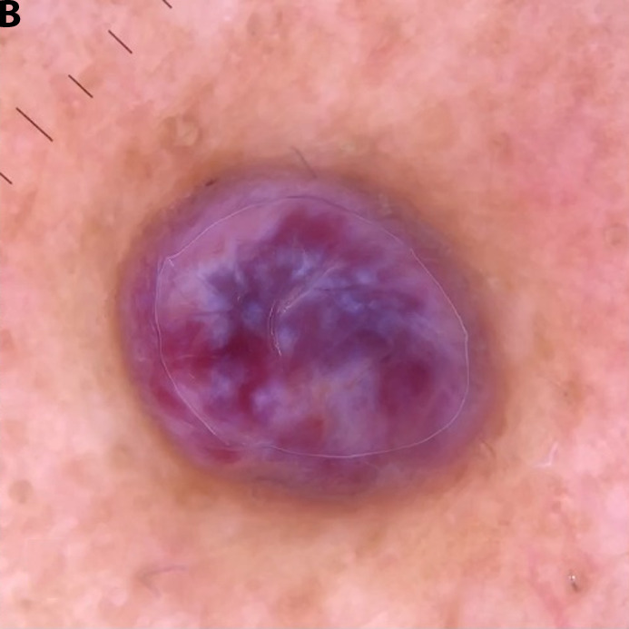

(A) Clinical presentation of a case of Kaposi sarcoma. (B) Iridescence or ‘rainbow pattern’ observed during cross-polarized dermoscopy at a specific angle. (C) Modified iridescence shown after rotating the dermoscope 90°.
Teaching point
The metaphoric term ‘rainbow pattern’ is used to describe polychromatic structureless areas only seen with polarized dermoscopy. It has been described in several different skin tumors (eg Kaposi sarcoma, hemosiderotic dermatofibroma, angiokeratoma, aneurysmal atypical fibroxanthoma, melanoma and basal cell carcinoma) and other skin conditions (eg stasis dermatitis, lichen planus and scars) [1]. It is surmised that polarized light passing through slits created by parallelly aligned collagen bundles or vessels may cause the light to separate into different wavelengths [2]. Although the exact structures that actually cause the optical interference are unknown, the observed phenomenon is called iridescence. By applying dynamic cross-polarized dermoscopy, we demonstrate that the iridescence changes with the angle of polarization. This angular dependence of cross-polarized light has also been demonstrated previously by Marghoob et al on shiny white lines, which are only visible with the dermoscope positioned at specific angles.
Supplementary Video Legend
The iridescence or ‘rainbow pattern’ changes substantially while rotating the dermoscope relative to the smartphone camera (dynamic cross-polarized dermoscopy), thus demonstrating the angular dependence of polarization.
Footnotes
Competing interests: None.
Authorship: All authors have contributed significantly to this publication
Funding: None.
References
- 1.Elmas OF, Mayisoglu H, Celik M, Kilitci A, Akdeniz N. Dermoscopic rainbow pattern: A strong clue to malignancy or just a light show? North Clin Istanb. 2020;7(5):494–498. doi: 10.14744/nci.2020.32656. [DOI] [PMC free article] [PubMed] [Google Scholar]
- 2.Draghici C, Vajaitu C, Solomon I, Voiculescu VM, Popa MI, Lupu M. The Dermoscopic Rainbow Pattern - A Review of the Literature. Acta Dermatovenerol Croat. 2019;27(2):111–115. [PubMed] [Google Scholar]
Associated Data
This section collects any data citations, data availability statements, or supplementary materials included in this article.
Supplementary Materials
The iridescence or ‘rainbow pattern’ changes substantially while rotating the dermoscope relative to the smartphone camera (dynamic cross-polarized dermoscopy), thus demonstrating the angular dependence of polarization.