

Case Presentation
A 12-year-old boy presented with a 1-year history of slowly growing, 1,5 cm x 2 cm, livedoid, hard and depressed livedoid lesion on his back (Figure 1, A and B). Dermoscopic evaluation showed homogenous gray-whitish and livedoid large clods and crystalline structures (Figure 1C). The patient was referred for total excision. Histologically, fibro-histiocytic benign tumoral lesion located in a dense collagenized stroma separated from the epidermis by a cell-poor zone was observed. A few scattered lymphocytes were observed in between (Figure 1, D and E).
Figure 1.


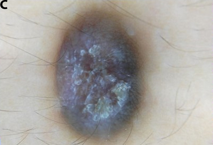


(A and B) Clinical presentation of a 1,5 cm x 2 cm, hard and depressed livedoid lesion on the back. (C) Gray-whitish and livedoid large clods with a homogenous distribution seen at dermoscopy. (D and E) Correlated histological images: fibro-histiocytic benign tumoral lesion located in a dense collagenized stroma separated from the epidermis by a cell-poor zone (H&E, x100, x200).
Teaching point
Atrophic dermatofibroma is a rare variant of dermatofibroma which was first described by Page and Assaad in 1987 [1]. It is commonly seen on the upper back of middle-aged women, and the mean age of the patients affected is 49.7 years, according to literature [1]. We present this uncommon entity, which is in the differential diagnosis with many benign and malignant lesions including atrophic scars, anetodermas, morphea, atrophic dermatofibrosarcoma protuberans and sclerosing basal cell carcinomas, as it is rare in the pediatric age group [2].
Footnotes
Competing interests: None.
Authorship: Both authors have contributed significantly to this publication.
Funding: None.
References
- 1.Ohnishi T, Sasaki M, Nakai K, Watanabe S. Atrophic dermatofibroma. J Eur Acad Dermatol Venereol. 2004;18(5):580–583. doi: 10.1111/j.1468-3083.2004.00975.x. [DOI] [PubMed] [Google Scholar]
- 2.Mota AN, Tortelly VD, Obadia DL, Silva RS. Atrophic dermatofibroma. An Bras Dermatol. 2013;88(5):793–795. doi: 10.1590/abd1806-4841.20132234. [DOI] [PMC free article] [PubMed] [Google Scholar]