Abstract
An inverse PCR strategy based on degenerate primers has been used to identify new genes of the cold shock protein family in Lactobacillus plantarum. In addition to the two previously reported cspL and cspP genes, a third gene, cspC, has been cloned and characterized. All three genes encode small 66-amino-acid proteins with between 73 and 88% identity. Comparative Northern blot analyses showed that the level of cspL mRNA increases up to 17-fold after a temperature downshift, whereas the mRNA levels of cspC and cspP remain unchanged or increase only slightly (about two- to threefold). Cold induction of cspL mRNA is transient and delayed in time as a function of the severity of the temperature downshift. The cold shock behavior of the three csp mRNAs contrasts with that observed for four unrelated non-csp genes, which all showed a sharp decrease in mRNA level, followed in one case (bglH) by a progressive recovery of the transcript during prolonged cold exposure. Abundance of the three csp mRNAs was also found to vary during growth at optimal temperature (28°C). cspC and cspP mRNA levels are maximal during the lag period, whereas the abundance of the cspL transcript is highest during late-exponential-phase growth. The differential expression of the three L. plantarum csp genes can be related to sequence and structural differences in their untranslated regions. It also supports the view that the gene products fulfill separate and specific functions, under both cold shock and non-cold shock conditions.
When exposed to abrupt temperature downshifts, microorganisms undergo severe physiological disturbances such as a reduction in membrane fluidity, changes in the level of DNA supercoiling, and the formation of stable secondary structures in DNA and RNA that impair replication, transcription, and protein synthesis. To overcome the deleterious effects of cold shock and ensure that cellular activity will resume or be maintained at low temperature, bacteria have developed a transient adaptive response, the cold shock response, during which the expression of a subset of specific proteins is induced (reviewed in references 19, 36, and 42). The most strongly induced proteins include a family of closely related small (∼7.5-kDa) single-stranded DNA- and RNA-binding proteins termed cold shock proteins (Csp). These proteins have been proposed to function as general RNA or DNA chaperones that stabilize single-stranded regions in RNA and DNA and thereby contribute to efficient translation, transcription, and DNA replication at low temperature (19, 24, 50). Their structure, a β barrel made up of five antiparallel β strands, is the archetype of a very common protein domain referred to as the oligonucleotide/oligosaccharide-binding fold (19, 38, 39).
Csp proteins are widespread among bacteria, and many species have been found to contain multiple variants of this large family of proteins (19, 50). Although the specific functions of these iso-Csp proteins remain unclear, most recent findings indicate that they not only are required for the cold shock adaptative response but also play a more general role in adapting cellular functions to various growth conditions. Of the nine Csp proteins from Escherichia coli (CspA to CspI), only four (CspA, CspB, CspG, and CspI) are cold inducible, with CspA being induced at the highest level and over the broadest temperature range (45, 50). Recent work has shown that CspA is also highly expressed during early exponential growth at optimal temperature (6), whereas expression of CspD is triggered at the onset of stationary phase and upon nutrient starvation (49). CspC and CspE are constitutively produced at 37°C and have been implicated in cellular processes taking place during normal growth, such as transcriptional regulation and chromosome condensation (3, 21, 48). The expression patterns and roles of CspH and CspF, the two most distant members of the E. coli Csp family, remain unexplored (50).
In contrast to E. coli, Bacillus subtilis contains three Csp proteins (CspB, CspC, and CspD) which are all induced after a temperature downshift (17). Like synthesis of E. coli CspD, synthesis of B. subtilis CspB and CspC is found to increase upon entry into stationary phase (20). Single and combined deletion of the three csp genes in B. subtilis has shown that the presence of at least one Csp protein is essential for cell viability and that the three Csp variants individually and complementarily contribute to efficient cell growth and survival at both low and optimal temperatures (17, 18).
Regulation of the expression of the E. coli and B. subtilis Csp proteins is complex, taking place at the transcriptional and posttranscriptional levels (18, 27, 36, 42). Furthermore, several observations indicate that iso-Csp proteins belonging to the same species self- and/or cross-regulate their own synthesis (2, 3, 10, 18).
Csp proteins are also found in different species of lactic acid bacteria (28). For example, a family of five csp genes (cspA to cspE) is present in Lactococcus lactis. Their expression is strongly induced upon cold shock, except for CspE, which is already abundant at optimal temperature (46, 47).
Lactobacillus plantarum is one of the most widespread lactic acid bacteria in the environment. This bacterium is also largely used as a starter for the production of fermented products of animal and vegetal origin (43). Strains that are used in fermentation and ripening processes undergo multiple stresses including drastic changes in temperature, pH, and carbon source. However, little is known about the mechanisms used by L. plantarum to adapt to environmental fluctuations. A better understanding of these mechanisms should provide important insight into how to improve current industrial starter strains.
We have previously reported the presence of two csp genes, cspL and cspP, in L. plantarum (32). In the present study, an inverse PCR strategy was used to isolate new members of the csp gene family from strain NC8. A new gene, cspC, has been cloned and characterized. Comparative transcriptional analyses of the three genes show that expression of cspL mRNA differs from that of cspC and cspP, both after a temperature downshift and during growth under optimal conditions. The cold shock behavior of the three csp transcripts also contrasts with that of non-csp genes, indicating that regulation of the csp genes involves cold-specific mechanisms acting at the level of transcription and/or stability of the mRNA. The data are consistent with the view that the three csp gene products of L. plantarum fulfill separate and specific functions in cold adaptation, as well as during normal growth.
MATERIALS AND METHODS
Bacterial strains and growth conditions.
The L. plantarum silage strain NC8 (1) was grown in MRS broth (Difco Laboratories, Detroit, Mich.) without shaking. Cold shocks were performed by transferring NC8 cultures grown at 28°C into water baths precooled at the appropriate temperature. The E. coli strain TG1 was grown in Luria broth at 37°C (37). When necessary, ampicillin was added to a final concentration of 250 μg/ml.
DNA amplification, cloning, and sequencing.
PCR amplifications were carried out using Taq (Advanced Biotechnologies, Leatherhead, United Kingdom) or Dynazyme (Finnzymes Oy, Espoo, Finland) DNA polymerase. Chromosomal DNA from NC8 was prepared as previously described (4). NC8 DNA was partially digested with MboI and then circularized by ligation to be used as a template in inverse PCR amplification. The degenerate primers used to amplify borders of the NC8 csp genes are CSPIF2 (5′-GGITWCAAAWCIYTRCAIGAAGGYCA-3′) and PIR1 (5′-GCYYTGRATMGCTGAGAARTGWAC-3′). Inverse PCR was carried out with 40 amplification cycles of 0.25 min at 95°C, 6 min at 50°C, and 4 min at 72°C.
The central region of the NC8 cspC gene was amplified using the degenerate primer CSPU3 (5′-GGTTACGTTASCWGCTKSHGGDCC-3′) (14) and the specific primer LPCF (nucleotides [nt] 290 to 310 of the cspC sequence). The resulting PCR fragment was radiolabeled with [α-32P]ATP (Amersham, Buckinghamshire, United Kingdom) by random-primer DNA labeling (GIBCO-BRL, Gaithersburg, Md.) and used as a probe in Southern blot analysis of NC8 DNA cut by different restriction enzymes. The same probe was used to screen a genomic library of L. plantarum Lp80 (partial Sau3AI restriction fragments cloned into the BamHI site of pGI4010) (26) transformed into TG1. Colony hybridization identified a plasmid (pGIS003) carrying a copy of the Lp80 cspC gene that was sequenced using CSPIF2, PIR1, and a set of specific primers. Two primers, CSPCG (5′-CACCGCTCAAGATTGGAC-3′) and PECA (5′GCCTTCAAGCAAGTCGCAAT-3′), were then derived from this sequence in order to amplify and sequence the NC8 cspC gene. Southern blot analysis and colony hybridization were performed using standard procedures (37).
Pairs of primers used to amplify and sequence the NC8 cspL (PEL91, nt 1 to 22; PEL17, nt 1263 to 1284) and cspP (PEP22, nt 1 to 19; PEPA, nt 687 to 703) genes were chosen based on the Lp80 cspL and C3.8 cspC sequences, respectively. Standard conditions used for direct PCR amplification are 30 cycles of 1 min at 92°C, 1 min at 50°C, and 1 min at 72°C, followed by an elongation step of 10 min at 72°C. Amplified products were extracted from agarose gels using a Qiaquick gel extraction kit (Qiagen GmbH, Hilden, Germany). DNA sequencing of purified PCR fragments and plasmid dsDNA was performed by Eurogentec S.A., Seraing, Belgium.
RNA hybridization and primer extension analysis.
NC8 total RNA was prepared from 25-ml cultures using a Tri reagent kit (Sigma, St. Louis, Mo.). Cells were harvested by centrifugation and mechanically broken with 0.18-mm-diameter glass beads in a Braun homogenizer (four 1-min periods of homogenization with 1-min intervals on ice). Primer extensions were performed as previously described (15). Oligonucleotides used to map the 5′ termini of L. plantarum csp mRNAs are PEC5 (nt 357 to 381) and PECC (nt 443 to 463) for cspC, PEL60 (nt 554 to 575) for cspL, and PEP40 (nt 447 to 465), PEP1 (nt 404 to 430), and PEP2 (nt 420 to 444) for cspP. Radiolabeled elongation products were analyzed on 7% (wt/vol) polyacrylamide–urea sequencing gels, next to DNA sequencing reactions performed with the same primers. Single-stranded DNA templates were generated by λ exonuclease-mediated digestion of the 5′-phosphorylated strand of PCR products (Pharmacia Biotech, Uppsala, Sweden). Sequencing reactions were performed using T7 DNA polymerase (Pharmacia Biotech).
For quantitative Northern blot analysis, RNA samples were separated by electrophoresis in 1.2% (wt/vol) agarose–0.6% (wt/vol) formaldehyde gels in MOPS (morpholinepropanesulfonic acid) buffer. Gels were stained with ethidium bromide to ensure that equivalent amounts of total RNA were loaded in each sample. RNA was transferred to Hybond-N nylon membranes (Amersham) and fixed by heat treatment (80°C, 2 h). Hybridization was performed as previously reported (15), using α-32P-radiolabeled PCR fragments as specific probes for cspC (nt 290 to 549), cspL (nt 317 to 1284), cspP (nt 1 to 703), alr (nt 653 to 1295), bglH (nt 661 to 1262), cbh (nt 116 to 945), and lldh (nt 654 to 1115). Radioactive bands were quantified with an Instant Imager (Packard Instruments, Meriden, Conn.) and visualized by autoradiography. When necessary, the relative amounts of transcript were standardized by hybridization with an L. plantarum 16S rRNA-specific probe (nt 87 to 1102).
Nucleotide sequence accession numbers.
The EMBL accession numbers for the L. plantarum NC8 strain sequences reported here are Y19217 (cspC), Y19218 (cspL), and Y19219 (cspP). The nucleotide sequences for Lp80 cspL and C3.8 cspP can be found in the GenBank database with accession no. Y08940 and Y08760, respectively. The GenBank accession numbers for L. plantarum alr, bglH, cbh, lldh, and 16S rRNA sequences are Y08941, Y15954, M96175, X70926, and D79210, respectively.
RESULTS
Cloning of cspC, a new member of the csp gene family in L. plantarum.
We have recently reported the cloning of cspL and cspP, two cold shock protein genes from L. plantarum Lp80 and C3.8, respectively (32). In the present study, an inverse PCR approach based on degenerate primers was used with the aim of amplifying new members of the csp family from strain NC8. This strain was chosen as a model for genetic studies because it displays interesting features such as being plasmid free and highly transformable (1). The nucleotide sequences of the degenerate primers PIR1 and CSPIF2 (see Materials and Methods) were determined based on highly conserved regions in csp genes from more than 30 different bacterial species (14; K. P. Francis, unpublished data). Total DNA from NC8 was partially digested with MboI, and the resulting fragments were circularized by ligation to serve as a template in PCR amplification. Six different amplification products were obtained and sequenced. One of them contained a sequence identical to the previously identified cspL gene of Lp80, while two other amplicons carried DNA fragments corresponding to the cspP gene from strain C3.8. The remaining three PCR products were found to contain a sequence belonging to a new member of the csp family that we named cspC. Although the primers and conditions used in the amplification were chosen so as to amplify the largest number of specific fragments, the possibility that L. plantarum contains additional and more distant Csp variants cannot be totally ruled out.
The central region of the NC8 cspC gene was amplified using a specific primer complementary to the 5′ end of the gene and a degenerate primer corresponding to a conserved region at the 3′ end (U3CSP) (14). This fragment was used as a probe to screen an Lp80 genomic library (26) by colony hybridization. A clone carrying a complete copy of the cspC gene was identified. PCR primers were then derived from the sequence of this clone to amplify and determine the complete sequence of the NC8 cspC gene. Southern blot analysis demonstrated that this gene is present as a single copy in the NC8 genome (data not shown). PCR amplification and DNA sequencing of the two other csp genes from NC8 showed that the cspL gene is 100% identical to that of Lp80, whereas 19 nucleotide differences were found between the NC8 and C3.8 sequences in the noncoding regions of cspP (data not shown).
The three csp genes encode small and closely related 66-amino-acid proteins, with a calculated pI value close to 4 (Fig. 1). They contain two RNA-binding motifs called RNP1 and RNP2 (30, 34, 40), together with additional conserved residues that are important for the formation of the β-barrel core of the protein and for nucleic acid binding activity (Fig. 1) (12, 34, 40). The newly identified CspC protein is the most distant member of the family since it displays 73 and 74% identity with CspL and CspP, respectively, while CspL and CspP are 88% identical.
FIG. 1.
Comparison of L. plantarum CspC, CspL, and CspP sequences. Amino acids residues that are identical in all three or just two Csp proteins are shaded in dark and light grey, respectively. The RNA-binding motifs RNP1 and RNP2 are boxed. Conserved residues that are critical for forming the hydrophobic core of the protein (asterisks) or for nucleic acid binding (dots) are indicated.
The three csp genes exhibit long 5′ untranslated leader regions (5′ UTRs).
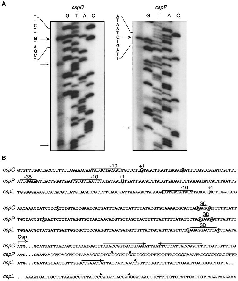
Primer extension analysis was performed on NC8 total RNA in order to map the transcription start point of cspC. Independent elongation reactions were performed using two different primers to selectively identify products arising from specific hybridization. Multiple signals were obtained with either primer individually, but only three of them were common to both reactions (Fig. 2A). A similar analysis of the NC8 cspL transcript identified a single 5′ end mapping at the same position as previously reported for the Lp80 cspL gene (Fig. 2B) (32), whereas two specific elongation products were detected for cspP (Fig. 2A). The smallest product corresponds to the cspP transcription start initially mapped in C3.8 (32). However, in light of the present results, it appears more likely that the minor signals observed for both cspC and cspP arise from specific degradation of the mRNA and that the actual transcription initiation sites of these two genes correspond to the 5′ ends of the longest and more abundant extension products (Fig. 2A).
FIG. 2.
(A) Primer extension analysis of the cspC (left) and cspP (right) transcripts. RNA samples were extracted from cold-shocked cultures (8°C). Bold and thin arrows indicate the positions of major and minor extension products, respectively, obtained with independent primers. Additional signals are nonspecific products that are detected only with individual primers. (B) Nucleotide sequences of the 3′ and 5′ regions of L. plantarum NC8 cspC, cspL, and cspP genes. Only the first and last codons of the Csp-coding sequence are shown. Transcription starts (+1) are circled, as are the 5′ ends of shorter and less abundant primer extension products detected for cspC and cspP. The promoter −35 and −10 boxes and the Shine-Dalgarno sequences (SD) are boxed and circled, respectively. Horizontal arrows indicate the positions of putative transcription terminators.
The proposed transcription start of the three csp genes is preceded by a sequence that is 67% (cspP) or 89% (cspC and cspL) identical to the extended −10 box consensus sequence (TNTGNTATAAT) of the ςA promoters from gram-positive bacteria (22). The presence of TG dinucleotides in this box has been found to increase contacts between the RNA polymerase and DNA (44). In cspP, which has the less well conserved −10 box, a putative −35 box sequence is found at the expected position of the promoter region, while no obvious −35 boxes are found in cspC and cspL (Fig. 2B).
This analysis reveals that cspC and cspP have similarly long predicted 5′ UTRs that are 49% identical, whereas cspL displays a slightly shorter 5′ UTR that is only 22% identical to that of cspC and 27% identical to that of cspP (Fig. 2B and data not shown). The sequence similarities in the 5′ UTRs contrast with the degree of homology between the proteins since, as mentioned above, CspL and CspP are closer to each other than CspC and CspP. In addition, cspC and cspP genes exhibit a typical monocistronic structure containing a single transcription terminator at the 3′ end, while two consecutive putative terminators are found downstream of the cspL coding region, as previously reported for the Lp80 cspL gene (Fig. 2B) (32).
Changes in csp and non-csp mRNA abundance upon cold shock.
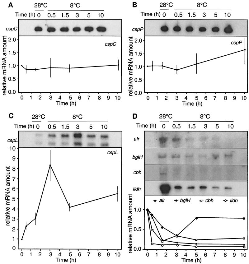
Quantitative Northern blot analysis was used to monitor the levels of cspC, cspL, and cspP mRNA after transferring exponentially growing NC8 cultures (optical density at 600 nm [OD600] = 0.8) from 28°C to 8°C (Fig. 3). Those values were set as optimal and medium cold shock temperatures, respectively, on the basis of the L. plantarum NC8 Arrhenius plot of growth (S. Derzelle, B. Hallet, T. Ferain, J. Delcour, and P. Hols, submitted for publication). The expression patterns of the csp genes were compared to those obtained for four non-csp genes which fulfill separate and unrelated functions in L. plantarum. These include the alanine racemase gene (alr), which is responsible for the production of d-alanine, an essential component of the bacterial cell wall (23); the phospho-β-glucosidase gene (bglH), involved in utilization of specific aromatic β-glucosides as a carbon source (31); the l-lactate dehydrogenase gene (ldhL), which catalyzes the last step of l-lactic acid production by fermentation (13); and the cbh gene, which encodes a conjugated bile acid hydrolase (7).
FIG. 3.
Comparative Northern blot analysis of csp and non-csp genes upon cold shock. Exponentially growing cultures of L. plantarum NC8 (OD600 = 0.8) were transferred from 28°C to 8°C, and RNA was prepared at the indicated times before (0 h) and after the temperature downshift. Equal amounts of each RNA sample were run on 1.2% (wt/vol) agarose–0.6% (wt/vol) formaldehyde gels and hybridized with specific radioactive probes for cspC (A), cspP (B), cspL (C), and the four non-csp genes alr, bglH, cbh, and lldh (D). The same membranes were rehybridized with the different probes. One representative result is shown for each gene. Relative mRNA amounts were calculated from the radioactivity measured in the transcript bands at each time point with respect to that found at 0 h. cspL quantification was performed by summing the radioactivities of the short and long transcripts. Data presented for cspC, cspL, and cspP are from three independent experiments.
We identified a single transcript of the expected size (330 nt) for both cspC and cspP, whereas two signals were observed for cspL, as previously reported for the Lp80 cspL gene (Fig. 3). The shorter transcript (330 nt) has been shown to contain the cspL open reading frame whereas the longer transcript (760 nt) extends further downstream to encompass a putative 77-amino-acid open reading frame bearing no similarity to known proteins (32). It remains unclear whether these two transcripts arise from alternative transcriptional termination (Fig. 1B) or whether they arise through posttranscriptional processing of the mRNA.
The amount of cspC and cspP transcripts remained unchanged after the temperature downshift (Fig. 3A and B). In contrast, the level of cspL mRNAs was found to increase up to eightfold after 3 h at 8°C. The amount of transcripts subsequently decreased to a steady-state level that remained five times higher than observed before the cold shock (Fig. 3C). When taken separately, the long and short transcripts displayed similar kinetics (Fig. 3C and data not shown). These results clearly differentiate the csp genes from the four non-csp genes examined, since all of them showed an abrupt diminution in their mRNA level immediately after cold shock (Fig. 3D). However, in the case of bglH, the amount of transcript increased again after 3 h of cold exposure to about the same level as before the temperature shift (Fig. 3D).
Cold induction of cspL mRNA is delayed according to the severity of the temperature downshift.
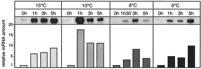
To further characterize the influence of cold shock on the expression of cspL, Northern blot analysis was performed on exponentially growing NC8 cultures shifted to different temperatures (Fig. 4). According to the Arrhenius plot of L. plantarum NC8 growth (Derzelle et al., submitted), these temperatures correspond to nonstressed suboptimal growth conditions (15°C) or to mild (10°C), medium (8°C), and severe (6°C) cold shocks. The level of the large cspL transcript was found to increase at all temperatures tested. The small transcript in this set of experiments was barely detectable for unknown reasons. However, a delay in induction was observed with decreasing temperatures, the highest level of the cspL transcript being detected after 1 h at 10°C, after 3 h at 8°C, and after 5 h at 6°C (Fig. 4). After 1 h at 15°C, the amount of the cspL transcript was already six to seven times higher than in precooled cells, and it stayed at about the same level after 3 h and 5 h of cold exposure. These findings indicate that there is a dose effect of cold on expression of the cspL transcript. Hybridization of the same RNA samples with cspC and cspP probes showed that their mRNA levels are only slightly (two- to threefold) higher at 10 and 15°C than at 6 and 8°C (data not shown).
FIG. 4.
Delayed cold induction of cspL mRNA level with decreasing temperatures. RNA was extracted from an exponentially growing NC8 culture (OD600 = 0.8) before and after different times following a temperature downshift from 28°C to 15, 10, 8, or 6°C. The resulting RNA samples were analyzed by hybridization with a cspL probe as described for Fig. 3. As the smaller transcript was barely detectable, quantification was performed only on the large transcript.
Changes in cspC, cspP, and cspL mRNA abundance during growth at optimal temperature.
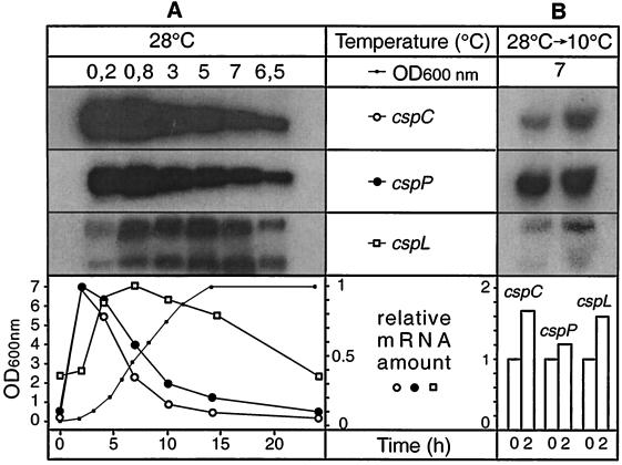
The level of cspC, cspL, and cspP transcripts was examined over a complete culture cycle performed at 28°C (Fig. 5A). This analysis revealed that early growing cells contain substantial amounts of cspC and cspP mRNAs, which rapidly decline to become 10 times less abundant at the entry into stationary phase (Fig. 5A). A very similar behavior has recently been reported for CspA, the major Csp protein in E. coli, and to some extent for CspE, which is not cold induced (3, 6). By contrast, a different pattern is observed for the L. plantarum cspL gene (Fig. 5A). The amount of cspL transcripts increases to reach a plateau during exponential growth and then decreases during stationary phase to return to its initial level (Fig. 5A). This pattern also distinguishes cspL from the cspD gene of E. coli, or the cspB and cspC genes of B. subtilis, the expression of which is induced at the beginning of stationary phase (20, 49).
FIG. 5.
(A) Growth phase-dependent expression of cspC, cspL, and cspP mRNAs. Stationary-phase NC8 cells were diluted with fresh MRS medium and allowed to grow for 25 h at 28°C. RNA samples were taken at different times of culture and analyzed by Northern blot hybridization with cspC, cspP, and cspL probes, as indicated. Results obtained after hybridization of the same membrane with the three different probes are shown. Relative mRNA amount is expressed with respect to the highest level of each transcript during the culture. Radioactivities in the short and large cspL transcripts were summed. (B) Increase of cspC, cspP, and cspL mRNA levels in cold-shocked stationary-phase cells. Relative amounts of cspC, cspP, and cspL transcripts were examined 2 h after transferring stationary-phase NC8 cultures (OD600 = 7) from 28°C to 10°C.
In E. coli, the extent of cspA cold induction is inversely proportional to the preexisting concentration of CspA protein at the time of the cold shock (6). To see whether a similar effect can be observed in L. plantarum, the amount of cspC, cspL, and cspP transcripts was quantified before and after transferring a stationary-phase culture (OD600 = 7) from 28°C to 10°C. After 2 h of cold exposure, all three genes showed a very limited (between 1.1- and 1.6-fold) increase, if any, of their transcript levels (Fig. 5B). For cspL, this contrasts with the strong induction observed after a cold shock of exponentially growing cells.
DISCUSSION
Using an inverse PCR approach based on degenerate primers, we demonstrate that strain NC8 of L. plantarum contains at least three Csp-encoding genes. Two of them, cspL and cspP, were previously identified from separate strains (32). The newly isolated cspC gene represents the most distant member of the csp family identified thus far in L. plantarum.
Comparative Northern blot analysis reveals that the relative abundance of cspC, cspL, and cspP transcripts differentially varies after a temperature downshift as well as during growth in optimal conditions. Upon cold shock of exponentially growing cells, cspL undergoes a significant and transient induction, whereas the amount of cspC and cspP mRNAs remains unchanged or increases slightly. These two different patterns distinguish the csp genes from non-csp genes, the mRNA level of which is found to rapidly decrease after cold shock. This is a key result of the present study, as it demonstrates that csp transcripts may be more stable and/or more efficiently expressed than other cellular messengers, even if their absolute level does not significantly increase during cold exposure.
Rapid disappearance of non-csp transcripts is likely to arise from the general inhibition of both RNA and protein synthesis that follows an abrupt temperature downshift (29, 41). In particular, impairment of translation initiation would leave RNA molecules unprotected, and consequently these become more sensitive to degradation although cellular RNAse activity itself is reduced at low temperature (35). However, the bglH gene can be distinguished from the three other non-csp genes examined since its mRNA level rises again after 3 h of cold exposure. Interestingly, this corresponds to the time at which cspL mRNA is most abundant. One may therefore suggest that resumption of bglH mRNA expression could be a consequence of cold adaptation mechanisms involving CspL and presumably other cold-induced proteins. As this enzyme provides a means of using a broader range of carbon sources, its early expression during cold acclimation may represent a valuable contribution to cell survival and growth at low temperature. This raises the possibility that separate classes of genes respond differently to continuous growth at cold temperatures as a function of their biological roles.
It becomes clear that Csp proteins are not only involved in cold adaptation but also play an important role during growth at optimal temperature. Recent reports have shown that cold-inducible Csp proteins may also be expressed at specific stages of the normal growth cycle. CspA, the major cold shock protein of E. coli, is transiently and massively expressed during the early phase of the growth curve at 37°C, whereas expression of CspB and CspC from B. subtilis is triggered at the onset of stationary phase (6, 20). Here, we demonstrated that the relative amounts of L. plantarum cspC, cspL, and cspP transcripts strongly fluctuate during growth at 28°C, indicating that specific regulation mechanisms operate on the three genes to direct their expression at different times of the culture. The growth phase is also found to determine the extent of cspL cold induction, since a cold shock of exponentially growing cells results in a much stronger increase in cspL transcript level than a cold shock performed in stationary phase. This suggests that stationary-phase cells could adapt to cold without requiring further expression of cspL.
Based on (i) the results presented here, (ii) our analysis of the L. plantarum cspL, cspP, and cspC sequences, and (iii) what is known about cold shock regulation in other bacteria, we can make a series of predictions on the regulation mechanisms that are experimentally testable. In E. coli and B. subtilis, although a moderate increase in the transcription of csp genes is observed, Csp protein expression is regulated primarily at a posttranscriptional level. In particular, cold-induced csp genes usually contain a long 5′ UTR which, in the case of E. coli cspA, has been found to destabilize the mRNA at 37°C and to enhance its stability and translation efficiency after cold shock (11, 16, 33, 51). Comparison of the L. plantarum csp genes shows that the 5′ UTRs of cspC and cspP are longer and more similar to each other than to the 5′ UTR of cspL. Furthermore, cspC and cspP 5′ UTRs are predicted to form similar hairpin structures, whereas the 5′ UTR of cspL exhibits a distinctive Y-like secondary structure (data not shown). Additionally, both cspC and cspP display minor extension signals ending close to bulge loops in the predicted hairpin structure of their 5′ UTRs (data not shown), suggesting an increased susceptibility to cleavage by a specific RNase and a higher instability of respective mRNAs compared to cspL. These differences in the csp gene 5′ UTRs relate to the finding that the mRNA expression patterns of cspC and cspP diverge from that of cspL, both at low and at optimal temperatures. The fact that the degree of similarity between the 5′ UTRs differs from that of the protein sequences suggests that the coding and untranslated regulatory regions of the csp genes have evolved independently in order to allow functionally distinct Csp proteins to be expressed in different conditions.
Cold induction of CspA and other E. coli Csp proteins requires the presence of a 14-nt sequence, referred to as the downstream box (DB), located 12 nt downstream of the initiation codon (8, 33). More recently, a second cis-acting element, the upstream box (UB), has been identified within the 5′ UTR, 14 nt upstream of the Shine-Dalgarno sequence (51). Although the actual roles of the DB and UB sequences remain unclear (for recent debates in the literature, see references 5 and 9), both boxes are complementary to specific regions of the 16S rRNA 3′ end, and it is proposed that they thereby contribute to translation initiation by increasing the affinity of the ribosome for mRNA. Enhanced translation may in turn contribute to the stabilization of the transcripts by protecting them from degradation (35).
Close examination of the L. plantarum csp transcripts reveals that each contains a DB-like box at the N terminus of the coding region (consensus, 5′-uGGuACAGUaAAAUGGUU-3′) that is complementary to the L. plantarum 16S rRNA 3′-end sequence (nt 1472 to 1487). Interestingly, a highly related DB-like sequence (5′-GUUCCCAGAAGGAU-3′) is also found in the coding region of the bglH gene, although in this case, the N-terminal amino acid sequence of the protein is totally divergent from that of the Csp proteins. As for the csp genes, the presence of this box may enhance the translation efficiency, and therefore the stability, of bglH mRNA compared to other cellular transcripts.
An upstream sequence that is complementary to a different region of the 16S rRNA 3′ end (nt 1049 to 1061) is also found in the 5′ UTR of cspL (5′-CCCAAGGUUAACG-3′) but not in cspC or in cspP. This UB-like sequence is entrapped within the arms of the proposed Y-like structure of the cspL 5′ UTR (data not shown). Therefore, we would predict that formation of this structure must be prevented to give access to the UB region and to permit translation-mediated stabilization of the transcript. This postulated mechanism would explain the temperature-dependent delay observed for cspL cold induction, since decreasing cold shock temperatures are expected to enhance the stability of the RNA duplex. Destabilization of the 5′-UTR structure could be promoted by specific cold-inducible RNA helicases analogous to the E. coli CsdA protein (27). It may also require the RNA chaperone activity of the Csp proteins themselves.
Whichever mechanisms are responsible for regulation of the csp genes in L. plantarum, their differential expression at low and optimal temperatures is consistent with the view that they fulfill specific and perhaps complementary functions in adapting cell physiology to different conditions. It remains to be determined whether fluctuations in the mRNA level observed for the three genes correlate with variations in the relative amounts of their products. The deletion or the constitutive overexpression of L. plantarum csp genes should also provide important insight on their respective contribution to cell survival and growth.
ACKNOWLEDGMENTS
K. Josson is acknowledged for providing the Lp80 genomic library.
S.D. holds a fellowship from the Fonds pour la Formation à la Recherche dans l'Industrie et dans l'Agriculture (FRIA). B.H. is a postdoctoral researcher at the FNRS.
REFERENCES
- 1.Aukrust T, Blom H. Transformation of Lactobacillus strains used in meat and vegetable fermentations. Food Res Int. 1992;25:253–261. [Google Scholar]
- 2.Bae W, Jones P G, Inouye M. CspA, the major cold shock protein of Escherichia coli, negatively regulates its own gene expression. J Bacteriol. 1997;179:7081–7088. doi: 10.1128/jb.179.22.7081-7088.1997. [DOI] [PMC free article] [PubMed] [Google Scholar]
- 3.Bae W, Phadtare S, Severinov K, Inouye M. Characterization of Escherichia coli cspE, whose product negatively regulates transcription of cspA, the gene for the major cold shock protein. Mol Microbiol. 1999;31:1429–1441. doi: 10.1046/j.1365-2958.1999.01284.x. [DOI] [PubMed] [Google Scholar]
- 4.Bernard N, Ferain T, Garmyn D, Hols P, Delcour J. Cloning of the d-lactate dehydrogenase gene from Lactobacillus delbrueckii subsp. bulgaricus by complementation in Escherichia coli. FEBS Lett. 1991;290:61–64. doi: 10.1016/0014-5793(91)81226-x. [DOI] [PubMed] [Google Scholar]
- 5.Bläsi U, O'Connor M, Squires C L, Dahlberg A E. Misled by sequence complementarity: does the DB-anti-DB interaction withstand scientific scrutiny? Mol Microbiol. 1999;33:439–441. doi: 10.1046/j.1365-2958.1999.01488.x. [DOI] [PubMed] [Google Scholar]
- 6.Brandi A, Spurio R, Gualerzi C O, Pon C L. Massive presence of the Escherichia coli ‘major cold-shock protein’ CspA under non-stress conditions. EMBO J. 1999;18:1653–1659. doi: 10.1093/emboj/18.6.1653. [DOI] [PMC free article] [PubMed] [Google Scholar]
- 7.Christiaens H, Leer R J, Pouwels P H, Verstraete W. Cloning and expression of a conjugated bile acid hydrolase gene from Lactobacillus plantarum by using a direct plate assay. Appl Environ Microbiol. 1992;58:3792–3798. doi: 10.1128/aem.58.12.3792-3798.1992. [DOI] [PMC free article] [PubMed] [Google Scholar]
- 8.Etchegaray J-P, Inouye M. A sequence downstream of the initiation codon is essential for cold shock induction of cspB of Escherichia coli. J Bacteriol. 1999;181:5852–5854. doi: 10.1128/jb.181.18.5852-5854.1999. [DOI] [PMC free article] [PubMed] [Google Scholar]
- 9.Etchegaray J-P, Inouye M. DB or not DB in translation? Mol Microbiol. 1999;33:438–439. doi: 10.1046/j.1365-2958.1999.01487.x. [DOI] [PubMed] [Google Scholar]
- 10.Fang L, Hou Y, Inouye M. Role of the cold-box region in the 5′ untranslated region of the cspA mRNA in its transient expression at low temperature in Escherichia coli. J Bacteriol. 1998;180:90–95. doi: 10.1128/jb.180.1.90-95.1998. [DOI] [PMC free article] [PubMed] [Google Scholar]
- 11.Fang L, Xia B, Inouye M. Transcription of cspA, the gene for the major cold-shock protein of Escherichia coli, is negatively regulated at 37°C by the 5′-untranslated region of its mRNA. FEMS Microbiol Lett. 1999;176:39–43. doi: 10.1111/j.1574-6968.1999.tb13639.x. [DOI] [PubMed] [Google Scholar]
- 12.Feng W, Tejero R, Zimmerman D E, Inouye M, Montelione G T. Solution NMR structure and backbone dynamics of the major cold-shock protein (CspA) from Escherichia coli: evidence for conformational dynamics in the single-stranded RNA-binding site. Biochemistry. 1998;37:10881–10896. doi: 10.1021/bi980269j. [DOI] [PubMed] [Google Scholar]
- 13.Ferain T, Garmyn D, Bernard N, Hols P, Delcour J. Lactobacillus plantarum ldhL gene: overexpression and deletion. J Bacteriol. 1994;176:596–601. doi: 10.1128/jb.176.3.596-601.1994. [DOI] [PMC free article] [PubMed] [Google Scholar]
- 14.Francis K P, Stewart G S A B. Detection and speciation of bacteria through PCR using universal major cold-shock protein primer oligomers. J Ind Microbiol Biotechnol. 1997;19:286–293. doi: 10.1038/sj.jim.2900463. [DOI] [PubMed] [Google Scholar]
- 15.Garmyn D, Ferain T, Bernard N, Hols P, Delcour J. Cloning, nucleotide sequence, and transcriptional analysis of the Pediococcus acidilacticil-(+)-lactate dehydrogenase gene. Appl Environ Microbiol. 1995;61:266–272. doi: 10.1128/aem.61.1.266-272.1995. [DOI] [PMC free article] [PubMed] [Google Scholar]
- 16.Goldenberg D, Azar I, Oppenheim A B, Brandi A, Pon C L, Gualerzi C O. Role of Escherichia coli cspA promoter sequences and adaptation of translational apparatus in the cold shock response. Mol Gen Genet. 1997;256:282–290. doi: 10.1007/s004380050571. [DOI] [PubMed] [Google Scholar]
- 17.Graumann P, Schröder K, Schmid R, Marahiel M A. Cold shock stress-induced proteins in Bacillus subtilis. J Bacteriol. 1996;178:4611–4619. doi: 10.1128/jb.178.15.4611-4619.1996. [DOI] [PMC free article] [PubMed] [Google Scholar]
- 18.Graumann P, Wendrich T M, Weber M H W, Schröder K, Marahiel M A. A family of cold shock proteins in Bacillus subtilis is essential for cellular growth and for efficient protein synthesis at optimal and low temperature. Mol Microbiol. 1997;25:741–756. doi: 10.1046/j.1365-2958.1997.5121878.x. [DOI] [PubMed] [Google Scholar]
- 19.Graumann P L, Marahiel M A. A superfamily of proteins that contain the cold-shock domain. Trends Biochem Sci. 1998;23:286–290. doi: 10.1016/s0968-0004(98)01255-9. [DOI] [PubMed] [Google Scholar]
- 20.Graumann P L, Marahiel M A. Cold shock proteins CspB and CspC are major stationary-phase-induced proteins in B. subtilis. Arch Microbiol. 1999;171:135–138. doi: 10.1007/s002030050690. [DOI] [PubMed] [Google Scholar]
- 21.Hanna M M, Liu K. Nascent RNA in transcription complexes interacts with CspE, a small protein in E. coli implicated in chromatin condensation. J Mol Biol. 1998;282:227–239. doi: 10.1006/jmbi.1998.2005. [DOI] [PubMed] [Google Scholar]
- 22.Helmann J D. Compilation and analysis of Bacillus subtilis ςA-dependent promoter sequences: evidence for extended contact between RNA polymerase and upstream promoter DNA. Nucleic Acids Res. 1995;23:2351–2360. doi: 10.1093/nar/23.13.2351. [DOI] [PMC free article] [PubMed] [Google Scholar]
- 23.Hols P, Defrenne C, Ferain T, Derzelle S, Delplace B, Delcour J. Alanine racemase gene is essential for growth in Lactobacillus plantarum. J Bacteriol. 1997;179:3804–3807. doi: 10.1128/jb.179.11.3804-3807.1997. [DOI] [PMC free article] [PubMed] [Google Scholar]
- 24.Jiang W, Hou Y, Inouye M. CspA, the major cold-shock protein of Escherichia coli, is an RNA chaperone. J Biol Chem. 1997;272:196–202. doi: 10.1074/jbc.272.1.196. [DOI] [PubMed] [Google Scholar]
- 25.Jones P G, Mitta M, Kim Y, Jiang W, Inouye M. Cold shock induces a major ribosomal-associated protein that unwinds double-stranded RNA in Escherichia coli. Proc Natl Acad Sci USA. 1996;93:76–80. doi: 10.1073/pnas.93.1.76. [DOI] [PMC free article] [PubMed] [Google Scholar]
- 26.Josson K, Scheirlinck T, Michiels F, Platteeuw C, Stanssens P, Joos H, Dhaese P, Zabeau M, Mahillon J. Characterization of a gram-positive broad-host-range plasmid isolated from Lactobacillus hilgardii. Plasmid. 1989;21:9–20. doi: 10.1016/0147-619x(89)90082-6. [DOI] [PubMed] [Google Scholar]
- 27.Kaan T, Jürgen B, Schweder T. Regulation of the expression of the cold shock proteins CspB and CspC in Bacillus subtilis. Mol Gen Genet. 1999;262:351–354. doi: 10.1007/s004380051093. [DOI] [PubMed] [Google Scholar]
- 28.Kim W S, Khunajakr N, Ren J, Dunn N W. Conservation of the major cold shock protein in lactic acid bacteria. Curr Microbiol. 1998;37:333–336. doi: 10.1007/s002849900387. [DOI] [PubMed] [Google Scholar]
- 29.Kunclova D, Liska V, Svoboda P, Svoboda J. Cold-shock response of protein, RNA, DNA and phospholipid synthesis in Bacillus subtilis. Folia Microbiol. 1995;40:627–632. [Google Scholar]
- 30.Landsman D. RNP-1, an RNA-binding motif is conserved in the DNA-binding cold shock domain. Nucleic Acids Res. 1992;20:2861–2864. doi: 10.1093/nar/20.11.2861. [DOI] [PMC free article] [PubMed] [Google Scholar]
- 31.Marasco R, Muscariello L, Varcamonti M, De Felice M, Sacco M. Expression of the bglH gene of Lactobacillus plantarum is controlled by carbon catabolite repression. J Bacteriol. 1998;180:3400–3404. doi: 10.1128/jb.180.13.3400-3404.1998. [DOI] [PMC free article] [PubMed] [Google Scholar]
- 32.Mayo B, Derzelle S, Fernandez M, Leonard C, Ferain T, Hols P, Suarez J E, Delcour J. Cloning and characterization of cspL and cspP, two cold-inducible genes from Lactobacillus plantarum. J Bacteriol. 1997;179:3039–3042. doi: 10.1128/jb.179.9.3039-3042.1997. [DOI] [PMC free article] [PubMed] [Google Scholar]
- 33.Mitta M, Fang L, Inouye M. Deletion analysis of cspA of Escherichia coli: requirement of the AT-rich UP element for cspA transcription and the downstream box in the coding region for its cold shock induction. Mol Microbiol. 1997;26:321–335. doi: 10.1046/j.1365-2958.1997.5771943.x. [DOI] [PubMed] [Google Scholar]
- 34.Newkirk K, Feng W, Jiang W, Tejero R, Emerson S D, Inouye M, Montelione G T. Solution NMR structure of the major cold shock protein (CspA) from Escherichia coli: identification of a binding epitope for DNA. Proc Natl Acad Sci USA. 1994;91:5114–5118. doi: 10.1073/pnas.91.11.5114. [DOI] [PMC free article] [PubMed] [Google Scholar]
- 35.Petersen C. Translation and mRNA stability in bacteria: a complex relationship. In: Belasco J G, Brawerman G, editors. Control of messenger RNA stability. San Diego, Calif: Academic Press; 1993. pp. 115–145. [Google Scholar]
- 36.Phadtare S, Alsina J, Inouye M. Cold-shock response and cold-shock proteins. Curr Opin Microbiol. 1999;2:175–180. doi: 10.1016/S1369-5274(99)80031-9. [DOI] [PubMed] [Google Scholar]
- 37.Sambrook J, Fritsch E F, Maniatis T. Molecular cloning: a laboratory manual. 2nd ed. Cold Spring Harbor, N.Y: Cold Spring Harbor Laboratory Press; 1989. [Google Scholar]
- 38.Schindelin H, Marahiel M A, Heinemann U. Universal nucleic acid-binding domain revealed by crystal structure of the B. subtilis major cold-shock protein. Nature. 1993;364:164–168. doi: 10.1038/364164a0. [DOI] [PubMed] [Google Scholar]
- 39.Schindelin H, Jiang W, Inouye M, Heinemann U. Crystal structure of CspA, the major cold shock protein of Escherichia coli. Proc Natl Acad Sci USA. 1994;91:5119–5123. doi: 10.1073/pnas.91.11.5119. [DOI] [PMC free article] [PubMed] [Google Scholar]
- 40.Schröder K, Graumann P, Schnuchel A, Holak T A, Marahiel M A. Mutational analysis of the putative nucleic acid-binding surface of the cold-shock domain, CspB, revealed an essential role of aromatic and basic residues in binding of single-stranded DNA containing the Y-box motif. Mol Microbiol. 1995;16:699–708. doi: 10.1111/j.1365-2958.1995.tb02431.x. [DOI] [PubMed] [Google Scholar]
- 41.Shaw M K, Ingraham J L. Synthesis of macromolecules by Escherichia coli near the minimal temperature for growth. J Bacteriol. 1967;94:157–164. doi: 10.1128/jb.94.1.157-164.1967. [DOI] [PMC free article] [PubMed] [Google Scholar]
- 42.Thieringer H A, Jones P G, Inouye M. Cold shock and adaptation. Bioessays. 1998;20:49–57. doi: 10.1002/(SICI)1521-1878(199801)20:1<49::AID-BIES8>3.0.CO;2-N. [DOI] [PubMed] [Google Scholar]
- 43.Vescoso M, Torriani S, Dellaglio F, Bottazzi V. Basic characteristics, ecology and application of Lactobacillus plantarum: a review. Ann Microbiol Enzymol. 1993;43:261–284. [Google Scholar]
- 44.Voskuil M, Voepel K, Chambliss G H. The −16 region, a vital sequence for the utilization of a promoter in Bacillus subtilis and Escherichia coli. Mol Microbiol. 1995;17:271–279. doi: 10.1111/j.1365-2958.1995.mmi_17020271.x. [DOI] [PubMed] [Google Scholar]
- 45.Wang N, Yamanaka K, Inouye M. CspI, the ninth member of the CspA family of Escherichia coli, is induced upon cold shock. J Bacteriol. 1999;181:1603–1609. doi: 10.1128/jb.181.5.1603-1609.1999. [DOI] [PMC free article] [PubMed] [Google Scholar]
- 46.Wouters J A, Sanders J-W, Kok J, de Vos W M, Kuipers O P, Abee T. Clustered organization and transcriptional analysis of a family of five csp genes of Lactococcus lactis MG1363. Microbiology. 1998;144:2885–2893. doi: 10.1099/00221287-144-10-2885. [DOI] [PubMed] [Google Scholar]
- 47.Wouters J A, Jeynov B, Rombouts F M, de Vos W M, Kuipers O P, Abee T. Analysis of the role of 7 kDa cold-shock proteins of Lactococcus lactis MG1363 in cryoprotection. Microbiology. 1999;145:3185–3194. doi: 10.1099/00221287-145-11-3185. [DOI] [PubMed] [Google Scholar]
- 48.Yamanaka K, Mitani T, Ogura T, Niki H, Hiraga S. Cloning, sequencing, and characterization of multicopy suppressors of a mukB mutation in Escherichia coli. Mol Microbiol. 1994;13:301–312. doi: 10.1111/j.1365-2958.1994.tb00424.x. [DOI] [PubMed] [Google Scholar]
- 49.Yamanaka K, Inouye M. Growth-phase-dependent expression of cspD, encoding a member of the CspA family in Escherichia coli. J Bacteriol. 1997;179:5126–5130. doi: 10.1128/jb.179.16.5126-5130.1997. [DOI] [PMC free article] [PubMed] [Google Scholar]
- 50.Yamanaka K, Fang L, Inouye M. The CspA family in Escherichia coli: multiple gene duplication for stress adaptation. Mol Microbiol. 1998;27:247–255. doi: 10.1046/j.1365-2958.1998.00683.x. [DOI] [PubMed] [Google Scholar]
- 51.Yamanaka K, Mitta M, Inouye M. Mutational analysis of the 5′ untranslated region of the cold shock cspA mRNA of Escherichia coli. J Bacteriol. 1999;181:6284–6291. doi: 10.1128/jb.181.20.6284-6291.1999. [DOI] [PMC free article] [PubMed] [Google Scholar]


