Abstract
Background
Heart failure (HF) is one of the diseases that seriously threaten human health today and its mechanisms are very complex. Our study aims to confirm the optimal dose ISO-induced chronic heart failure mice model for better study of HF-related mechanisms and treatments in the future.
Methods
C57BL/6 mice were used to establish mice model of chronic heart failure. We injected isoproterenol subcutaneously in a dose gradient of 250 mg/kg, 200 mg/kg, 150 mg/kg, 100 mg/kg and 50 mg/kg. Echocardiography and ELISA were performed to figure out the occurrence of HF. We also supplemented the echocardiographic changes in mice over 30 days.
Results
Except group S and group E, echocardiographic abnormalities were found in other groups, suggesting a decrease in cardiac function. Except group S, myofibrolysis were found in the hearts of mice in other groups. Brain natriuretic peptide was significantly increased in groups B and D, and C-reactive protein was significantly increased in each group.
Conclusion
Our research finally found that the HFrEF mice model created by injection at a dose of 100 mg/kg for 7 days was the most suitable and a relatively stable chronic heart failure model could be obtained by placing it for 21 days.
Supplementary Information
The online version contains supplementary material available at 10.1186/s12872-022-02852-x.
Keywords: Heart failure, Animal model, Isoproterenol, Dosage standards, Modeling success criteria
Introduction
Heart failure(HF) is a group of complex clinical syndromes caused by abnormal changes of cardiac structure and/or function, resulting in dysfunction of ventricular systolic and/or diastolic function [1]. The main manifestations are dyspnea, fatigue, fluid retention (pulmonary congestion, systemic blood stasis and peripheral edema) and so on [2]. Brain natriuretic peptide (BNP) and N-terminal proBNP(NT-proBNP) are widely used as diagnostic biomarkers for HF and cardiac dysfunction in clinical medicine [3]. It was long considered as an incurable disease with little hope of recovery [4]. With the development of hemodynamics, neurohormones and effective treatments, heart failure has been transformed into a chronic disease. Chronic heart failure (CHF) is a kind of clinical cardiovascular disease seriously endangering human health and its morbidity and mortality are increasing year by year. The high incidence, poor prognosis and relapse of CHF lead to more and more hospitalizations, undertreatment and higher economic costs [5–9]. Basic animal experiments are essential for CHF research.
At present, the commonly used animal models for the study of CHF are mainly divided into four categories, mainly drug injection, surgery, hypertension model outcome and genetic technology. Each method has its own advantages and disadvantages: There are a variety of surgical modeling methods, mainly ischemic injury and LV pressure overload [10], which can simulate the mechanism of different etiologies to develop CHF. The representative method of ischemic HF model is coronary artery ligation, which was used to mimic myocardial infarction [11]. Ligation of the left anterior descending artery results in HF developing by 6 weeks after infarction. The representative method of pressure overload model is transverse aortic coarctation (TAC), which can simulate HF caused by hypertension [12]. TAC causes an increase in LV afterload, giving rise to concentric hypertrophy, interstitial fibrosis and increasing LV stiffness, eventually leading to systolic failure [13]. However, both methods have the disadvantages of high cost, high operator requirements, high postoperative infection rate and high mortality rate [14]. Genetic techniques are suitable for exploring the etiology of HF but the price is high. More importantly, it cannot reflect the actual disease process of patients [15]. The two most popular methods to generate whole-body gene deletions and conditional knockouts are Cre/loxP and Flippase/FRT-mediated recombination methods [16]. Hypertension-induced CHF model does not require additional interventions but the modeling time is too long [17, 18].Drug induction mainly includes doxorubicin and isoproterenol, which have the advantages of easy operation, short modeling time and low infection rate [19, 20]. For doxorubicin, its mortality rate is higher and it is not in line with the etiology of most HF patients while for isoproterenol, it is affected by animal batch, drug batch, route of administration, etc. [21]. In addition, mice after doxorubicin injection experience toxic adverse effects in their bone marrow and gastrointestinal systems, making this model less than ideal for the investigation of immunologic impacts on HF [22]. There are many different doses in existing articles and this study aims to address the standardization of isoproterenol-induced CHF animal models [23].
Methods
Animal procedures
Male C57BL/6 mice were purchased from Nanjing Qingzilan Biotechnology Co.,Ltd. The mice were randomly divided into 6 groups with 6 mice in each group: Group A(250 mg/kg subcutaneous injection) [24], Group B(200 mg/kg subcutaneous injection) [25], Group C(150 mg/kg subcutaneous injection) [26], Group D(100 mg/kg subcutaneous injection) [27], Group E(50 mg/kg subcutaneous injection) and Group S(normal saline group) [28]. According to the above groups, mice were injected with different doses of ISO (concentration: 100 mg/ml,dissolved in normal saline). Animals were randomized per cage, with all in the same cage receiving the same treatment. Investigators were not blinded to treatment group allocation. Housing and procedure rooms were under specific pathogen-free conditions. The mice had a 12-h day/night cycle, with daytime being from 7 am to 7 pm. All animals in this study received humane care in compliance with the “Guide for the Care and Use of Laboratory Animals” published by the US National Institutes of Health (NIH Publication No. 85–23, revised 1996). All animal experiments were performed at the Key Laboratory of Acupuncture and Medicine, Ministry of Education, Nanjing University of Chinese Medicine and were approved by the Animal Ethics Committee, Laboratory Animal Center, Nanjing University of Chinese Medicine(procedure protocol:202103A039). This study involved no human subject research.
Echocardiography
Mice were anesthetized with 5% isoflurane with high purity oxygen and maintained at concentration of 1%-2%. The mice were placed supine and tilted 30° to the right and the chest hair was culled. Apply ultrasound coupling agent to the chest and place the ultrasound probe on the left side of the sternum. Record test results including left ventricular ejection fraction (LVEF), fraction shortening (FS) and heart rate (HR).
Histological measurements
After surgical removal of mice hearts, the hearts were flushed with PBS and fixed with 4% paraformaldehyde. The hearts were paraffin embedded and cross sectioned at 5-μm thickness for haematoxylin and eosin staining (H&E staining). The specimens were photographed by a microscope and the relevant sites were collected and analyzed.
Enzyme linked immunosorbent assay (ELISA)
Mice serum was collected for the measurement of cytokines secretion using ELISA kits (mlbio) according to the manufacturer’s instructions.
Data and statistical analysis
The data and statistical analysis comply with the recommendations of the British Journal of Pharmacology on experimental design and analysis in pharmacology. All the animal experiments were designed to generate groups of equal size, using randomization and blinded analysis. Data are expressed as the mean ± SEM and GraphPad Prism software was used for statistical analysis. One-way ANOVA followed by post-hoc test adjustments using Bonferroni correction for comparisons among more than two groups. Post-hoc tests were run only if F achieved P < 0.05 and there was no significant variance inhomogeneity. P < 0.05 was considered to represent a significant difference between group means.
Materials
Inhalation anesthesia machine for small animals (Shenzhen Reward Life Technology Co., Ltd.), Doppler Ultrasound (Esaote), Desktop high-speed refrigerated centrifuge (Shanghai Anting Scientific Instrument Factory), Microscope (Nikon), Electronic analytical balance (Shanghai Precision Scientific Instrument Co., Ltd.), high purity oxygen (Nanjing Chuangda Special Gas Co., Ltd.), Isoflurane (Shenzhen Reward Life Technology Co., Ltd.).
Results
Primary outcome measures
ISO causes echocardiographic changes in mice, mainly decreased ejection fraction and changes in heart rate
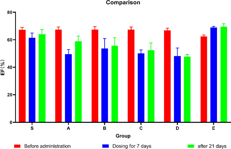
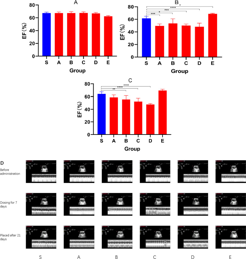
The main diagnostic criterion for HFrEF is decreased ejection fraction, which is closely related to cardiac function. Our study found that group A, group B, group C and group D all had decreased ejection fraction. In Fig. 1, the graph shows the changes in EF after different events. Figure 2A shows that there was no statistical difference in the EF of the initially healthy mice (P > 0.05). After 7 injections (Fig. 2B), the EF of the E group increased and the others decreased. After 21 days of placement (Fig. 2C), the other model groups also showed fluctuations in EF. Figure 2D is a recorded echocardiogram and the original images have been in Additional file 1.
Fig. 1.
The figure showing the EF before administration (Red), EF of dosing for 7 days (Blue) and EF after 21 days (Green) in groups
Fig. 2.
Bar graphs showing the EF before administration (A), EF after dosing for 7 days (B), EF after 21 days (C). (D)Echocardiogram showing FS and EF at 3 times in each group. Data shown are means ± SD; n = 6 in each group. *P < 0.05, significantly different as indicated
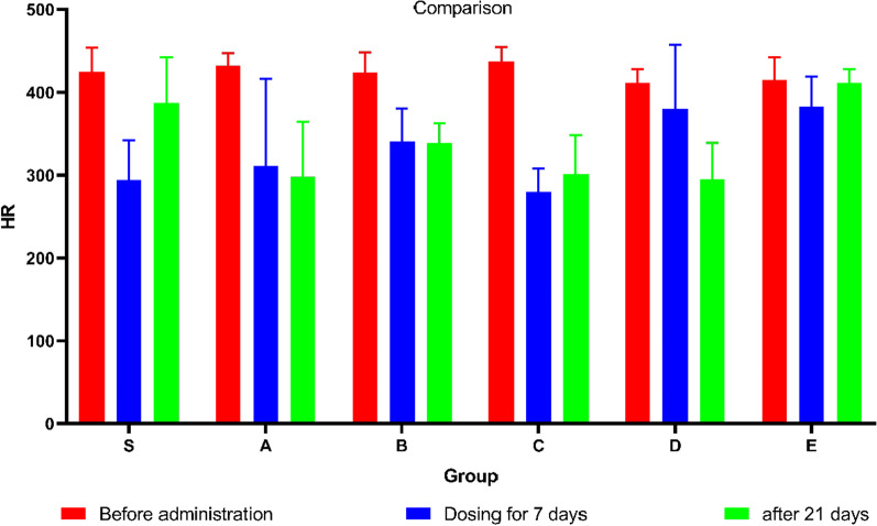
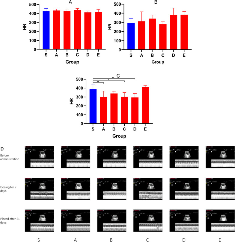
Important indicators of echocardiography also include HR reflecting changes in cardiac function. Figure 3 shows the changes in HR after different events. Figure 4A shows that there was no statistical difference in the HR of the initial healthy mice (P > 0.05). After 7 injections, the HR (Fig. 4B) did not change significantly. After 21 days of placement, the HR (Fig. 4C) of Group A, Group C and Group D decreased significantly (P < 0.05). Figure 4D is a recorded echocardiogram and the original images have been in Additional file 1.
Fig. 3.
The figure showing the HR before administration (Blue bar graph), HR of dosing for 7 days (Red bar graph) and HR after 21 days (Green bar graph) in groups
Fig. 4.
Bar graphs showing the HR before administration (A), HR after dosing for 7 days (B), HR after 21 days (C). (D) Echocardiogram showing HR at 3 times in each group. Data shown are means ± SD; n = 6 in each group. *P < 0.05,significantly different as indicated
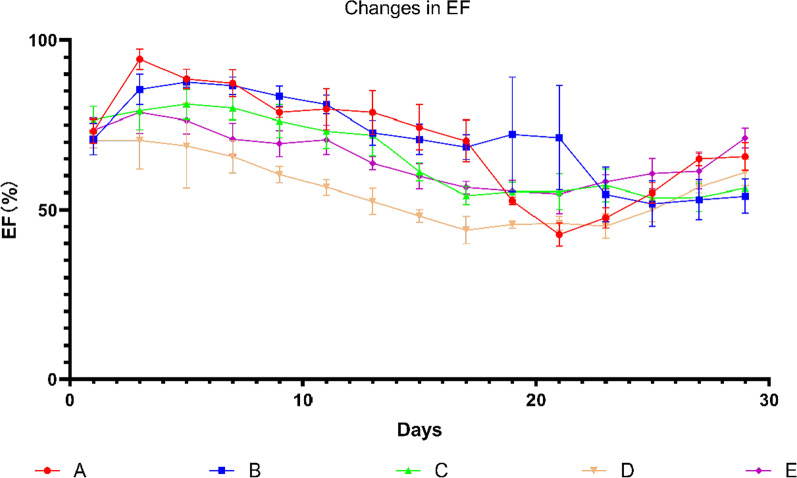
Changes of the three parameters of echocardiography with the change of days. (7 days for modeling, 21 days for placement)
In order to explore the changes of EF, FS and HR during the modeling process, we added 6 mice to each group repeated the experimental operation and recorded the changes of these three indicators.
There was no significant difference in initial EF. Group A and Group B showed significant increase in EF at the initial stage of drug injection, then decreased and finally showed the damage of cardiac function. The EF of Group C and Group D decreased gradually from the initial stage and finally showed the damage of cardiac function. There was no obvious change of EF in Group E and there was no significant difference between the initial EF and the final EF (Fig. 5).
Fig. 5.
Graph showing changes in EF over days. Data shown are means ± SD; n = 6 in each group. The red line represents Group A, the blue line represents Group B, the green line represents Group C, the yellow line represents Group D, and the purple line represents Group E. Group S was not recorded in this experiment
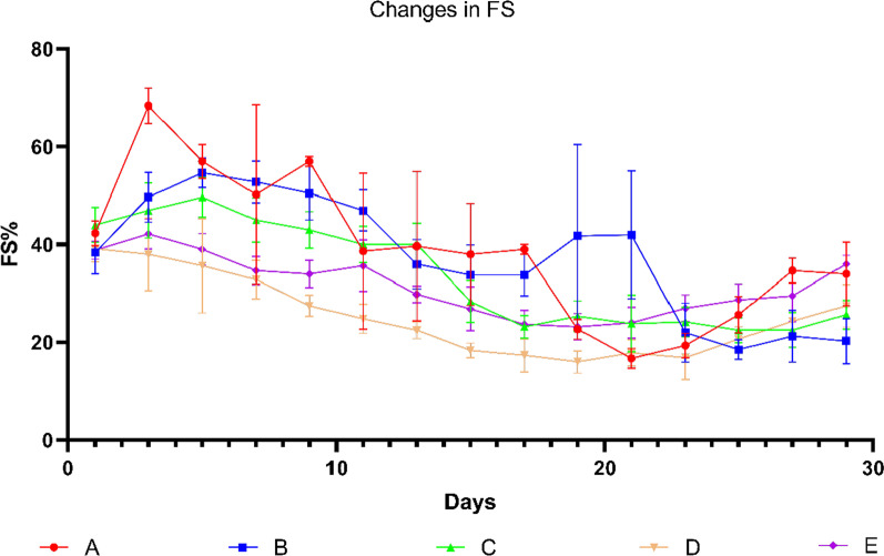
The changes of FS had no obvious regularity but besides the Group E, others also showed different degrees of cardiac function damage, which was close to the results of EF (Fig. 6).
Fig. 6.
Graph showing changes in FS over days. Data shown are means ± SD; n = 6 in each group. The red line represents Group A, the blue line represents Group B, the green line represents Group C, the yellow line represents Group D, and the purple line represents Group E. Group S was not recorded in this experiment
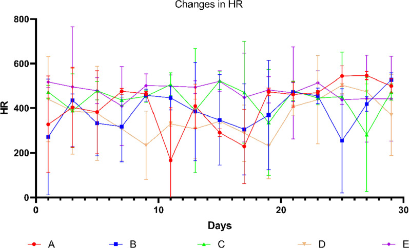
The change of HR was messy, but we still keep track (Fig. 7).
Fig. 7.
Graph showing changes in EF over days. Data shown are means ± SD; n = 6 in each group. The red line represents Group A, the blue line represents Group B, the green line represents Group C, the yellow line represents Group D, and the purple line represents Group E. Group S was not recorded in this experiment
It was found that except for the Group S, the mice in each group had different degrees of BNP and CRP elevation by ELISA
We found by echocardiography that there was no impaired cardiac function in Group E (no significant changes in EF, FS and HR), so Group E was canceled in the following-up study.
BNP is a key marker for the diagnosis of HF and the detection of BNP is more helpful for us to determine the standard of ISO modelling. BNP was significantly increased in Group S compared with Group B and Group D (P < 0.05), which confirmed the occurrence of HF (Fig. 8).
Fig. 8.
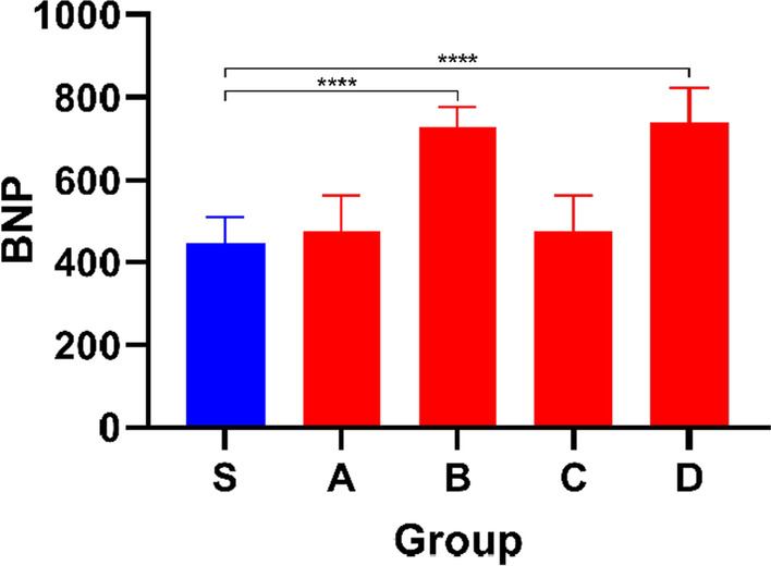
Bar graph showing BNP levels in different groups. Data shown are means ± SD; n = 6 in each group. *P < 0.05, significantly different as indicated
Inflammation plays an important role in the development of HF and we measured C-reactive protein (CRP). Compared with Group S, the CRP of the mice in other groups was significantly increased (P < 0.05), which also confirmed the occurrence of inflammatory response (Fig. 9).
Fig. 9.
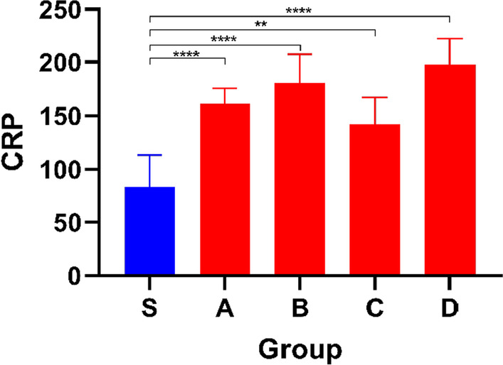
Bar graph showing CRP levels in different groups.Data shown are means ± SD; n = 6 in each group. *P < 0.05, significantly different as indicated
Secondary outcome measures
Secondary outcomes included echocardiographically recorded FS
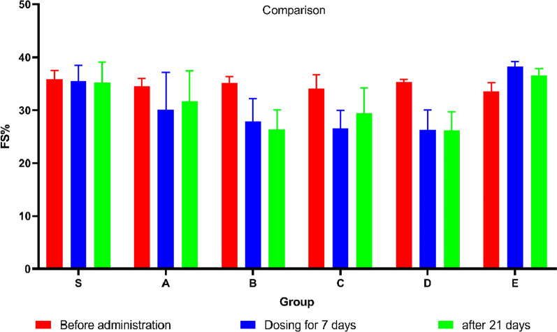
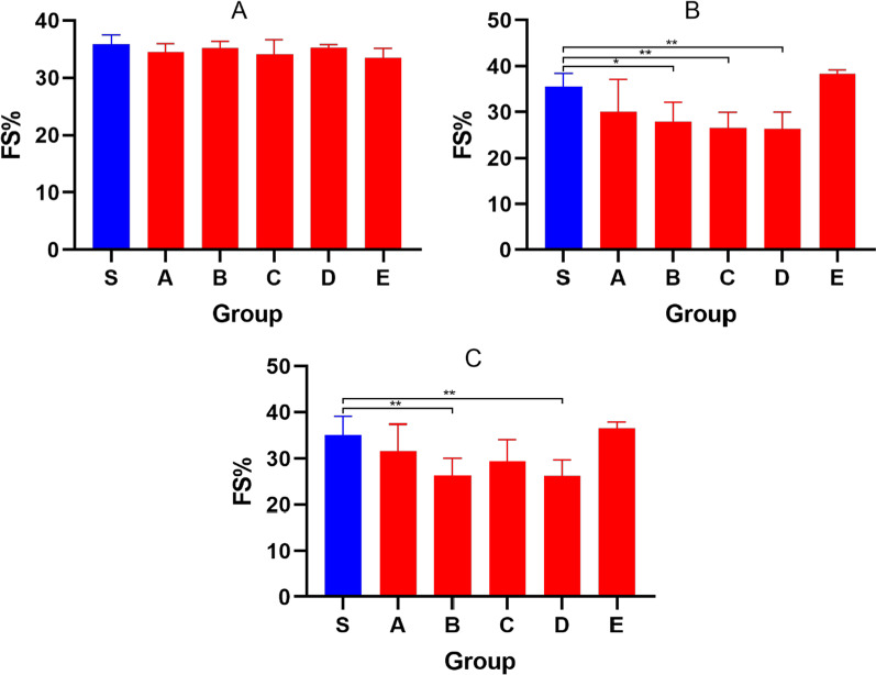
FS can reflect changes in cardiac function too. Figure 10 shows the changes in FS after different events. Figure 11A shows that there was no statistical difference in the FS of initial healthy mice (P > 0.05). After 7 injections, the FS (Fig. 11 B) of Group B, Group C and Group D decreased significantly (P < 0.05). After 21 days of placement, the FS (Fig. 11C) of Group B and Group D was still significantly different from that of Group S (P < 0.05).
Fig. 10.
The figure showing the FS before administration (Blue), FS of dosing for 7 days (Red) and FS after 21 days (Green) in groups
Fig. 11.
Bar graphs showing the FS before administration(A), FS after dosing for 7 days(B), FS after 21 days(C). Data shown are means ± SD; n = 6 in each group. *P < 0.05, significantly different as indicated
After injection of ISO, inflammatory cells increased in the heart tissue of mice in each group
HE staining results revealed the degree of myocardial tissue in mice models. Through HE staining, we observed myofibrolysis and nuclear enlargement in the other groups compared with Group S (Fig. 12).
Fig. 12.
Representative images of HE staining showing changes in inflammatory cells in cardiac sections from each group of mice. Green arrows indicate exceptions
Discussion
CHF causes myocardial tissue injury and cardiac systolic and diastolic dysfunction due to a variety of reasons [29], but it is not limited to myocardial infarction, hypertension, myocarditis and so on [30]. All studies on heart failure require an appropriate heart failure model. The ideal model should be able to reproduce all aspects of the progression of naturally-occurring congestive heart failure [31]. The CHF model protocol of ISO‐induced mice has the advantages of simplicity and easy operation but it is difficult to replicate for the sake of ambiguous administration dose and nonuniform modeling time. Our study aims to establish a stable and reliable CHF model that meets the experimental purpose to provide a basic choice of models in practical studies, thus to facilitate the development of new treatment strategies for patients with heart failure.
In this study, the criteria of model formation and the related indexes analysis were clarified through studying the previous methods of ISO preparation of C57BL / 6 mice chronic heart failure model and performing group experiments that explore the models of different ISO doses, combining with the current conditions and partial experience of our own laboratory and the characteristics of the CHF model preparation protocol [32]. We found that Group A, Group B, Group C and group D can all cause CHF but the mortality rate of Group A and Group B is higher and the mice with higher doses will have skin ulceration and other phenomena that may affect the following research. Group D has a high success rate of modeling and should be the recommended dose for CHF mouse model.
To establish a recommended and stable animal model of heart failure is of great significance for relevant researches. Over the past few decades, many small animal models have been generated to mimic various pathological mechanisms resulting in heart failure. Despite some limitations, these animal models have greatly advanced our understanding of the pathogenesis of heart failure in etiology and have paved the way for understanding the underlying mechanisms and the development of successful therapies [33]. Although close to humans, the cardiac structure of large animals has relatively few applications due to the high modeling cost and complicated operation [34]. Relatively, small animal models are more commonly used when performing relevant medical and pharmacological studies [33].C57BL/6 mice are commonly used experimental animals for related medical and pharmacological research. They have become one of the most commonly used animal models of heart failure because of their short time of reproduction, easy genetic modification, good stability and low cost [30].
ISO has been widely reported to cause heart failure in animals [35, 36] and the ISO induced CHF model is applicable to various inbred strains of mice [37, 38]. Literature reports that the ISO induced myocardial injury model has been widely used to research the beneficial effects of drugs on cardiac dysfunction [39], playing a crucial role in the pathogenesis of HF [40].
The injection doses were divided into three intervals: 30–10 mg/kg, 10–120 mg/kg, 150–400 mg/kg [41]. The first type of long-term intervention resulted in myocardial hypertrophy, causing myocardial overload and increased mortality. Up to 80–90% of prolonged ISO access resulted in advanced hypertrophy characterized by pathological hypertrophy with extensive confluent cardiomyopathy. The second dose resulted in changes in cardiomyocyte energy metabolism. Injection of large doses of ISO resulted in acute myocardial injury, similar to acute myocardial infarction but with a higher mortality rate.
The pathophysiological and morphological abnormalities produced in experimental models of ISO induced CHF are comparable to those occurring in humans. Experimental animal models established by ISO, including pathological myocardial injury, myocardial infarction, cardiac hypertrophy and heart failure, are beneficial to our understanding β- Pathological changes and pathological mechanisms under adrenergic receptor stimulation and finding the best way to treat sympathetic overactivation.
Chronic stimulation of ISO to G-protein-coupled ß-adrenergic receptor causes cardiomyocyte hypertrophy and fibrosis in mice and rats. It resembles the development of progressive heart failure aroused by cardiac specific overexpression of the β1-ADR in mice, suggesting that mice can model aspects of the pathogenesis of CHF. Underlying pathogenic mechanisms of the disease can be explained by the model [33]. The ISO induced CHF model is non-invasive, easy to operate and highly reproducible and can well reflect the natural pathological process of CHF. Though it is simple to operate and easy to handle, it is affected by different batches of animals, different drug lots, different routes of administration. Pre-experiments must be performed before testing to determine the final experimental protocol.
CHF models are usually prepared using favorable surgical procedures, chemical induction, genetic modification, genetic techniques and hypertension induction. Since the most important part of the etiology of CHF is ischemic origin [42], most studies use myocardial infarction models to explore CHF, usually by ligating the left anterior descending artery, resulting in CHF 4 weeks after operation [43]. This model is an acute ischemic injury model [44], generally used to evaluate the remodeling of cells and extracellular matrix after myocardial infarction but not suitable for the long-term exploration of neuroendocrine function [31].
ISO is a synthetic catecholamine and non-selective β- Adrenergic agonist [45], which agonizes the heart β1 receptor to exerts a positive effect on the myocardium, leading to a marked drop in diastolic blood pressure caused by intense vasodilation and in turn giving rise to coronary hypoperfusion, persistently producing cardiac dysfunction and left ventricular dilation [46]. Studies have shown that adrenergic receptors are involved in regulating physiological and pathological processes in the myocardium and their increasing drive plays an essential role in compensating for the decline in cardiac function [47].
β-Adrenergic receptors (β-ARS) chronic hyperactivity of signal transduction is the interface between sympathetic nerve fibers and deterioration of cardiac function [48]. As an independent risk factor for cardiovascular mortality [49–51], cardiac fibrosis is an adaptive remodeling process during cardiac injury [52],whose underlying reasons include mechanical stress, inflammation, ischemia and neurohormonal overactivation. It is characterized by the production of excessive extracellular matrix (ECM) due to the accumulation of inflammatory cells and activated cardiac fibroblasts (CFS). Many lines of evidence indicate that the progression of heart failure manifested by ventricular diastolic and systolic dysfunction is characterized by a marked cardiac hypertrophic and myocardial fibrotic response [53–56] and ISO can stimulate adrenaline and promote cardiac inflammation and fibrosis, causing decompensation and left ventricular remodeling featured by cell death and the generation of inflammation [57], contributing to the development of myocardial injury and cardiac remodeling models [58, 59].
However, this study has some shortcomings. We did not experimentally compare the differences in CHF caused by subcutaneous injection of ISO and other methods, which do not fully reflect the characteristics of each model. We only divided injection doses into 5 groups, so there may be better modeling doses. Last but not least, ISO-induced myocardial injury is usually a variable method that some animals develop more, some less injury. We did not consider this issue, we will continue to explore improvements in follow-up research.
Conclusion
Our research finally found that the HFrEF mice model created by injection at a dose of 100 mg/kg for 7 days was the most suitable and a relatively stable chronic heart failure model could be obtained by placing it for 21 days. Our study aimed to confirm the criteria for a mice model of CHF and will continue to explore the development and treatment mechanisms of CHF in subsequent studies.
Supplementary Information
Additional file 1. Recorded echocardiogram and the original images.
Acknowledgements
Not applicable.
Abbreviations
- HF
Heart failure
- CHF
Chronic heart failure
- BNP
Brain natriuretic peptide
- CRP
C-reactive protein
- TAC
Transverse aortic coarctation
- LV
Left ventricular
- EF
Ejection fraction
- FS
Fraction shortening
- HR
Heart rate
- ISO
Isoproterenol
Author contributions
YP and JG are co-first authors. HZ initiated the project and provided critical suggestions for the project; HC initiated the project and designed the experiments and provided critical suggestions for the project; YP and JG wrote and revised the manuscript, performed the animal experiments, analyzed data and prepare the figures; RG and YG provide consultation and advice on the project; RG analyzed data; WS, HL and JW performed the animal experiments. All authors approved the submission of the manuscript.
Funding
This work was supported in part by the National Natural Science Foundation of China (81674063,81974583,81704169), Jiangsu Province Traditional Chinese Medicine Leading Talent Project (SLJ0226), Phase III Project of Nursing Advantage Discipline of Nanjing University of Chinese Medicine (2019YSHL051), Youth Project of Natural Science Foundation of Nanjing University of Chinese Medicine (NZY8170419).
Availability of data and materials
The datasets used and/or analyzed during the current study are available from the corresponding author on reasonable request. The data that support the findings of this study are available from the corresponding author upon reasonable request. Some data may not be made available because of privacy or ethical restrictions.
Declarations
Ethics approval and consent to participate
All animal experiments were executed conforming to the “Guide for the Care and Use of Laboratory Animals” published by the US National Institutes of Health (NIH Publication No. 85–23, revised 1996). The study is reported in accordance with ARRIVE guidelines. The study was approved by the Animal Ethics Committee, Laboratory Animal Center, Nanjing University of Chinese Medicine.
Consent for publication
Not applicable.
Competing interests
The authors declare that they have no competing interest.
Footnotes
Publisher's Note
Springer Nature remains neutral with regard to jurisdictional claims in published maps and institutional affiliations.
Yujing Pan and Jin Gao contributed equally to this work and share the first authorship
Hao Chen and Hongru Zhang contributed equally to this work and share the corresponding authorship
Contributor Information
Hao Chen, Email: chenhao@njucm.edu.cn.
Hongru Zhang, Email: zhr5001@vip.163.com.
References
- 1.van der Meer P, Gaggin HK, Dec GW. ACC/AHA versus ESC guidelines on heart failure: JACC guideline comparison. J Am Coll Cardiol. 2019;73(21):2756–2768. doi: 10.1016/j.jacc.2019.03.478. [DOI] [PubMed] [Google Scholar]
- 2.Bozkurt B, Coats AJ, Tsutsui H, Abdelhamid M, Adamopoulos S, Albert N, Anker SD, Atherton J, Böhm M, Butler J, Drazner MH. Universal definition and classification of heart failure: a report of the heart failure society of America, heart failure association of the European society of cardiology, Japanese heart failure society and writing committee of the universal definition of heart failure. J Card Fail. 2021;27(4):387–413. doi: 10.1016/j.cardfail.2021.01.022. [DOI] [PubMed] [Google Scholar]
- 3.Cao Z, Zhu J. BNP and NT-proBNP as diagnostic biomarkers for cardiac dysfunction in both clinical and forensic medicine. Int J Mol Sci. 2019;20(8):1820. doi: 10.3390/ijms20081820. [DOI] [PMC free article] [PubMed] [Google Scholar]
- 4.Mudd JO, Kass DA. Tackling heart failure in the twenty-first century. Nature. 2008;451(7181):919–928. doi: 10.1038/nature06798. [DOI] [PubMed] [Google Scholar]
- 5.Seferovic PM, et al. Organization of heart failure management in European society of cardiology member countries: survey of the heart failure association of the European society of cardiology in collaboration with the heart failure national societies/working groups. Eur J Heart Fail. 2013;15(9):947–959. doi: 10.1093/eurjhf/hft092. [DOI] [PubMed] [Google Scholar]
- 6.Akwo EA, et al. Heart failure incidence and mortality in the southern community cohort study. Circ Heart Fail. 2017 doi: 10.1161/CIRCHEARTFAILURE.116.003553. [DOI] [PMC free article] [PubMed] [Google Scholar]
- 7.Farré N, et al. Real world heart failure epidemiology and outcome: a population-based analysis of 88,195 patients. PLoS ONE. 2017;12(2):e0172745. doi: 10.1371/journal.pone.0172745. [DOI] [PMC free article] [PubMed] [Google Scholar]
- 8.Korda RJ, et al. Variation in readmission and mortality following hospitalisation with a diagnosis of heart failure: prospective cohort study using linked data. BMC Health Serv Res. 2017;17(1):220. doi: 10.1186/s12913-017-2152-0. [DOI] [PMC free article] [PubMed] [Google Scholar]
- 9.Piccinni C, et al. The burden of chronic heart failure in primary care in Italy. High Blood Press Cardiovasc Prev. 2017;24(2):171–178. doi: 10.1007/s40292-017-0193-4. [DOI] [PubMed] [Google Scholar]
- 10.Chen J, et al. Ischemic model of heart failure in rats and mice. Methods Mol Biol. 2018;1816:175–182. doi: 10.1007/978-1-4939-8597-5_13. [DOI] [PubMed] [Google Scholar]
- 11.Sawall S, et al. In vivo quantification of myocardial infarction in mice using micro-CT and a novel blood pool agent. Contrast Media Mol Imaging. 2017;2017:2617047. doi: 10.1155/2017/2617047. [DOI] [PMC free article] [PubMed] [Google Scholar]
- 12.Bosch L, et al. The transverse aortic constriction heart failure animal model: a systematic review and meta-analysis. Heart Fail Rev. 2021;26(6):1515–1524. doi: 10.1007/s10741-020-09960-w. [DOI] [PMC free article] [PubMed] [Google Scholar]
- 13.Benjamin EJ, et al. Heart disease and stroke statistics-2018 update: a report from the american heart association. Circulation. 2018;137(12):e67–e492. doi: 10.1161/CIR.0000000000000558. [DOI] [PubMed] [Google Scholar]
- 14.Ma D, et al. Effects of ivabradine hydrochloride combined with trimetazidine on myocardial fibrosis in rats with chronic heart failure. Exp Ther Med. 2019;18(3):1639–1644. doi: 10.3892/etm.2019.7730. [DOI] [PMC free article] [PubMed] [Google Scholar]
- 15.Wang QD, Bohlooly YM, Sjöquist PO. Murine models for the study of congestive heart failure: Implications for understanding molecular mechanisms and for drug discovery. J Pharmacol Toxicol Methods. 2004;50(3):163–174. doi: 10.1016/j.vascn.2004.05.005. [DOI] [PubMed] [Google Scholar]
- 16.Kanisicak O, et al. Genetic lineage tracing defines myofibroblast origin and function in the injured heart. Nat Commun. 2016;7:12260. doi: 10.1038/ncomms12260. [DOI] [PMC free article] [PubMed] [Google Scholar]
- 17.Kemuriyama T, et al. Endogenous angiotensin II has fewer effects but neuronal nitric oxide synthase has excitatory effects on renal sympathetic nerve activity in salt-sensitive hypertension-induced heart failure. J Physiol Sci. 2009;59(4):275–281. doi: 10.1007/s12576-009-0034-x. [DOI] [PMC free article] [PubMed] [Google Scholar]
- 18.Dixon DL, et al. Pulmonary effects of chronic elevation in microvascular pressure differ between hypertension and myocardial infarct induced heart failure. Heart Lung Circ. 2015;24(2):158–164. doi: 10.1016/j.hlc.2014.08.009. [DOI] [PubMed] [Google Scholar]
- 19.Spivak M, et al. Doxorubicin dose for congestive heart failure modeling and the use of general ultrasound equipment for evaluation in rats. Longitudinal Vivo Study Med Ultrason. 2013;15(1):23–28. doi: 10.11152/mu.2013.2066.151.ms1ddc2. [DOI] [PubMed] [Google Scholar]
- 20.Gao L, et al. Schisandrin a protects against isoproterenol‑induced chronic heart failure via miR‑155. Mol Med Rep. 2021 doi: 10.3892/mmr.2021.12540. [DOI] [PMC free article] [PubMed] [Google Scholar]
- 21.Kazachenko AA, et al. Comparative characteristics of some pharmacological models of chronic heart failure. Eksp Klin Farmakol. 2008;71(6):16–19. [PubMed] [Google Scholar]
- 22.Noll NA, Lal H, Merryman WD. Mouse models of heart failure with preserved or reduced ejection fraction. Am J Pathol. 2020;190(8):1596–1608. doi: 10.1016/j.ajpath.2020.04.006. [DOI] [PMC free article] [PubMed] [Google Scholar]
- 23.Adamo L, et al. Reappraising the role of inflammation in heart failure. Nat Rev Cardiol. 2020;17(5):269–285. doi: 10.1038/s41569-019-0315-x. [DOI] [PubMed] [Google Scholar]
- 24.Liang J. Research progress of animal models of myocardial injury induced by isoproterenol. Acta Lab Anim Sci Sin. 2019;27(1):110–114. [Google Scholar]
- 25.Wallner M, et al. Acute catecholamine exposure causes reversible myocyte injury without cardiac regeneration. Circ Res. 2016;119(7):865–879. doi: 10.1161/CIRCRESAHA.116.308687. [DOI] [PMC free article] [PubMed] [Google Scholar]
- 26.Li X, Zhang ZL, Wang HF. Fusaric acid (FA) protects heart failure induced by isoproterenol (ISP) in mice through fibrosis prevention via TGF-β1/SMADs and PI3K/AKT signaling pathways. Biomed Pharmacother. 2017;93:130–145. doi: 10.1016/j.biopha.2017.06.002. [DOI] [PubMed] [Google Scholar]
- 27.Lu H, Guo J, Xu C. Cardioprotective efficacy of hispidulin on isoproterenol-induced heart failure in wistar rats. Int J Pharmacol. 2019;15(7):816–822. doi: 10.3923/ijp.2019.816.822. [DOI] [Google Scholar]
- 28.Zhang X, et al. Matrine attenuates pathological cardiac fibrosis via RPS5/p38 in mice. Acta Pharmacol Sin. 2021;42(4):573–584. doi: 10.1038/s41401-020-0473-8. [DOI] [PMC free article] [PubMed] [Google Scholar]
- 29.Bo L, et al. Research on the function and mechanism of survivin in heart failure mice model. Eur Rev Med Pharmacol Sci. 2017;21(16):3699–3704. [PubMed] [Google Scholar]
- 30.Bacmeister L, et al. Inflammation and fibrosis in murine models of heart failure. Basic Res Cardiol. 2019;114(3):19. doi: 10.1007/s00395-019-0722-5. [DOI] [PubMed] [Google Scholar]
- 31.Monnet E, Chachques JC. Animal models of heart failure: what is new? Ann Thorac Surg. 2005;79(4):1445–1453. doi: 10.1016/j.athoracsur.2004.04.002. [DOI] [PubMed] [Google Scholar]
- 32.Xiao H, et al. IL-18 cleavage triggers cardiac inflammation and fibrosis upon β-adrenergic insult. Eur Heart J. 2018;39(1):60–69. doi: 10.1093/eurheartj/ehx261. [DOI] [PubMed] [Google Scholar]
- 33.Riehle C, Bauersachs J. Small animal models of heart failure. Cardiovasc Res. 2019;115(13):1838–1849. doi: 10.1093/cvr/cvz161. [DOI] [PMC free article] [PubMed] [Google Scholar]
- 34.Sag CM, et al. Calcium/calmodulin-dependent protein kinase II contributes to cardiac arrhythmogenesis in heart failure. Circ Heart Fail. 2009;2(6):664–675. doi: 10.1161/CIRCHEARTFAILURE.109.865279. [DOI] [PMC free article] [PubMed] [Google Scholar]
- 35.Yang J, Wang Z, Chen DL. Shikonin ameliorates isoproterenol (ISO)-induced myocardial damage through suppressing fibrosis, inflammation, apoptosis and ER stress. Biomed Pharmacother. 2017;93:1343–1357. doi: 10.1016/j.biopha.2017.06.086. [DOI] [PubMed] [Google Scholar]
- 36.Zhang MX, et al. Exchange-protein activated by cAMP (EPAC) regulates L-type calcium channel in atrial fibrillation of heart failure model. Eur Rev Med Pharmacol Sci. 2019;23(5):2200–2207. doi: 10.26355/eurrev_201903_17267. [DOI] [PubMed] [Google Scholar]
- 37.Park S, et al. Genetic regulation of fibroblast activation and proliferation in cardiac fibrosis. Circulation. 2018;138(12):1224–1235. doi: 10.1161/CIRCULATIONAHA.118.035420. [DOI] [PMC free article] [PubMed] [Google Scholar]
- 38.Wang JJ, et al. Genetic dissection of cardiac remodeling in an isoproterenol-induced heart failure mouse model. PLoS Genet. 2016;12(7):e1006038. doi: 10.1371/journal.pgen.1006038. [DOI] [PMC free article] [PubMed] [Google Scholar]
- 39.Fan C, et al. Qi-Li-Qiang-Xin alleviates isoproterenol-induced myocardial injury by inhibiting excessive autophagy via activating AKT/mTOR pathway. Front Pharmacol. 2019;10:1329. doi: 10.3389/fphar.2019.01329. [DOI] [PMC free article] [PubMed] [Google Scholar]
- 40.Ahmed LA, et al. Boosting Akt pathway by rupatadine modulates Th17/Tregs balance for attenuation of isoproterenol-induced heart failure in rats. Front Pharmacol. 2021;12:651150. doi: 10.3389/fphar.2021.651150. [DOI] [PMC free article] [PubMed] [Google Scholar]
- 41.Periasamy S, Chen SY, Liu MY. The study of ISO induced heart failure rat model. Exp Mol Pathol. 2010;88:299–304. doi: 10.1016/j.yexmp.2009.10.011. [DOI] [PubMed] [Google Scholar]
- 42.Batkai S, et al. CDR132L improves systolic and diastolic function in a large animal model of chronic heart failure. Eur Heart J. 2021;42(2):192–201. doi: 10.1093/eurheartj/ehaa791. [DOI] [PMC free article] [PubMed] [Google Scholar]
- 43.van den Borne SW, et al. Mouse strain determines the outcome of wound healing after myocardial infarction. Cardiovasc Res. 2009;84(2):273–282. doi: 10.1093/cvr/cvp207. [DOI] [PubMed] [Google Scholar]
- 44.Fiedler LR, Maifoshie E, Schneider MD. Mouse models of heart failure: cell signaling and cell survival. Curr Top Dev Biol. 2014;109:171–247. doi: 10.1016/B978-0-12-397920-9.00002-0. [DOI] [PubMed] [Google Scholar]
- 45.Ren S. Implantation of an isoproterenol mini-pump to induce heart failure in mice. J f Vis Exp. 2019 doi: 10.3791/59646. [DOI] [PMC free article] [PubMed] [Google Scholar]
- 46.Shukla SK, Sharma SB, Singh UR. β-Adrenoreceptor agonist isoproterenol alters oxidative status, inflammatory signaling, injury markers and apoptotic cell death in myocardium of rats. Indian J Clin Biochem. 2015;30(1):27–34. doi: 10.1007/s12291-013-0401-5. [DOI] [PMC free article] [PubMed] [Google Scholar]
- 47.LaRocca TJ, et al. CXCR4 cardiac specific knockout mice develop a progressive cardiomyopathy. Int J Mol Sci. 2019;20(9):2267. doi: 10.3390/ijms20092267. [DOI] [PMC free article] [PubMed] [Google Scholar]
- 48.Hartupee J, Mann DL. Neurohormonal activation in heart failure with reduced ejection fraction. Nat Rev Cardiol. 2017;14(1):30–38. doi: 10.1038/nrcardio.2016.163. [DOI] [PMC free article] [PubMed] [Google Scholar]
- 49.Iles L, et al. Evaluation of diffuse myocardial fibrosis in heart failure with cardiac magnetic resonance contrast-enhanced T1 mapping. J Am Coll Cardiol. 2008;52(19):1574–1580. doi: 10.1016/j.jacc.2008.06.049. [DOI] [PubMed] [Google Scholar]
- 50.Moore-Morris T, et al. Cardiac fibroblasts: from development to heart failure. J Mol Med (Berl) 2015;93(8):823–830. doi: 10.1007/s00109-015-1314-y. [DOI] [PMC free article] [PubMed] [Google Scholar]
- 51.Chen G, et al. Sca-1(+) cardiac fibroblasts promote development of heart failure. Eur J Immunol. 2018;48(9):1522–1538. doi: 10.1002/eji.201847583. [DOI] [PMC free article] [PubMed] [Google Scholar]
- 52.Schafer S, et al. IL-11 is a crucial determinant of cardiovascular fibrosis. Nature. 2017;552(7683):110–115. doi: 10.1038/nature24676. [DOI] [PMC free article] [PubMed] [Google Scholar]
- 53.Kong P, Christia P, Frangogiannis NG. The pathogenesis of cardiac fibrosis. Cell Mol Life Sci. 2014;71(4):549–574. doi: 10.1007/s00018-013-1349-6. [DOI] [PMC free article] [PubMed] [Google Scholar]
- 54.Ma ZG, et al. Cardiac fibrosis: new insights into the pathogenesis. Int J Biol Sci. 2018;14(12):1645–1657. doi: 10.7150/ijbs.28103. [DOI] [PMC free article] [PubMed] [Google Scholar]
- 55.Liu QH, et al. I(K1) channel agonist zacopride alleviates cardiac hypertrophy and failure via alterations in calcium dyshomeostasis and electrical remodeling in rats. Front Pharmacol. 2019;10:929. doi: 10.3389/fphar.2019.00929. [DOI] [PMC free article] [PubMed] [Google Scholar]
- 56.Metra M. February 2017 at a glance: fibrosis, acute heart failure and neurologic abnormalities. Eur J Heart Fail. 2017;19(2):165–166. doi: 10.1002/ejhf.760. [DOI] [PubMed] [Google Scholar]
- 57.Park SW, et al. CRABP1 protects the heart from isoproterenol-induced acute and chronic remodeling. J Endocrinol. 2018;236(3):151–165. doi: 10.1530/JOE-17-0613. [DOI] [PMC free article] [PubMed] [Google Scholar]
- 58.Benjamin IJ, et al. Isoproterenol-induced myocardial fibrosis in relation to myocyte necrosis. Circ Res. 1989;65(3):657–670. doi: 10.1161/01.RES.65.3.657. [DOI] [PubMed] [Google Scholar]
- 59.Wang M, et al. GHSR deficiency exacerbates cardiac fibrosis: role in macrophage inflammasome activation and myofibroblast differentiation. Cardiovasc Res. 2020;116(13):2091–2102. doi: 10.1093/cvr/cvz318. [DOI] [PubMed] [Google Scholar]
Associated Data
This section collects any data citations, data availability statements, or supplementary materials included in this article.
Supplementary Materials
Additional file 1. Recorded echocardiogram and the original images.
Data Availability Statement
The datasets used and/or analyzed during the current study are available from the corresponding author on reasonable request. The data that support the findings of this study are available from the corresponding author upon reasonable request. Some data may not be made available because of privacy or ethical restrictions.


