Abstract
Background
This study aims to investigate the effects of different direct oral anticoagulants on experimental renal injury induced by temporary infrarenal aortic occlusion.
Methods
A total of 35 male Wistar rats (250 to 350 g) were randomly allocated to any of the five groups: sham, ischemia-reperfusion, rivaroxaban, dabigatran, and apixaban groups. Sham group underwent median laparotomy. Ischemia-reperfusion group was given saline gavage for one week. Animals in the other groups received rivaroxaban (3 mg/kg), dabigatran (15 mg/kg), or apixaban (10 mg/kg) daily once for one week via oral gavage. The infrarenal abdominal aorta was clamped for 60 min, and reperfusion was maintained for 120 min in the ischemia-reperfusion, rivaroxaban, dabigatran, and apixaban groups. At the end of reperfusion, kidneys were harvested for biochemical and histopathological analysis.
Results
Renal total antioxidant capacity was reduced, and total oxidant status, interleukin-1 beta, and tumor necrosis factor-alpha were elevated in the ischemia-reperfusion group, compared to the sham group (p<0.005). Histological damage scores were also higher in the ischemia-reperfusion group (p<0.005). Administration of direct oral anticoagulants caused an increase of total antioxidant capacity and reduction of total oxidant status, tumor necrosis factor-alpha, and interleukin-1 beta in the rivaroxaban, dabigatran, and apixaban groups compared to the ischemia-reperfusion group (p<0.005). Histological damage scores were lower in the rivaroxaban and dabigatran groups than the ischemia-reperfusion group scores (p<0.005).
Conclusion
Direct oral anticoagulants reduce aortic clamping-induced renal tissue oxidation and inflammation. Rivaroxaban and dabigatran attenuate ischemia-reperfusion-related histological damage in kidneys.
Keywords: Apixaban, dabigatran, inflammation, ischemia-reperfusion, kidney injury, rivaroxaban
Introduction
Arterial embolism, intravascular thrombus formation, rupture of atherosclerotic plaques, hypercoagulability, propagation of the previously present mural thrombus, or vascular graft thrombosis may cause acute arterial occlusion. The maintenance of the perfusion through endovascular or surgical techniques is essential in preventing tissue damage.
However, reperfusion after the ischemic phase leads to the depletion of cellular antioxidant defense mechanisms and the formation of reactive oxidant species (ROS) in tissues. Cytokines are also released into circulation upon reperfusion, and these may mediate inflammation and organ damage. Inflammatory cytokines such as tumor necrosis factoralpha (TNF-α) and interleukin-1 (IL-1β) are the major mediators of ischemia-reperfusion (IR) injury.[1]
Direct oral anticoagulants (DOACs) are agents used for direct inhibition of factor Xa (FXa) or factor II (thrombin) of the coagulation cascade. These agents can pharmacologically prevent target clot formation and fibrin accumulation.[2] Direct oral anticoagulants have emerged as reliable alternatives to vitamin K antagonists, as they reduce drug interactions, lower the risk of major bleeding and eliminate the need for routine laboratory monitoring. Dabigatran, rivaroxaban, and apixaban are DOACs that are commonly used to prevent and treat thrombotic and embolic complications in cases of non-valvular atrial fibrillation and venous thromboembolism.[3]
As well as their primary effects on the coagulation cascade, it is postulated that DOACs may also have some effects on the modulation of oxidative and inflammatory responses. The evidence gained from experimental studies suggests a possible protective role of FXa and thrombin inhibition in organ damage triggered by IR.[4]
The kidneys are one of the important targets of ischemia-mediated damage, and although many treatment modalities have been studied to reduce this damage, it is still a clinically important cause of acute renal failure.[5] To the best of our knowledge, the comparative effects of DOACs on aortic IR-induced renal tissue damage in the same experimental setting have not been investigated previously.
In this experimental study, we aimed to investigate the effects of different DOACs on temporary infrarenal aortic occlusion induced renal injury by the examination of renal histology, kidney TNF-α, IL-1β, total antioxidant capacity, and total antioxidant status levels.
Patients and Methods
In the study, 35 male Wistar albino rats (250 to 350 g) were used. The animals were simply randomized into five groups, each of seven animals. The rats were kept in cages of four animals per cell. Prior to the experiment, the animals were housed in humidity (45 to 50%) and temperature (22±2°C) controlled room. The rats were fed a daily diet and had unlimited access to tap water, but the food and water access were restricted 12 h before the experimental surgery.
Ischemia-reperfusion and surgical procedures
A mixture of the ketamine (90 mg/kg) (Ketalar®, Parker-Dawis, Pfizer, Istanbul, Türkiye) and xylazine (10 mg/kg) (Rompun®, Bayer AG, Leverkusen, Germany) was administered by intraperitoneal (i.p.) injection for anesthesia induction. The abdominal hair was shaved, scrubbed with povidone-iodine solution, and covered in a sterile manner. The skin was opened with a No. 15 scalpel and a median laparotomy was performed with Mayo scissors. Intestines were positioned to reach the abdominal aorta. Traction by 4/0 silk sutures was applied to the abdominal aorta at the level of the renal arteries and, then, occluded with an atraumatic clamp (Vascu-Statt II-Scanlan, St. Paul, MN, USA) for 60 min. At the end of the ischemic period, the clamp was removed, and the rats remained in the reperfusion phase for 120 min.[6]
Direct oral anticoagulant agents
Rivaroxaban (3 mg/kg) (Xarelto®, Bayer HealthCare AG, Leverkusen, Germany), dabigatran (15 mg/kg) (Pradaxa®, Boehringer Ingelheim, Ingelheim, Germany), and apixaban (10 mg/kg) (Eliquis®, Bristol-Myers Squibb Company, Princeton, USA) were given to rats by oral gavage in a homogeneous distribution in 2 mL distilled water. The drug doses were adjusted based on previous studies.[7-9]
Study groups
Sham group (n=7), A median laparotomy was performed, and the aortic clamping was not applied.
IR group (n=7), The IR model was applied after median laparotomy.
Rivaroxaban group (n=7), Rivaroxaban was given for one week before the induction of IR.
Dabigatran group (n=7), Dabigatran was given for one week before the induction of IR.
Apixaban group (n=7), Apixaban was given for one week before the induction of IR.
Kidney tissue harvesting
After the reperfusion period, the chest cavities were opened. Animals were sacrificed by cardiac puncture exsanguination under deep general anesthesia. Right and left kidneys were harvested, and the collected tissues were allowed to be fixed for 48 h in 10% buffered formalin. Samples were embedded in paraffin, 3 to 4-µm sections were obtained, and stained with hematoxylin and eosin (H&E).
Biochemical analysis
Tissue samples were placed in the Eppendorf tubes, cooled in an ice-cold container, and stored at -80°C for biochemical analysis. On the day of analysis, the frozen tissue samples were thawed, weighed, and homogenized in 50 mM pH 7.4 phosphate buffer (PBS) (PRO 250 Scientology Inc., Monroe, CT, USA) and, then, centrifuged at 20,000 g for 15 min. The supernatants were obtained, and assays were performed.
Tissue levels of total antioxidant capacity (TAC) and total oxidant status (TOS) are biochemical tests that were performed based on the Fenton reaction.[10]
The TAC and TOS levels were measured by Erel's colorimetric method by using commercially available kits (Rel Assay Diagnostics; Mega Tıp, Gaziantep, Türkiye). Tissue TAC levels were expressed in terms of Trolox equivalents per gram protein (μmol Trolox Eq/g protein) and TOS levels were expressed in micromole hydrogen peroxide equivalents per gram protein (µmol H2O2 Eq/g protein).
The TNF-alpha and IL-1β concentrations in tissue samples were determined via enzymelinked immunosorbent assay (ELISA). Rat-specific EE-EL-R0019, ELISA kits (Elabscience Biotechnology Co. Wuhan, PRC) were used to quantify inflammatory mediators in samples. For measurements, an ELISA microplate reader (DAR 800, Diagnostic Automation/ Cortez Diagnostic, Inc., CA, USA) was used, and the results were expressed as picograms per milligram of protein (pg/mg protein).
Histopathological evaluation of kidney tissue samples
Kidney tissue samples were harvested and were kept in a 10% buffered formalin solution for pathological assessment. Samples were embedded in paraffin, and 3 to 4-µm section blocks were obtained by using a microtome (Thermo Shandon HM 430 Sliding Microtome, Thermo Fisher Scientific Inc., MA, USA). Tissue sections were stained with H-E for histopathological examination. The examinations were conducted by a single pathologist who was blinded to the experimental groups. A light microscope was used (Olympus BX53, Olympus Co., Tokyo, Japan) and Image Analysis Software (Olympus DP-22 Microscope digital camera software program, Tokyo, Japan) was used to examine the samples.
Kidney tissue samples were analyzed using the semi-quantitative method described by Koçyiğit et al.[11] A four-point scale was used to determine focal glomerular necrosis, Bowman capsule dilatation, tubular epithelial degeneration, tubular epithelial necrosis, tubular dilatation, and interstitial inflammatory infiltration: (0) no change, (1) focal, mild changes, (2) multifocal intermediate changes and (3) prominent extensive changes. Following the examinations, each histopathological characteristic score was summed to yield a total histological damage score for each study group.
Statistical analysis
Statistical analysis was performed using the IBM SPSS version 25.0 software (IBM Corp., Armonk, NY, USA). Descriptive data were expressed in median (minmax). The Shapiro-Wilk test was used to analyze the normality distribution. The Kruskal-Wallis variance analysis was used to compare variables between groups. The Mann-Whitney U test was used for a paired analysis of the groups and Bonferroni correction was utilized for multiple comparisons using a p value of 0.005. For statistical analysis, a p value of <0.05 was considered statistically significant.
Results
Tissue antioxidant capacity and tissue oxidant status
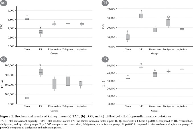
The TAC and TOS kidney tissue levels are shown in Figure 1a and b, respectively. Accordingly, TAC levels were significantly higher in the sham group compared to the other groups (p<0.005). In the IR group, TAC levels were significantly reduced than the levels in the rivaroxaban, apixaban, and dabigatran groups (p<0.005). In the sham group, TOS levels were found to be significantly lower than the other study groups (p<0.005). The TOS levels were significantly increased in the IR group compared to the levels in the rivaroxaban, apixaban, and dabigatran groups (p<0.005). In the dabigatran group, TOS levels were found to be higher than the levels in the rivaroxaban and apixaban groups (p<0.005).
Figure 1. Biochemical results of kidney tissue (a) TAC, (b) TOS, and (c) TNF-a, (d) IL-1b, proinflammatory cytokines.<br> TAC: Total antioxidant capacity; TOS: Total oxidant status; TNF-a: Tumor necrosis factor-alpha; IL-1b: Interleukin-1 beta; † p<0.005 compared to IR, rivaroxaban, dabigatran, and apixaban groups; ¥ p<0.005 compared to rivaroxaban, dabigatran, and apixaban groups; Ω p<0.005 compared to rivaroxaban and apixaban groups; ∑ p<0.005 compared to dabigatran and apixaban groups.
Tumor necrosis factor-α and interleukin-1β tissue levels
The kidney tissue TNF-α levels are shown in Figure 1c and IL-1β levels are shown in Figure 1d. Accordingly, TNF-α and IL-1β were found to be significantly lower in the sham group compared to the other study groups (p<0.005). In the IR group, TNF-α and IL-1β were significantly increased compared to the other study groups (p<0.005). There were no significant in-group differences of TNF-α levels among rivaroxaban, apixaban, and dabigatran groups. However, IL-1β was significantly reduced in the rivaroxaban group compared to the apixaban and the dabigatran groups (p<0.005).
Histopathological damage scores
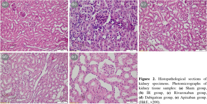
The results of histopathological damage scores are presented in Table 1. The total damage score in the sham group was significantly lower than the scores of the other study groups (p<0.005). In the rivaroxaban and dabigatran groups, the total damage scores were found to be significantly reduced compared to the IR and the apixaban groups (p<0.005). There was no significant difference in total damage scores between the apixaban and IR groups.
Table 1. Histopathological damage scores.
| Focal glomerular necrosis | Bowman's capsule dilation | Degeneration of tubular epithelium | Tubular dilatation | Interstitial inflammatory infiltration | Total score | |||||||
| Median | IQR | Median | IQR | Median | IQR | Median | IQR | Median | IQR | Median | IQR | |
| Sham | 0 | 0 | 0 | 0 | 0 | 0 | 0 | 0 | 0 | 0 | 0 | 1† |
| IR | 3 | 0 | 2 | 0.75 | 1 | 0.75 | 3 | 1.5 | 1 | 0.75 | 9.5 | 3δ |
| Rivaroxaban | 1.5 | 1 | 1 | 0 | 0 | 0 | 1 | 1 | 0 | 0 | 3.5 | 2 |
| Dabigatran | 1 | 1 | 1.5 | 1 | 0 | 1 | 1 | 0.75 | 0 | 0 | 4 | 3 |
| Apixaban | 2 | 1 | 1 | 1 | 1 | 0 | 3 | 1 | 0 | 0 | 6 | 3δ |
| IQR: Interquartile range; † p<0.005 compared to the IR, rivaroxaban, dabigatran, and apixaban groups; δ p<0.005 compared to the rivaroxaban and dabigatran groups. | ||||||||||||
Figure 2. Histopathological sections of kidney specimens. Photomicrographs of kidney tissue samples: (a) Sham group, (b) IR group, (c) Rivaroxaban group, (d) Dabigatran group, (e) Apixaban group. (H&E, x200).
Discussion
In this experimental study, we used temporary infrarenal aortic clamping to form distant organ damage in the kidneys. Aortic IR provoked a considerable amount of tissue oxidation and utilization of antioxidant defenses as evidenced by increased renal TOS and decreased TAC concentrations in our animal model. We also observed that the levels of kidney tissue proinflammatory cytokines (TNF-α and IL-1β) were increased as a result of IR. The histopathological examination in animals subjected to IR revealed severe damage to renal tissue. Our findings showed that administrations of rivaroxaban, dabigatran, and apixaban before temporary infrarenal aortic clamping in rats prevented the consumption of renal antioxidant defenses. These anticoagulant agents also attenuated experimental IR injury by limiting the oxidative burst in kidneys. We observed that anticoagulant administration effectively limited the renal tissue cytokine responses induced by temporary aortic occlusion in our model. Rivaroxaban and dabigatran, but not the apixaban, application resulted in the reduction of histological damage in kidneys.
The kidneys are not subject to the direct effect of cessation of blood flow during infrarenal aortic clamping nevertheless they are majorly affected. In many studies, it has been observed that transient infrarenal aortic occlusion disrupts TAC/TOS balance, triggers an inflammatory response via pro-inflammatory cytokines including the IL-1β and TNF-α, and forms histological damage in renal tissue.[6] The findings of the present study are also similar to these studies.
Coagulation and inflammation cascades interact through some key elements which can play a major role in IR-induced tissue damage.[4] The generation of ROS during IR can potentially induce the release of tissue factor (TF) from adventitial fibroblasts, smooth muscle cells, and the circulating monocytes. Upon its release into the blood, TF triggers the activations of coagulation cascade factors including the elements of the common pathway (FXa and thrombin). Other than participating in the coagulation cascade, these factors can act as proteases and can activate the proteaseactivated receptors (PARs).[12] These receptors are a large family of G-protein coupled membrane receptors that are located on various cell types such as platelets, endothelium, and myocytes. Activated PARs can increase the generation of ROS and play a major role in the production of pro-inflammatory cytokines.[13-15] It has been suggested that inhibition of PARs can be a therapeutic target for the reduction of inflammation in chronic diseases.[16] Therefore, DOACs may also be beneficial in reducing inflammatory responses during the thrombotic conditions, by the inhibition of coagulation cascade triggered PARs activation.
In the current study, we observed that rivaroxaban, which is an FXa inhibitor, blocked the infra-renal aortic IR-induced renal oxidation and inflammation and attenuated the histological damage. In similar experimental models of aortic and femoral artery clamping, rivaroxaban pre-treatment has been previously reported to decrease the levels of biochemical markers of oxidation in renal tissue.[17,18] Rivaroxaban has also been shown to decrease the in vitro ROS production in renal tubular cells that have been exposed to the advanced glycation end-product via the thrombin/PAR-2 system.[19] In cisplatin-induced renal toxicity, rivaroxaban was observed to reduce the histological damage to kidneys by modulating the coagulation cascade activation.[20] Furthermore, rivaroxaban has attenuated angiotensin-II triggered hypertensive renal inflammatory damage in rats via inhibition of PAR-2 signaling.[21] We believe that the antioxidant and anti-inflammatory potentials of rivaroxaban are related to the renal protective effects in our animal model.
Apixaban is another FXa inhibitor investigated in this study. There are limited data regarding the biological effects of apixaban in experimental settings of oxidative and inflammatory conditions. Ishibashi et al.[22] showed that a pixaban effectively inhibited the oxidative stress and inflammatory reactions in plasma-exposed renal mesangial cells via blockade of FXa and suppression of the thrombin-PAR-1 interaction. As well as through the antioxidant effect, apixaban reduced synovial PAR-2 expression and decreased serum IL-6 levels in an FXa induced arthritis model in rats.[23] In our study, although apixaban showed antioxidant and anti-inflammatory effects similar to the previous studies, it did not significantly prevent renal histological damage. We believe that, if the sample size of the experiment was larger, the effects of apixaban in terms of histological damage may have been more apparent. However, there may be other reasons to explain this result, and the effects of apixaban in IR-induced organ damage require further investigation.
Dabigatran is a direct thrombin inhibitor that has been reported to exert antioxidant and anti-inflammatory effects in addition to anticoagulant action.[24] Yazici et al.[8] indicated that dabigatran reduced tissue malondialdehyde and prolidase levels, which were increased in response to experimental renal IR. Recently, in another study, dabigatran was shown to inhibit PAR-1 activation, tubulointerstitial fibrosis, and renal inflammation induced by ureteral obstruction in rats.[25] In the current study, we found that dabigatran had a renal protective property in IR injury which was associated with antioxidant and anti-inflammatory effects.
There can be other mechanisms of the renoprotective effect of DOACs which we observed in the current study. During IR, various released factors and mediators may trigger endothelial injury and initiate microvascular thrombosis in many organ systems including the kidney. This thrombotic process may adversely affect tissue perfusion. We believe that some of the renal protection, which we observed in our IR model, can be related to the primary effect of the anticoagulant agents which prevents thrombosis and microvascular complications.
In this study, we investigated the antioxidant and anti-inflammatory effects of the agents, regardless of their primary effects. We believe that these pleiotropic effects may vary depending on the different molecular structures and mode of action of these agents independent from their anticoagulant activities in vivo. Some of the results we obtained in this study may also be related to the structural differences of the agents, but since we do not have enough data, it is not possible to conclude.
Certain limitations of our study should be considered while interpreting the results. First, physiological functions of renal tissue could have been assessed by more detailed tests, such as glomerular filtration rate and urinalysis. Second, determination of the levels of renal tissue PAR activity could have yielded important knowledge on the mechanism of protection provided by DOACs in this study. In addition, this study is a hypothetical animal model conducted in a limited number of subjects and, therefore, the translation of these results into clinical conditions necessitates confirmation in humans.
In conclusion, pre-treatment with direct oral anticoagulants before infrarenal aortic clamping in rats decreases inflammation by reducing oxidative stress and proinflammatory cytokine levels in the kidney. Rivaroxaban and dabigatran also attenuate the detrimental histological effects of IR in renal tissue. Direct oral anticoagulants may have potential benefits in limiting IR injury and distant tissue damage during acute ischemic clinical conditions, such as vascular stent or graft occlusion and acute coronary syndrome. We believe that more research is needed to determine the potential effects of direct oral anticoagulants on inflammatory responses in ischemic conditions.
Footnotes
Conflict of Interest: The authors declared no conflicts of interest with respect to the authorship and/or publication of this article.
Author Contributions: Study design, acquisition and analysis of data, drafting and approval of the final manuscript - S.D.; Study design, acquisition and analysis of data, drafting and approval of the final manuscript - T.K.; Acquisition and analysis of data, drafting and approval of the final manuscript - Ö.F.R.; Analysis of data, drafting and approval of the final manuscript - C.T.; Analysis of data, drafting and approval of the final manuscript - M.Y.; Analysis of data, drafting and approval of the final manuscript - E.B.; Analysis of data, drafting and approval of the final manuscript - M.H.E.
Financial Disclosure: The authors received no financial support for the research and/or authorship of this article.
References
- 1.Kalogeris T, Baines CP, Krenz M, Korthuis RJ. Cell biology of ischemia/reperfusion injury. Int Rev Cell Mol Biol. 2012;298:229–317. doi: 10.1016/B978-0-12-394309-5.00006-7. [DOI] [PMC free article] [PubMed] [Google Scholar]
- 2.Kustos SA, Fasinu PS. Direct-acting oral anticoagulants and their reversal agents-an update. Medicines (Basel) 2019;6:103–103. doi: 10.3390/medicines6040103. [DOI] [PMC free article] [PubMed] [Google Scholar]
- 3.Mekaj YH, Mekaj AY, Duci SB, Miftari EI. New oral anticoagulants: Their advantages and disadvantages compared with vitamin K antagonists in the prevention and treatment of patients with thromboembolic events. Ther Clin Risk Manag. 2015;11:967–977. doi: 10.2147/TCRM.S84210. [DOI] [PMC free article] [PubMed] [Google Scholar]
- 4.Göbel K, Eichler S, Wiendl H, Chavakis T, Kleinschnitz C, Meuth SG. The coagulation factors fibrinogen, thrombin, and factor XII in inflammatory disorders-a systematic review. Front Immunol. 2018;9:1731–1731. doi: 10.3389/fimmu.2018.01731. [DOI] [PMC free article] [PubMed] [Google Scholar]
- 5.Devarajan P. Update on mechanisms of ischemic acute kidney injury. J Am Soc Nephrol. 2006;17:1503–1520. doi: 10.1681/ASN.2006010017. [DOI] [PubMed] [Google Scholar]
- 6.Adali F, Gonul Y, Aldemir M, Hazman O, Ahsen A, Bozkurt MF, et al. Investigation of the effect of crocin pretreatment on renal injury induced by infrarenal aortic occlusion. J Surg Res. 2016;203:145–153. doi: 10.1016/j.jss.2016.03.022. [DOI] [PubMed] [Google Scholar]
- 7.Caliskan A, Yavuz C, Karahan O, Yazici S, Guclu O, Demirtas S, et al. Factor-Xa inhibitors protect against systemic oxidant damage induced by peripheral-ischemia reperfusion. J Thromb Thrombolysis. 2014;37:464–468. doi: 10.1007/s11239-013-1019-4. [DOI] [PubMed] [Google Scholar]
- 8.Yazici S, Karahan O, Oral MK, Bayramoğlu Z, Unal M, Caynak B, et al. Comparison of renoprotective effect of dabigatran with low-molecular-weight heparin. Clin Appl Thromb Hemost. 2016;22:361–365. doi: 10.1177/1076029615571629. [DOI] [PubMed] [Google Scholar]
- 9.Kono S, Yamashita T, Deguchi K, Omote Y, Yunoki T, Sato K, et al. Rivaroxaban and apixaban reduce hemorrhagic transformation after thrombolysis by protection of neurovascular unit in rat. Stroke. 2014;45:2404–2410. doi: 10.1161/STROKEAHA.114.005316. [DOI] [PubMed] [Google Scholar]
- 10.Erel O. A novel automated direct measurement method for total antioxidant capacity using a new generation, more stable ABTS radical cation. Clin Biochem. 2004;37:277–285. doi: 10.1016/j.clinbiochem.2003.11.015. [DOI] [PubMed] [Google Scholar]
- 11.Koçyiğit A, Gülmen Ş, Kurtoğlu T, Kumbul Doğuç D, Meteoğlu İ, Okutan H, et al. The effect of ozone treatment on remote organ myocardial injury in an aortic ischemia-reperfusion model. Turk Gogus Kalp Dama. 2018;26:207–213. doi: 10.5606/tgkdc.dergisi.2018.15484. [DOI] [PMC free article] [PubMed] [Google Scholar]
- 12.Foley JH, Conway EM. Cross talk pathways between coagulation and inflammation. Circ Res. 2016;118:1392–1408. doi: 10.1161/CIRCRESAHA.116.306853. [DOI] [PubMed] [Google Scholar]
- 13.Bolton SJ, McNulty CA, Thomas RJ, Hewitt CR, Wardlaw AJ. Expression of and functional responses to proteaseactivated receptors on human eosinophils. J Leukoc Biol. 2003;74:60–68. doi: 10.1189/jlb.0702351. [DOI] [PubMed] [Google Scholar]
- 14.Ossovskaya VS, Bunnett NW. Protease-activated receptors: Contribution to physiology and disease. Physiol Rev. 2004;84:579–621. doi: 10.1152/physrev.00028.2003. [DOI] [PubMed] [Google Scholar]
- 15.Posma JJ, Grover SP, Hisada Y, Owens AP 3rd, Antoniak S, Spronk HM, et al. Roles of coagulation proteases and PARs (Protease-Activated Receptors) in mouse models of inflammatory diseases. Arterioscler Thromb Vasc Biol. 2019;39:13–24. doi: 10.1161/ATVBAHA.118.311655. [DOI] [PMC free article] [PubMed] [Google Scholar]
- 16.Heuberger DM, Schuepbach RA. Protease-activated receptors (PARs): Mechanisms of action and potential therapeutic modulators in PAR-driven inflammatory diseases. Thromb J. 2019;17:4–4. doi: 10.1186/s12959-019-0194-8. [DOI] [PMC free article] [PubMed] [Google Scholar]
- 17.Demirtas S, Karahan O, Yazıcı S, Guclu O, Calıskan A, Tezcan O, et al. Investigation of possible prophylactic, renoprotective, and cardioprotective effects of thromboprophylactic drugs against ischemia-reperfusion injury. Kaohsiung J Med Sci. 2015;31:115–122. doi: 10.1016/j.kjms.2014.12.005. [DOI] [PubMed] [Google Scholar]
- 18.Özbudak E, Eraldemir FC, Arıkan AA, Şahin D, Maral Kır H, Kurt T, et al. An evaluation of rivaroxaban and clopidogrel in a rat lower extremity ischemia-reperfusion model: An experimental study. Turk Gogus Kalp Dama. 2019;27:513–520. doi: 10.5606/tgkdc.dergisi.2019.18061. [DOI] [PMC free article] [PubMed] [Google Scholar]
- 19.Ishibashi Y, Matsui T, Fukami K, Ueda S, Okuda S, Yamagishi S. Rivaroxaban inhibits oxidative and inflammatory reactions in advanced glycation end product-exposed tubular cells by blocking thrombin/protease-activated receptor-2 system. Thromb Res. 2015;135:770–773. doi: 10.1016/j.thromres.2015.01.023. [DOI] [PubMed] [Google Scholar]
- 20.Ewees MG, Messiha BAS, Abo-Saif AA, Bayoumi AMA, Abdel-Bakky MS. Interference with coagulation cascade as a novel approach to counteract cisplatin-induced acute tubular necrosis; an experimental study in rats. Front Pharmacol. 2018;9:1155–1155. doi: 10.3389/fphar.2018.01155. [DOI] [PMC free article] [PubMed] [Google Scholar]
- 21.Ichikawa H, Shimada M, Narita M, Narita I, Kimura Y, Tanaka M, et al. Rivaroxaban, a direct factor xa inhibitor, ameliorates hypertensive renal damage through inhibition of the inflammatory response mediated by protease-activated receptor pathway. e012195J Am Heart Assoc. 2019;8 doi: 10.1161/JAHA.119.012195. [DOI] [PMC free article] [PubMed] [Google Scholar]
- 22.Ishibashi Y, Matsui T, Yamagishi S. Apixaban exerts anti-inflammatory effects in mesangial cells by blocking thrombin/protease-activated receptor-1 system. Thromb Res. 2014;134:1365–1367. doi: 10.1016/j.thromres.2014.09.028. [DOI] [PubMed] [Google Scholar]
- 23.El-Ghafar OAMA, Helal GK, Abo-Youssef AM. Apixaban exhibits anti-arthritic effects by inhibiting activated factor X-mediated JAK2/STAT3 and MAPK phosphorylation pathways. Inflammopharmacology. 2020;28:1253–1267. doi: 10.1007/s10787-020-00693-8. [DOI] [PubMed] [Google Scholar]
- 24.Song K, Wang Y, Sheng J, Ma C, Li H. Effects of dabigatran regulates no-reflow phenomenon in acute myocardial infarction mice through anti-inflammatory and anti-oxidative activities and connective tissue growth factor expression. Mol Med Rep. 2018;17:580–585. doi: 10.3892/mmr.2017.7861. [DOI] [PubMed] [Google Scholar]
- 25.Saifi MA, Annaldas S, Godugu C. A direct thrombin inhibitor, dabigatran etexilate protects from renal fibrosis by inhibiting protease activated receptor-1. Eur J Pharmacol. 2021;893:173838–173838. doi: 10.1016/j.ejphar.2020.173838. [DOI] [PubMed] [Google Scholar]


