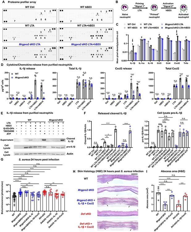
Figure 7. Defensin-Mrgpra2 interaction triggered neutrophil release of IL-1β and Cxcl2.
(A) Proteome profiler array analysis of culture medium of bone marrow neutrophils purified from WT (black) or Mrgpra2 dKO (blue) animals stimulated by control medium, hBD3 only, S. aureus lipoteichoic acid (LTA), or LTA+hBD3. Numbers: 1=IL-1β, 2=IL-1ra, 3=IL-16, 4=Ccl3, 5=Ccl4, 6=Cxcl2, 7=Tnfα. Quantifications are shown in (C).
(B) Schematic illustration of experimental design. Neutrophils are first primed with LTA (“signal 1”), then activated by hBD3 (“signal 2”).
(C) Quantification of mean pixel intensity of proteome profiler arrays shown in (A). Arrows point to IL-1β (1 in A) and Cxcl2 (6 in A), whose releases were enhanced by hBD3. Pixel intensities were normalized to positive controls on the same array.
(D) ELISA quantification of IL-1β and Cxcl2 release from neutrophils. WT neutrophils pretreated with LTA released IL-1β and Cxcl2 in response to hBD3 or mBD14 stimuli (black). Neutrophils purified from Mrgpra2 dKO animals failed to respond to hBD3 (blue). Total IL-1β and Cxcl2 was calculated as the sum of secreted and intracellular amounts. hBD3 or mBD14 alone did not induce the synthesis of IL-1β or Cxcl2. n=5
(E) Western blot showing the release of mature IL-1β (17kDa) released into the supernatant by WT neutrophils when stimulated with hBD3 (5μM). IL-1β release was inhibited by pan-caspase inhibitor Q-VD-OPh and Gq inhibitor YM-254890 and was abolished in the Mrgpra2 mutant neutrophils. ATP (5mM) was used as positive control.
(F) Quantification of western blots of released IL-1β (17kDa) and cellular pro-IL-1β (31kDa) normalized to Actin. n=3
(G) Bacteria bioluminescence 24 hour post S. aureus infection. Cxcl2 alone did not rescue Mrgpra2 or Def KO phenotypes, whereas IL-1β or IL-1β+Cxcl2 together rescued the anti-S. aureus defect of Mrgpra2 dKO and Def cKO mice. n=8-36
(H) H&E staining showing that injecting IL-1β and Cxcl2 restored abscess formation in Mrgpra2 dKO (blue) and Def cKO (red) animals 24 hours post S. aureus infection. Arrow heads point to neutrophil abscesses. Scale bar=1mm
(I) Quantification of abscess areas based on H&E staining shown in (H). n=5
Results are presented as mean ± SEM from at least three independent experiments. *p < 0.05, **p < 0.01, ***p < 0.001, ****p < 0.0001, n.s. not significant by one-way ANOVA.
See also Figure S7.

