PLOS ONE logoLink to PLOS ONE
. 2022 Sep 15;17(9):e0274181. doi: 10.1371/journal.pone.0274181

Performance evaluation of the Roche Elecsys® Anti-SARS-CoV-2 immunoassays by comparison with neutralizing antibodies and clinical assessment

Satomi Takei 1,2, Tomohiko Ai 2, Takamasa Yamamoto 1, Gene Igawa 1, Takayuki Kanno 3, Minoru Tobiume 3, Makoto Hiki 4,5, Kaori Saito 2, Abdullah Khasawneh 2, Mitsuru Wakita 1, Shigeki Misawa 1, Takashi Miida 2, Atsushi Okuzawa 6,7, Tadaki Suzuki 3, Kazuhisa Takahashi 6,8, Toshio Naito 6,9, Yoko Tabe 2,6,*
Editor: Gheyath K Nasrallah10
PMCID: PMC9477342  PMID: 36107911

Abstract

Quantitative measurement of SARS-CoV-2 neutralizing antibodies is highly expected to evaluate immune status, vaccine response, and antiviral therapy. The Elecsys® Anti-SARS-CoV-2 S (Elecsys® anti-S) was developed to measure anti-SARS-CoV-2 S proteins. We sought to investigate whether Elecsys® anti-S can be used to predict neutralizing activities in patients’ serums using an authentic virus neutralization assay. One hundred forty-six serum samples were obtained from 59 patients with COVID-19 at multiple time points. Of the 59 patients, 44 cases were included in Group M (mild 23, moderate 21) and produced 84 samples (mild 35, moderate 49), while 15 cases were included in Group S (severe 11, critical 4) and produced 62 samples (severe 43, critical 19). The neutralization assay detected 73% positive cases, and Elecsys® anti-S and Elecsys® Anti-SARS-CoV-2 (Elecsys® anti-N) showed 72% and 66% positive cases, respectively. A linear correlation between the Elecsys® anti-S assay and the neutralization assay were highly correlated (r = 0.7253, r2 = 0.5261) than a linear correlation between the Elecsys® anti-N and neutralization assay (r = 0.5824, r2 = 0.3392). The levels of Elecsys® anti-S antibody and neutralizing activities were significantly higher in Group S than in Group M after 6 weeks from onset of symptoms (p < 0.05). Conversely, the levels of Elecsys® anti-N were comparable in both groups. Three immunosuppressed patients, including cancer patients, showed low levels of anti-S and anti-N antibodies and neutralizing activities throughout the measurement period, indicating the need for careful follow-up. Our data indicate that Elecsys® anti-S can predict the neutralization antibodies in COVID-19.

Introduction

Coronavirus disease 2019 (COVID-19), caused by SARS-CoV-2, has been mainly diagnosed by reverse transcription-polymerase chain reaction (RT-PCR) that can directly detect the viral genomes [1]. Antigen testing has been also developed to detect pathogens rapidly without complicated procedures [2]. However, these tests cannot detect SARS-CoV-2 in certain periods after infection [3]. On the other hand, serological tests are essential tools to evaluate neutralizing antibody titers upon vaccination and to assess SARS-CoV-2 seroprevalence in cohorts [4, 5]. Neutralizing antibodies targeting the receptor-binding domain (RBD) of the spike (S) protein can reduce viral infectivity by binding to the surface epitopes of viral particles which blocks virus entry [6]. Although the authentic virus neutralization assays can directly measure the neutralizing activities of the SARS-CoV-2 virus, those methods need to be performed in Biosafety Level 3 facilities, which limits their application [7]. Therefore, there is a need for safer, high-throughput, and widely available measurement methods that correlate well with neutralizing activities.

Recently, the Elecsys® Anti-SARS-CoV-2 S (Elecsys® anti-S) has been developed to quantitatively measure total antibodies against the S protein RBD, and the Elecsys® Anti-SARS-CoV-2 (Elecsys® anti-N) has been developed to semi-quantitatively measure total antibodies against SARS-CoV-2 N proteins that regulates viral replication [6] (Roche Diagnostics International Ltd, Rotkreuz, Switzerland). The aim of this study was to evaluate the feasibility and usefulness of these immunoassays by comparing the chronological seroprevalences in patients with various severity of COVID-19 along with the neutralizing activities measured by an authentic virus neutralization assay.

Materials and methods

Clinical characteristics

This study complied with all relevant national regulations and institutional policies and was conducted in accordance with the tenets of the Declaration of Helsinki and was approved by the Institutional Review Board (IRB) at Juntendo University Hospital (IRB # 20–036). The need for informed consent from individual patients was waived because all samples were de-identified in line with the Declaration of Helsinki. From April to August 2020, 146 serum samples were collected from 59 patients with symptomatic COVID-19 confirmed by RT-PCR at multiple time points (number of samples per patient, median 2, IQR [2, 3]). The periods between different time points were 2 to 58 days. Of the 59 COVID-19 patients, including 58 inpatients and one outpatient, 44 cases were included Group M (mild 23, moderate 21) and produced 84 samples (mild 35, moderate 49), while 15 cases were included Group S (severe 11, critical 4) and produced 62 samples (severe 43, critical 19). Clinical data were retrospectively collected from patients’ charts. All samples were from the unvaccinated patients.

Laboratory assays

Elecsys® Anti-SARS-CoV-2 S and Elecsys® Anti-SARS-CoV-2 immunoassays

Serum samples were tested with the automated serological immunoassays, Elecsys ® Anti-SARS-CoV-2 S (Elecsys® anti-S, Cat # 0928926750) and the Elecsys® Anti-SARS-CoV-2 (Elecsys® anti-N, Cat # 09203095501), by detecting antibodies to the receptor-binding domain (RBD) of S protein and antibodies to the N protein of SARS-CoV-2 (Roche Diagnostics) [8]. These assays received emergency use authorization approval from the US Food and Drug Administration (https://www.fda.gov/media/137605 (2020)). All samples were processed according to the manufacturer’s instructions.

The results by Elecsys® anti-S were quantitatively shown in units of U/mL with the cut-off point of 0.80 U/mL to differentiate samples as reactive (≥ 0.80 U/mL) and non-reactive (< 0.80 U/mL). The values between 0.40–250 U/mL represented a linear range, and the results below this range were set to 0.4 U/mL. The samples above 250 U/mL were diluted into the linear range of the assay (1:10 or 1:100) with Diluent Universal reagent (Roche Diagnostics, Rotkreuz, Switzerland). Thus, the applied setting enabled an upper limit of quantification of 25000 U/mL for these analyses before dilution. The Elecsys® anti-N is a semi-quantitative assay, and the results were interpreted as follows: cutoff index (COI) <1.0 was non-reactive, and ≥1.0 was reactive.

Neutralization assay

The SARS-CoV-2 ancestral strain, WK-521 (lineage A, GISAID ID: EPI_ISL_408667), was used for the authentic virus neutralization assay that has been performed at the National Institute of Infectious Diseases (NIID) with ethics approval by the medical research ethics committee of NIID for the use of human subjects (#1178). Authentic virus neutralization assay has been performed as described previously [7]. Briefly, serially diluted serum samples (2-fold serial dilutions starting at 1:5 dilution, diluted with high glucose Dulbecco’s Modified Eagle Medium supplemented with 2% Fetal Bovine Serum and 100 U/mL penicillin/streptomycin, from Fujifilm Wako Pure Chemicals, Japan) were mixed with the virus from 100 Median Tissue Culture Infectious Dose (TCID50) and incubated at 37°C for 1 hour. The mixture was subsequently incubated with VeroE6/TMPRSS2 cells (JCRB1819, JCRB Cell Bank, Japan) and seeded in 96-well flat-bottom plates for 4–6 days at 37°C in a chamber supplied with 5% CO2. Then the cells were fixed with 20% formalin (Fujifilm Wako Pure Chemicals) and stained with crystal violet solution (Sigma-Aldrich, St Louis, MO). Each sample was assayed in 2–4 wells and the average cut-off dilution index of >50% cytopathic effect was presented as a neutralizing titer. Neutralizing titer of the sample below the detection limit (1:5 dilution) was set as 2.5. Neutralizing antibody titer of <5 is considered negative and >5 is considered positive.

Statistical analysis

Correlation studies were performed using Spearman’s coefficient. Assay performance, linear regression, and curve fitting calculations were performed using Prism 9 (GraphPad Software, LLC, San Diego, CA, USA). For experiments involving two group comparisons, Wilcoxon signed-rank test was performed. The following notation was used to show statistical significance: * p value <0.05, ** p value <0.01, and *** p value <0.001.

Results

As shown in Table 1, patients were classified into two groups according to the WHO criteria [WHO. Clinical management of COVID-19. Available from: https://www.who.int/publications/i/item/clinical-management-of-covid-19]: Group M included mild and moderate cases and Group S included severe and critical cases.

Table 1. Clinical characteristics of patients with COVID-19.

Disease severitya Group M Group S
Mild Moderate Severe Critical
Patient number (n = 59) 39% (23/59) 36% (21/59) 19% (11/59) 7% (4/59)
Male, % 78% (18/23) 57% (12/21) 100% (11/11) 75% (3/4)
Age range (average) 24–82 (43.3) 18–80 (54.9) 6–86 (66.5) 67–79 (75.3)
Past medical history
Hypertension 9% (2/23) 14% (3/21) 9% (1/11) 50% (2/4)
Hyperlipidemia 9% (2/23) 5% (1/21) 0% (0/11) 0% (0/4)
Diabetes 0% (0/23) 14% (3/21) 18% (2/11) 25% (1/4)
Cancer 9% (2/23) 0% (0/21) 18% (2/11) 25% (1/4)
Renal failure 0% (0/23) 0% (0/21) 9% (1/11) 25% (1/4)
Others, None known 83% (19/23) 76% (16/21) 64% (7/11) 50% (2/4)
Sample percentage (n = 146) 24% (35/146) 34% (49/146) 29% (43/146) 13% (19/146)
Days from onset
0–6 (n = 25) 7 8 8 2
7–13 (n = 28) 7 11 10 0
14–20 (n = 26) 9 8 7 2
21–27 (n = 24) 4 8 6 6
28–34 (n = 15) 3 5 5 2
35–41 (n = 13) 4 3 4 2
>42 (n = 15) 1 6 3 5

aWHO criteria.

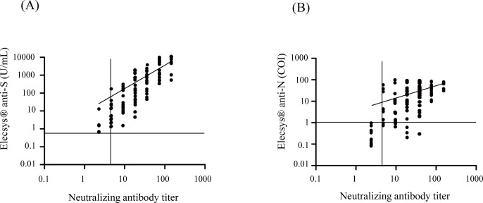
We first compared the results of Elecsys® anti-S and Elecsys® anti-N to those of the authentic virus neutralizing assay. Of the 146 samples, the neutralization assay detected 73% (106/146) positives, and Elecsys® anti-S and Elecsys® anti-N showed 72% (105/146) and 66% (97/146) positives, respectively. These results were plotted, and the positive results were fitted with a linear regression. Fig 1A shows a linear correlation between the Elecsys® anti-S assay and the neutralization assay (r = 0.7253, r2 = 0.5261), and Fig 1B shows a linear correlation between the Elecsys® anti-N and neutralization assay (r = 0.5824, r2 = 0.3392).

Fig 1. Correlations of Elecsys® Anti-SARS-CoV-2 S and Elecsys®Anti-SARS-CoV-2 assays results to neutralization assay.

Fig 1

One hundred forty-six serum samples from COVID-19 patients were tested by Elecsys® anti-S, Elecsys® anti-N, and neutralization assay and were examined for correlations. (A) Correlation of Elecsys® anti-S and neutralization assay. (B)Correlation of Elecsys® anti-N and neutralization assay. Dotted lines represent the manufacturer’s positive cutoff values: Elecsys® anti-S, 0.8 U/ml; Elecsys® anti-N, COI 1.0; neutralization assay, titer 5. The horizontal axis and the vertical axis are in logarithmic notations. Correlation studies were performed using Spearman’s coefficient.

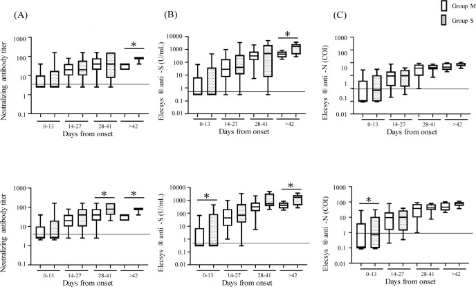
We next examined the neutralizing activities (Fig 2A) and levels of antibodies (Fig 2B and 2C) at various time points after onset of symptoms. Fig 2 shows that the levels of neutralizing activities and antibodies tended to increase over time. However, there was no significant difference between Groups M and S until the sixth week. After the seventh week, the neutralization assay and Elecsys® anti-S showed significantly higher values in Group S than in Group M (p < 0.05), which was not observed in the Elecsys® anti-N results. Table 2 summarizes the details.

Fig 2. Comparison of antibody levels between Group M (mild + moderate) and Group S (severe + critical).

Fig 2

One hundred forty-six serum samples from COVID-19 patients at various time points after the onset of symptoms were tested by neutralization assay, Elecsys® anti-S, and Elecsys® anti-N, and were examined for changes over time. All cases of antibody values of neutralization assay (A), Elecsys® anti-S (B), and Elecsys® anti-N (C) in Group M (n = 84) and S (n = 62) subjects were shown. Four cases receiving immunosuppressive therapy (1 in Group M; 3 in Group S) were excluded and the antibody values of neutralization assay (D), Elecsys® anti-S (E), and Elecsys® anti-N (F) were shown. Open bars represent Group M and dotted bars represent Group S. Wilcoxon signed-rank test was performed. The data were presented as median with interquartile ranges (IQR). *p < 0.05. The vertical axes are in logarithmic notation.

Table 2. Time course of seroprevalence by neutralization assay, Elecsys® Anti-SARS-CoV-2 S, and Elecsys® Anti-SARS-CoV-2.

Group M n = 84 (n = 80) Group S n = 62 (n = 47)
Days from onset sample number Virus neutralization assay Elecsys® Anti-SARS-CoV-2 S (Elecsys® anti-S) Elecsys® Anti-SARS-CoV-2 (Elecsys® anti-N) sample number Virus neutralization assay Elecsys® Anti-SARS-CoV-2 S (Elecsys® anti-S) Elecsys® Anti-SARS-CoV-2 (Elecsys® anti-N)
Positive % Positive % Positive % Positive % Positive % Positive %
0–6 15 (14) 3 (3) 20% (21%) 3 (3) 20% (21%) 2(2) 13% (14%) 10 (7) 1 (1) 10% (14%) 1 (1) 10% (14%) 1 (1) 10% (14%)
7–13 18 (17) 11 (11) 61% (65%) 10 (10) 56% (59%) 9 (9) 50% (53%) 10 (6) 5 (5) 50% (83%) 5 (5) 50% (83%) 5 (5) 50% (83%)
14–20 17 (16) 16 (16) 94% (100%) 16 (16) 94% (100%) 11 (11) 65% (69%) 9 (7) 8 (7) 89% (100%) 7 (7) 78% (100%) 8 (7) 89% (100%)
21–27 12 (11) 11 (11) 92% (100%) 11 (11) 92% (100%) 11 (11) 92% (100%) 12 (10) 11 (10) 92% (100%) 11 (10) 92% (100%) 10 (9) 83% (90%)
28–34 8 (8) 8 (8) 100% (100%) 8 (8) 100% (100%) 8 (8) 100% (100%) 7 (5) 6 (5) 86% (100%) 6 (5) 86% (100%) 6 (5) 86% (100%)
35–41 7 (7) 7 (7) 100% (100%) 7 (7) 100% (100%) 7 (7) 100% (100%) 6 (4) 5 (4) 83% (100%) 5 (4) 83% (100%) 5 (4) 83% (100%)
>42 7 (7) 7 (7) 100% (100%) 7 (7) 100% (100%) 7 (7) 100% (100%) 8 (8) 8 (8) 100% (100%) 8 (8) 100% (100%) 8 (8) 100% (100%)

Numbers in parenthesis indicate the sample numbers after removing the samples from 3 patients under immunosuppressive therapy.

Since immunosuppressive therapies may cause false-negative results in antibody tests [9], the results after removing three patients receiving immunosuppressive therapies are shown in Fig 2D–2F. Fig 2D shows that the levels of neutralizing activities were higher in Group S than M after the fourth week (p < 0.05). Fig 2E shows that the levels of anti-S antibodies were significantly higher in Group S compared to Group M within the first 2 weeks and after the seventh week (p < 0.05). Fig 2F shows that the levels of anti-N antibodies were higher in Group S than M only in the first 2 weeks (p < 0.05).

Chronological changes in the results of the neutralization assay, Elecsys® anti-S assay, and Elecsys® anti-N assay were examined simultaneously in 23 inpatients who were tested in three time points or more. Table 3 summarizes the clinical characteristics. Fig 3 shows the line plots of the results in the patients of Group M. One patient (Pt #1) on immunosuppressive treatment for rheumatoid arthritis showed suppressed antibody responses for all three tests. The other patients’ results became positive between 10 and 39 days from symptom onset.

Table 3. Clinical characteristics of inpatients with COVID-19.

Patient# Disease severitya Age (y) Sex Past medical history Therapy Outcome
1 Mild 65 M Hepatic cancer, Rheumatoid arthritis N/A N/A cure/discharge
2 Mild 63 M None known N/A N/A cure/discharge
3 Mild 72 M None known N/A Favipiravir, Ciclesonide, DEX cure/discharge
4 Mild 82 M Bile duct cancer N/A Favipiravir cure/discharge
5 Moderate 64 M post-Pancreatic Cancer N/A N/A cure/discharge
6 Moderate 78 M None known N/A Ciclesonide, Favipiravir cure/discharge
7 Moderate 41 M Lung sarcoidosis N/A Ciclesonide, Favipiravir cure/discharge
8 Moderate 76 M Prostatic hypertrophy N/A Ciclesonide, Favipiravir, Heparin cure/discharge
9 Moderate 37 M None known N/A Ciclesonide, Favipiravir cure/discharge
10 Moderate 59 M Hypertension, Hyperlipidemia N/A Ciclesonide, Favipiravir cure/discharge
11 Moderate 71 F Hyperlipidemia, Diabetes N/A Ciclesonide, Favipiravir, Heparin cure/discharge
12 Moderate 75 F Hypertension, Hyperlipidemia, Angina N/A Ciclesonide, Favipiravir cure/discharge
13 Moderate 18 F None known N/A N/A cure/discharge
14 Moderate 80 F Hypertension N/A Heparin cure/discharge
15 Severe 78 M Diabetes, Rheumatoid arthritis O2 Heparin cure/discharge
16 Severe 57 M Hyponatremia O2 Ciclesonide, Favipiravir cure/discharge
17 Severe 64 M Urinary stone N/A N/A cure/discharge
18 Severe 67 M Fatty liver, Kidney stones O2 Ciclesonide, Favipiravir, Heparin cure/discharge
19 Severe 46 M Diabetes, Angina, Stiff-person syndrome O2 Favipiravir, Heparin, mPSL, PSL, Remdesivir cure/discharge
20 Severe 84 M Colon cancer, Parkinson, Dementia O2 Heparin cure/discharge
21 Severe 84 M Hypertension, Lung cancer, Renal failure O2 CHDF, Ciclesonide, DEX, FFP, Favipiravir, mPSL, Plasmapheresis, PSL death
22 Critical 67 M Hypertension, Renal failure Ventilation CHDF, Ciclesonide, FFP, mPSL, Plasmapheresis death
23 Critical 77 M Hypertension, Diabetes, Prostate cancer Ventilation CHDF, Favipiravir, FFP, Heparin, mPSL, Plasmapheresis, PSL, Remdesivir death

Patients tested 3 times or more were included.

aWHO criteria.

Abbreviations: CHDF, Continuous hemodiafiltration; DEX, Dexamethasone; FFP, fresh frozen plasma; O2, Oxygen inhalation; PSL, prednisolone; N/A, not applicable.

Fig 3. Longitudinal change of antibodies in Group M.

Fig 3

Levels of SARS-CoV-2 antibodies in 14 mild to moderate cases (Group M) were tested. (A) Neutralizing antibody (B) Elecsys® anti-S (C) Elecsys® anti-N. The vertical axes are in logarithmic notation.

Fig 4 shows the line plots of the results in Group S. Three patients were treated with plasmapheresis. Regardless of the timing of plasmapheresis, one patient (Pt #21) remained negative, Pt #22 kept relatively low values, and Pt #23 showed high values in all assays. We observed no significant decrease of antibody levels of Pt #15 and Pt #23 which were continuously measured until 69 and 58 days after the onset of symptoms, respectively.

Fig 4. Longitudinal change of antibodies in Group S.

Fig 4

Levels of SARS-CoV-2 antibodies in 9 severe to critical cases (Group S) were tested. (A) Neutralizing antibody (B) Elecsys® anti-S (C) Elecsys® anti-N. Colored boxes represent the days of plasmapheresis. The vertical axes are in logarithmic notation.

Discussion

In this study, we demonstrated that the Elecsys® anti-S assay detected anti-spike (S) protein RBD antibodies at about the same time that the virus neutralization assay detected neutralizing activity: 2 weeks after onset of symptoms. These results are consistent with a recent analysis of naturally acquired SARS-CoV-2 infected individuals showing that neutralizing antibodies were almost detectable about 14 days after infection [10]. A previous study has further reported that production of neutralizing antibodies within 14 days of onset of symptoms is an important factor in recovery [11]. In this study, we detected that there is a humoral increase in S-specific antibodies with neutralizing activity. The measurement values of Elecsys® anti-S and neutralization assays were highly correlated, and the cases of Group S showed significantly higher values than Group M during the late phase of infection. The positive rate of the Elecsys® anti-N assay was lower than that of the Elecsys® anti-S and virus neutralization assays.

The measurement values of Elecsys® anti-S and neutralization assays were highly correlated, and the cases of Group S showed significantly higher values than Group M during the late phase of infection. When patients receiving immunosuppressive treatment were removed from the cohort, both the Elecsys® anti-S and Elecsys® anti-N assays detected significantly higher levels of antibody in Group S than in Group M in the early stages of infection. It has been shown that patients undergoing immunosuppressive treatment have higher incidence rates and serious adverse events of COVID-19 [12]. Antibody levels in patients receiving chemotherapy, radiation therapy, and other immunosuppressive therapies require careful evaluation. It has been shown that IgG levels of SARS-CoV-2 S and N antibodies begin to decrease 2–3 months after infection [13, 14] and neutralizing titers begin to decrease 8 months later [1517]. Regarding the effects of plasmapheresis treatment, a previous case report demonstrated that S-protein IgG increased after plasma exchange within 3 days, and moderately decreased from day 3 to day 7, without any change in N-protein IgG [18]. In this study, however, no significant changes in antibody levels caused by plasmapheresis were observed.

This study has several limitations: first, this study is a single-center study with limited sample size and short-term detections. Our results should be confirmed by additional assessments at other study sites. Second, variant determination has not been performed in the study. In terms of the SARS-CoV-2 strain that was used in the authentic virus neutralization assay, the utilized strain might have been different from the emerging variants. In Japan, lineage A and B variants were prevalent during the study period from April to August 2020 (1st and 2nd waves) [19]. We utilized the SARS-CoV-2 ancestral strain of lineage A for the authentic neutralization assay. On the other hand, the differences between the neutralizing antibody titers measured by the authentic neutralization assay and the commercially available immunoassays might depend on the viral variant, which is required to be verified in the future. Third, a comparably small sample size was utilized to measure agreement between the Elecsys® anti-N immunoassays and a neutralization assay; as such, these data would benefit from further evaluation and validation. Forth, this study does not include samples from asymptomatic patients nor from vaccinated patients. Therefore, the validity of our findings for patients with asymptomatic/mildly symptomatic SARS-CoV-2 infection after vaccination is yet to be shown and requires further study.

In conclusion, we found good correlation between SARS-CoV-2 viral S protein RBD levels measured by Elecsys® anti-S and neutralizing antibody titers detected by an authentic virus neutralization assay. The findings of this study indicate that the quantitative detection of anti-SARS-CoV2 S protein by Elecsys® anti-S assay reliably quantifies the antibody response to SARS-CoV-2, which is highly relevant to estimate protection.

Acknowledgments

The authors thank the Department of Research Support Utilizing Bioresource Bank, Juntendo University Graduate School of Medicine, for use of their facilities.

Data Availability

All relevant data are within the paper.

Funding Statement

This research was supported by AMED (JP20fk0108472 to TN) and by Japan Society for the Promotion of Science Grants-in Aid for Scientific Research (22K15675 to ST).

References

  • 1.Wölfel R, Corman VM, Guggemos W, Seilmaier M, Zange S, Müller MA, et al. Virological assessment of hospitalized patients with COVID-2019. Nature. 2020;581: 465–469. doi: 10.1038/s41586-020-2196-x [DOI] [PubMed] [Google Scholar]
  • 2.Saito K, Ai T, Kawai A, Matsui J, Fukushima Y, Kikukawa N, et al. Performance and usefulness of a novel automated immunoassay HISCL SARS-CoV-2 Antigen assay kit for the diagnosis of COVID-19. Scientific reports. 2021;11(23196). doi: 10.1038/s41598-021-02636-x [DOI] [PMC free article] [PubMed] [Google Scholar]
  • 3.Mina MJ, Parker R, Larremore DB. Rethinking Covid-19 Test Sensitivity—A Strategy for Containment. N Engl J Med. 2020; 383(e120). doi: 10.1056/NEJMp2025631 [DOI] [PubMed] [Google Scholar]
  • 4.Weitz JS, Beckett SJ, Coenen AR, Demory D, Dominguez-Mirazo M, Dushoff J, et al. Modeling shield immunity to reduce COVID-19 epidemic spread. Nat Med. 2020; 26: 849–854. doi: 10.1038/s41591-020-0895-3 [DOI] [PMC free article] [PubMed] [Google Scholar]
  • 5.Krammer F, Simon V. Serology assays to manage COVID-19. Science. 2020; 368:1060–1061. doi: 10.1126/science.abc1227 [DOI] [PubMed] [Google Scholar]
  • 6.Weisberg SP, Connors TJ, Farber DL. Distinct antibody responses to SARS-CoV-2 in children and adults across the COVID-19 clinical spectrum. Nat Immunol. 2021; 22: 25–31. doi: 10.1038/s41590-020-00826-9 [DOI] [PMC free article] [PubMed] [Google Scholar]
  • 7.Moriyama S, Adachi Y, Sato T, Tonouchi K, Sun L, Fukushi S, et al. Temporal maturation of neutralizing antibodies in COVID-19 convalescent individuals improves potency and breadth to circulating SARS-CoV-2 variants. Immunity. 2021; 54 (e1844): 1841–1852. doi: 10.1016/j.immuni.2021.06.015 [DOI] [PMC free article] [PubMed] [Google Scholar]
  • 8.Muench P, Jochum S, Wenderoth V, Ofenloch-Haehnle B, Hombach M, Strobl M, et al. Development and Validation of the Elecsys Anti-SARS-CoV-2 Immunoassay as a Highly Specific Tool for Determining Past Exposure to SARS-CoV-2. J Clin Microbiol. 2020; 58. doi: 10.1128/JCM.01694-20 [DOI] [PMC free article] [PubMed] [Google Scholar]
  • 9.Caturegli G, Materi J, Howard BM, Caturegli P. Clinical Validity of Serum Antibodies to SARS-CoV-2: A Case-Control Study. Ann Intern Med. 2020; 173: 614–622. doi: 10.7326/M20-2889 [DOI] [PMC free article] [PubMed] [Google Scholar]
  • 10.Yuce M, Filiztekin E, Ozkaya KG. COVID-19 diagnosis -A review of current methods. Biosens Bioelectron. 2021;172 (112752). doi: 10.1016/j.bios.2020.112752 [DOI] [PMC free article] [PubMed] [Google Scholar]
  • 11.Lucas C, Klein J, Sundaram M, Liu F, Wong P, Silva J, et al. Kinetics of antibody responses dictate COVID-19 outcome. medRxiv. 2020. doi: 10.1101/2020.12.18.20248331 [DOI] [PMC free article] [PubMed] [Google Scholar]
  • 12.Yang F, Shi S, Zhu J, Shi J, Dai K, Chen X. Clinical characteristics and outcomes of cancer patients with COVID-19. J Med Virol. 2020; 92:2067–2073. doi: 10.1002/jmv.25972 [DOI] [PubMed] [Google Scholar]
  • 13.Long QX, Tang XJ, Shi QL, Li Q, Deng HJ, Yuan J, et al. Clinical and immunological assessment of asymptomatic SARS-CoV-2 infections. Nat Med. 2020; 26: 1200–1204. doi: 10.1038/s41591-020-0965-6 [DOI] [PubMed] [Google Scholar]
  • 14.Wang X, Guo X, Xin Q, Pan Y, Hu Y, Li J, et al. Neutralizing Antibody Responses to Severe Acute Respiratory Syndrome Coronavirus 2 in Coronavirus Disease 2019 Inpatients and Convalescent Patients. Clin Infect Dis. 2020; 71:2688–2694. doi: 10.1093/cid/ciaa721 [DOI] [PMC free article] [PubMed] [Google Scholar]
  • 15.Wheatley AK, Juno JA, Wang JJ, Selva KJ, Reynaldi A, Tan HX, et al. Evolution of immune responses to SARS-CoV-2 in mild-moderate COVID-19. Nat Commun. 2021; 12 (1162). doi: 10.1038/s41467-021-21444-5 [DOI] [PMC free article] [PubMed] [Google Scholar]
  • 16.Gaebler C, Wang Z, Lorenzi JCC, Muecksch F, Finkin S, Tokuyama M, et al. Evolution of antibody immunity to SARS-CoV-2. Nature. 2021; 591: 639–644. doi: 10.1038/s41586-021-03207-w [DOI] [PMC free article] [PubMed] [Google Scholar]
  • 17.Dan JM, Mateus J, Kato Y, Hastie KM, Yu ED, Faliti CE, et al. Immunological memory to SARS-CoV-2 assessed for up to 8 months after infection. Science. 2021; 371 (6529). doi: 10.1126/science.abf4063 [DOI] [PMC free article] [PubMed] [Google Scholar]
  • 18.Xia X, Li K, Wu L, Wang Z, Zhu M, Haung B, et al. Improved clinical symptoms and mortality among patients with severe or critical COVID-19 after convalescent plasma transfusion. Blood. 2020; 136: 755–759. doi: 10.1182/blood.2020007079 [DOI] [PMC free article] [PubMed] [Google Scholar]
  • 19.Hassan AYI, Lamura G, Hagedoorn M. Predictors of digital support services use by informal caregivers: a cross-sectional comparative survey. BMJ. 2022; 12(4): e059897. doi: 10.1136/bmjopen-2021-059897 [DOI] [PMC free article] [PubMed] [Google Scholar]

Decision Letter 0

Gheyath K Nasrallah

21 Mar 2022

PONE-D-22-05585Performance evaluation of the Roche Elecsys Anti-SARS-CoV-2 immunoassays by comparison with neutralizing antibodies and clinical assessmentPLOS ONE

Dear Dr. Tabe

Thank you for submitting your manuscript to PLOS ONE. After careful consideration, we feel that it has merit but does not fully meet PLOS ONE’s publication criteria as it currently stands. Therefore, we invite you to submit a revised version of the manuscript that addresses the points raised during the review process. The table fonts are very small. The figures are hazy and blurry. The abstract should include detailed results with number. Please correct and resubmit

Please submit your revised manuscript by 20/3/2022. If you will need more time than this to complete your revisions, please reply to this message or contact the journal office at plosone@plos.org. When you're ready to submit your revision, log on to https://www.editorialmanager.com/pone/ and select the 'Submissions Needing Revision' folder to locate your manuscript file.

Please include the following items when submitting your revised manuscript:

  • A rebuttal letter that responds to each point raised by the academic editor and reviewer(s). You should upload this letter as a separate file labeled 'Response to Reviewers'.

  • A marked-up copy of your manuscript that highlights changes made to the original version. You should upload this as a separate file labeled 'Revised Manuscript with Track Changes'.

  • An unmarked version of your revised paper without tracked changes. You should upload this as a separate file labeled 'Manuscript'.

If you would like to make changes to your financial disclosure, please include your updated statement in your cover letter. Guidelines for resubmitting your figure files are available below the reviewer comments at the end of this letter.

If applicable, we recommend that you deposit your laboratory protocols in protocols.io to enhance the reproducibility of your results. Protocols.io assigns your protocol its own identifier (DOI) so that it can be cited independently in the future. For instructions see: https://journals.plos.org/plosone/s/submission-guidelines#loc-laboratory-protocols. Additionally, PLOS ONE offers an option for publishing peer-reviewed Lab Protocol articles, which describe protocols hosted on protocols.io. Read more information on sharing protocols at https://plos.org/protocols?utm_medium=editorial-email&utm_source=authorletters&utm_campaign=protocols.

We look forward to receiving your revised manuscript.

Kind regards,

Gheyath K. Nasrallah

Academic Editor

PLOS ONE

Journal Requirements:

When submitting your revision, we need you to address these additional requirements.

1. Please ensure that your manuscript meets PLOS ONE's style requirements, including those for file naming. The PLOS ONE style templates can be found at 

https://journals.plos.org/plosone/s/file?id=wjVg/PLOSOne_formatting_sample_main_body.pdf and 

https://journals.plos.org/plosone/s/file?id=ba62/PLOSOne_formatting_sample_title_authors_affiliations.pdf

2. We note that the grant information you provided in the ‘Funding Information’ and ‘Financial Disclosure’ sections do not match. 

When you resubmit, please ensure that you provide the correct grant numbers for the awards you received for your study in the ‘Funding Information’ section.

3. Thank you for stating the following in the Competing Interests section: 

"The reagents used in this study were provided by Roche, but the study was performed by scientifically proper methods without any bias.  "

Please confirm that this does not alter your adherence to all PLOS ONE policies on sharing data and materials, by including the following statement: "This does not alter our adherence to  PLOS ONE policies on sharing data and materials.” (as detailed online in our guide for authors http://journals.plos.org/plosone/s/competing-interests).  If there are restrictions on sharing of data and/or materials, please state these. Please note that we cannot proceed with consideration of your article until this information has been declared. 

Please include your updated Competing Interests statement in your cover letter; we will change the online submission form on your behalf.

4. In your Data Availability statement, you have not specified where the minimal data set underlying the results described in your manuscript can be found. PLOS defines a study's minimal data set as the underlying data used to reach the conclusions drawn in the manuscript and any additional data required to replicate the reported study findings in their entirety. All PLOS journals require that the minimal data set be made fully available. For more information about our data policy, please see http://journals.plos.org/plosone/s/data-availability.

"Upon re-submitting your revised manuscript, please upload your study’s minimal underlying data set as either Supporting Information files or to a stable, public repository and include the relevant URLs, DOIs, or accession numbers within your revised cover letter. For a list of acceptable repositories, please see http://journals.plos.org/plosone/s/data-availability#loc-recommended-repositories. Any potentially identifying patient information must be fully anonymized.

Important: If there are ethical or legal restrictions to sharing your data publicly, please explain these restrictions in detail. Please see our guidelines for more information on what we consider unacceptable restrictions to publicly sharing data: http://journals.plos.org/plosone/s/data-availability#loc-unacceptable-data-access-restrictions. Note that it is not acceptable for the authors to be the sole named individuals responsible for ensuring data access.

We will update your Data Availability statement to reflect the information you provide in your cover letter.

[Note: HTML markup is below. Please do not edit.]

Reviewers' comments:

[NOTE: If reviewer comments were submitted as an attachment file, they will be attached to this email and accessible via the submission site. Please log into your account, locate the manuscript record, and check for the action link "View Attachments". If this link does not appear, there are no attachment files.]

While revising your submission, please upload your figure files to the Preflight Analysis and Conversion Engine (PACE) digital diagnostic tool, https://pacev2.apexcovantage.com/. PACE helps ensure that figures meet PLOS requirements. To use PACE, you must first register as a user. Registration is free. Then, login and navigate to the UPLOAD tab, where you will find detailed instructions on how to use the tool. If you encounter any issues or have any questions when using PACE, please email PLOS at figures@plos.org. Please note that Supporting Information files do not need this step.

PLoS One. 2022 Sep 15;17(9):e0274181. doi: 10.1371/journal.pone.0274181.r002

Author response to Decision Letter 0


23 Mar 2022

March 23, 2022

Ms. Agatha Macaraig

Straive Editorial Assistant

RE: Resubmission of the revised manuscript titled “Performance evaluation of the Roche Elecsys Anti-SARS-CoV-2 immunoassays by comparison with neutralizing antibodies and clinical assessment” by Takei S., et al.

Ms. Macaraig,

We answer the requirements of the journal requirements as follows;

Reply (Answer; A) to the comments and questions (Comment; C):

Journal Requirements:

When submitting your revision, we need you to address these additional requirements.

C1. Please ensure that your manuscript meets PLOS ONE's style requirements, including those for file naming. The PLOS ONE style templates can be found at

https://journals.plos.org/plosone/s/file?id=wjVg/PLOSOne_formatting_sample_main_body.pdf and

https://journals.plos.org/plosone/s/file?id=ba62/PLOSOne_formatting_sample_title_authors_affiliations.pdf

A1. We corrected the manuscript following PLOS ONE's style requirements, including those for file naming.

C2. We note that the grant information you provided in the ‘Funding Information’ and ‘Financial Disclosure’ sections do not match.

When you resubmit, please ensure that you provide the correct grant numbers for the awards you received for your study in the ‘Funding Information’ section.

A2. We stated the financial disclosure in the cover letter.

C3. Thank you for stating the following in the Competing Interests section:

"The reagents used in this study were provided by Roche, but the study was performed by scientifically proper methods without any bias. "

Please confirm that this does not alter your adherence to all PLOS ONE policies on sharing data and materials, by including the following statement: "This does not alter our adherence to PLOS ONE policies on sharing data and materials.” (as detailed online in our guide for authors http://journals.plos.org/plosone/s/competing-interests). If there are restrictions on sharing of data and/or materials, please state these. Please note that we cannot proceed with consideration of your article until this information has been declared.

Please include your updated Competing Interests statement in your cover letter; we will change the online submission form on your behalf.

A3. We included our updated Competing Interests statement in the cover letter as follows;

"The reagents used in this study were provided by Roche, but the study was performed by scientifically proper methods without any bias. This does not alter our adherence to PLOS ONE policies on sharing data and materials.”

C4. In your Data Availability statement, you have not specified where the minimal data set underlying the results described in your manuscript can be found. PLOS defines a study's minimal data set as the underlying data used to reach the conclusions drawn in the manuscript and any additional data required to replicate the reported study findings in their entirety. All PLOS journals require that the minimal data set be made fully available. For more information about our data policy, please see http://journals.plos.org/plosone/s/data-availability.

"Upon re-submitting your revised manuscript, please upload your study’s minimal underlying data set as either Supporting Information files or to a stable, public repository and include the relevant URLs, DOIs, or accession numbers within your revised cover letter. For a list of acceptable repositories, please see http://journals.plos.org/plosone/s/data-availability#loc-recommended-repositories. Any potentially identifying patient information must be fully anonymized.

Important: If there are ethical or legal restrictions to sharing your data publicly, please explain these restrictions in detail. Please see our guidelines for more information on what we consider unacceptable restrictions to publicly sharing data: http://journals.plos.org/plosone/s/data-availability#loc-unacceptable-data-access-restrictions. Note that it is not acceptable for the authors to be the sole named individuals responsible for ensuring data access.

We will update your Data Availability statement to reflect the information you provide in your cover letter.

A4. We corrected the Data Availability statement as follows; “All relevant data are within the paper.“

Sincerely,

Yoko Tabe, M.D., Ph.D.

Professor

Department of Clinical Laboratory Medicine,

Juntendo University, School of Medicine

Attachment

Submitted filename: Response to Reviewers.docx

Decision Letter 1

Gheyath K Nasrallah

9 May 2022

PONE-D-22-05585R1Performance evaluation of the Roche Elecsys Anti-SARS-CoV-2 immunoassays by comparison with neutralizing antibodies and clinical assessmentPLOS ONE

Dear Dr. Tabe,

Thank you for submitting your manuscript to PLOS ONE. After careful consideration, we feel that it has merit but does not fully meet PLOS ONE’s publication criteria as it currently stands. Therefore, we invite you to submit a revised version of the manuscript that addresses the points raised during the review process. Bothe reviewers has serious concerns about the methodology. Further Rev2 believes that the manuscript is poorly written and organized. Please make sure that you address all of comments of the reviewers before you submit  a revised version.   

Please submit your revised manuscript by Jun 23 2022 11:59PM. If you will need more time than this to complete your revisions, please reply to this message or contact the journal office at plosone@plos.org. When you're ready to submit your revision, log on to https://www.editorialmanager.com/pone/ and select the 'Submissions Needing Revision' folder to locate your manuscript file.

Please include the following items when submitting your revised manuscript:

  • A rebuttal letter that responds to each point raised by the academic editor and reviewer(s). You should upload this letter as a separate file labeled 'Response to Reviewers'.

  • A marked-up copy of your manuscript that highlights changes made to the original version. You should upload this as a separate file labeled 'Revised Manuscript with Track Changes'.

  • An unmarked version of your revised paper without tracked changes. You should upload this as a separate file labeled 'Manuscript'.

If you would like to make changes to your financial disclosure, please include your updated statement in your cover letter. Guidelines for resubmitting your figure files are available below the reviewer comments at the end of this letter.

If applicable, we recommend that you deposit your laboratory protocols in protocols.io to enhance the reproducibility of your results. Protocols.io assigns your protocol its own identifier (DOI) so that it can be cited independently in the future. For instructions see: https://journals.plos.org/plosone/s/submission-guidelines#loc-laboratory-protocols. Additionally, PLOS ONE offers an option for publishing peer-reviewed Lab Protocol articles, which describe protocols hosted on protocols.io. Read more information on sharing protocols at https://plos.org/protocols?utm_medium=editorial-email&utm_source=authorletters&utm_campaign=protocols.

We look forward to receiving your revised manuscript.

Kind regards,

Gheyath K. Nasrallah

Academic Editor

PLOS ONE

[Note: HTML markup is below. Please do not edit.]

Reviewers' comments:

Reviewer's Responses to Questions

Comments to the Author

1. If the authors have adequately addressed your comments raised in a previous round of review and you feel that this manuscript is now acceptable for publication, you may indicate that here to bypass the “Comments to the Author” section, enter your conflict of interest statement in the “Confidential to Editor” section, and submit your "Accept" recommendation.

Reviewer #1: (No Response)

Reviewer #2: (No Response)

**********

2. Is the manuscript technically sound, and do the data support the conclusions?

The manuscript must describe a technically sound piece of scientific research with data that supports the conclusions. Experiments must have been conducted rigorously, with appropriate controls, replication, and sample sizes. The conclusions must be drawn appropriately based on the data presented.

Reviewer #1: Yes

Reviewer #2: Partly

**********

3. Has the statistical analysis been performed appropriately and rigorously?

Reviewer #1: Yes

Reviewer #2: No

**********

4. Have the authors made all data underlying the findings in their manuscript fully available?

The PLOS Data policy requires authors to make all data underlying the findings described in their manuscript fully available without restriction, with rare exception (please refer to the Data Availability Statement in the manuscript PDF file). The data should be provided as part of the manuscript or its supporting information, or deposited to a public repository. For example, in addition to summary statistics, the data points behind means, medians and variance measures should be available. If there are restrictions on publicly sharing data—e.g. participant privacy or use of data from a third party—those must be specified.

Reviewer #1: Yes

Reviewer #2: No

**********

5. Is the manuscript presented in an intelligible fashion and written in standard English?

PLOS ONE does not copyedit accepted manuscripts, so the language in submitted articles must be clear, correct, and unambiguous. Any typographical or grammatical errors should be corrected at revision, so please note any specific errors here.

Reviewer #1: Yes

Reviewer #2: No

**********

6. Review Comments to the Author

Please use the space provided to explain your answers to the questions above. You may also include additional comments for the author, including concerns about dual publication, research ethics, or publication ethics. (Please upload your review as an attachment if it exceeds 20,000 characters)

Reviewer #1: Takei et al. investigated the correlation between the neutralizing antibody titre measured by two commercially available immunoassays and a standard virus neutralization assay. The measurement values of Elecsys Anti-SARS-CoV-2 S and neutralization assays were highly correlated and significantly higher values has been measured in the group of severe and critically ill cases. The study is of interest as serological tests are essential tools to evaluate the neutralizing antibodies targeting the receptor binding domain of the SARS-CoV-2 spike protein.

The authors might consider some minor comments:

Section Materials and methods, Neutralization assay:

It would be interesting to read which strain of SARS-CoV-2 was used in the assay, as the neutralizing activity of the antibodies may be different against the original SARS-CoV-2 strain and the emerging variants of concern. There may be differences between the neutralizing antibody titer measured by the commercially available immunoassays and the protective immunity, depending on the viral variant.

Section Results, Table 3:

Is there any information available regarding the SARS-CoV-2 vaccination status of these patients? Or they were all immunologically naïve prior to the acute infection?

In addition to the diagnostic PCR assays, has a variant determination been performed in the study group?

Reviewer #2: In this study, the authors investigated whether measuring anti-SARS-CoV-2 Spike antibodies can predict neutralizing activities in patients’ sera. A commercial assay (Elecsys® Anti-SARS-CoV-2) was utilized for measuring anti-S and anti-N SARS-CoV-2 antibodies. This was then compared to neutralizing titers utilizing an authentic virus neutralization assay.

Major comments:

1- The article is poorly written. All sections need to be improved. There are missing info in the abstract and methods. The authors did not discuss similar studies nor compared their results to literature in their discussion.

2- A major concern in the study in the correlation analysis. The authors indicated that 146 serum samples were tested, with a positivity rate of >70% for both binding and neutralization assays. However, the correlation figures (Fig 1A and 1B) included only few data points (n=6). All data points (n=146) need to be plotted along with a properly drawn fitted line, in addition, r, and r2 values need to be both mentioned in order to assess the strength of the correlation.

3- The abstract needs to be improved. The numbers and percentages need to be added accordingly with each finding.

4- Patients sample details needs to be better described in the Methods section. There is no information on how many samples were collected per patient. What is the number of inpatients and outpatients? Also, what is the duration between different time-points?

5- Figures and legends are poorly presented. Please fix the following:

a.Figure 1 needs re-plotting after including all the results.

b.In figure 2, better to label the lower panels with other labels (E,F and G), better than referring to them as “the lower panel of panel A, B or C”

c.Figure 3 is not clear at all. It is better to use colors to differentiate between patients.

d.In figure 3, why did they choose patients who were tested more than 3 time points only? Patients with 3 time-points can be included in the analysis especially if there is a good time gap between each time-point.

e.All legends need to be expanded to clearly indicate the type of test, analysis, and sample numbers.

6- In line 260, it is mentioned: “no significant reduction in antibody levels tested was observed until 69 days after the onset of symptoms”. I believe this was observed in 2 patients only whose samples were available at that time-point. This is not enough to draw conclusions. The limitation of sample size need to be clearly mentioned in the discussion.

Minor comments:

1- In the abstract, change "levels of neutral antibodies in COVID-19" to "neutralization antibodies"

2- The catalog numbers of the used commercial assays need to be mentioned in the methods.

3- Kindly indicate the type/source of the real virus used in the neutralization assay.

4- In the methods section, it is better to change “Clinical Backgrounds” to “Clinical characteristics”.

5- Table 1. If age is represented as range, please add this to the label of the row “Age range (average)”

6- Whenever “Elecsys® Anti-SARS-CoV-2” is mentioned, it is better to add “anti-N”, so it does not confuse the reader. This applies to figures and tables too.

7- Table 1 and 3 have the same title. Kindly change the title of table 3 to “inpatients”.

**********

7. PLOS authors have the option to publish the peer review history of their article (what does this mean?). If published, this will include your full peer review and any attached files.

If you choose “no”, your identity will remain anonymous but your review may still be made public.

Do you want your identity to be public for this peer review? For information about this choice, including consent withdrawal, please see our Privacy Policy.

Reviewer #1: No

Reviewer #2: Yes: Maria K. Smatti

[NOTE: If reviewer comments were submitted as an attachment file, they will be attached to this email and accessible via the submission site. Please log into your account, locate the manuscript record, and check for the action link "View Attachments". If this link does not appear, there are no attachment files.]

While revising your submission, please upload your figure files to the Preflight Analysis and Conversion Engine (PACE) digital diagnostic tool, https://pacev2.apexcovantage.com/. PACE helps ensure that figures meet PLOS requirements. To use PACE, you must first register as a user. Registration is free. Then, login and navigate to the UPLOAD tab, where you will find detailed instructions on how to use the tool. If you encounter any issues or have any questions when using PACE, please email PLOS at figures@plos.org. Please note that Supporting Information files do not need this step.

PLoS One. 2022 Sep 15;17(9):e0274181. doi: 10.1371/journal.pone.0274181.r004

Author response to Decision Letter 1


24 Jun 2022

Reply (Answer; A) to Reviewer’s comments and questions (Comment; C):

Reviewer 1

COMMENTS by Reviewer #1:

Takei et al. investigated the correlation between the neutralizing antibody titer measured by two commercially available immunoassays and a standard virus neutralization assay. The measurement values of Elecsys Anti-SARS-CoV-2 S and neutralization assays were highly correlated and significantly higher values has been measured in the group of severe and critically ill cases. The study is of interest as serological tests are essential tools to evaluate the neutralizing antibodies targeting the receptor binding domain of the SARS-CoV-2 spike protein.

COMMENTS (continued):

The authors might consider some minor comments:

C1: Section Materials and methods, Neutralization assay:

It would be interesting to read which strain of SARS-CoV-2 was used in the assay, as the neutralizing activity of the antibodies may be different against the original SARS-CoV-2 strain and the emerging variants of concern. There may be differences between the neutralizing antibody titers measured by the commercially available immunoassays and the protective immunity, depending on the viral variant.

A1: Thank you for the valuable suggestions. We added the sentence in the Materials and Methods section and discussed about the differences between the neutralizing antibody titer and the protective immunity, which might depend on the viral variant (L120-123, L275-282).

C2: Is there any information available regarding the SARS-CoV-2 vaccination status of these patients? Or they were all immunologically naïve prior to the acute infection?

A2: This study targeted only pre-vaccinated patients, who were immunologically naive prior to infection. We added this information to the Materials and Methods section (L98).

C3: In addition to the diagnostic PCR assays, has a variant determination been performed in the study group?

A3: Unfortunately, no variant determination has been performed in the study group. We mentioned this as the study limitation and discussed the SARS-CoV-2 strain used in the neutralization assay and the SARS-CoV-2 variant prevalent in Japan during the study period (L275-282).

Reviewer #2:

In this study, the authors investigated whether measuring anti-SARS-CoV-2 Spike antibodies can predict neutralizing activities in patients’ sera. A commercial assay (Elecsys® Anti-SARS-CoV-2) was utilized for measuring anti-S and anti-N SARS-CoV-2 antibodies. This was then compared to neutralizing titers utilizing an authentic virus neutralization assay.

C1: The article is poorly written. All sections need to be improved. There are missing info in the abstract and methods. The authors did not discuss similar studies nor compared their results to literature in their discussion.

A1: Following the reviewer’s suggestion, we added more details in the Abstract and Materials and Method section (L44-55, L91-98). We also discussed and compared our results to previous literatures (L248-253, L263-269).

C2: A major concern in the study in the correlation analysis. The authors indicated that 146 serum samples were tested, with a positivity rate of >70% for both binding and neutralization assays. However, the correlation figures (Fig 1A and 1B) included only few data points (n=6). All data points (n=146) need to be plotted along with a properly drawn fitted line, in addition, r, and r2 values need to be both mentioned in order to assess the strength of the correlation.

A2: One hundred and forty-six samples were included in the correlation analysis. Although some of the plotted sample dots appear to overlap in the graphs in Fig 1A and 1B, all of 146 samples are included in these figures.

We appreciate the reviewer’s comments about the r2 values. We added the r2 along with the r values (L51-52, L155-156).

C3: The abstract needs to be improved. The numbers and percentages need to be added accordingly with each finding.

A3: Following the reviewer’s suggestion, we added the numbers and percentages in the Abstract section (L44-55).

C4: Patients sample details needs to be better described in the Methods section. There is no information on how many samples were collected per patient. What is the number of inpatients and outpatients? Also, what is the duration between different time-points?

A4: We added the detailed information of sample numbers and duration in the Materials and Methods section (L91-97).

C5: Figures and legends are poorly presented. Please fix the following:

a. Figure 1 needs re-plotting after including all the results.

b. In figure 2, better to label the lower panels with other labels (E,F and G), better than referring to them as “the lower panel of panel A, B or C”

c. Figure 3 is not clear at all. It is better to use colors to differentiate between patients.

d. In figure 3, why did they choose patients who were tested more than 3 time points only? Patients with 3 time-points can be included in the analysis especially if there is a good time gap between each time-point.

e. All legends need to be expanded to clearly indicate the type of test, analysis, and sample numbers.

A5:

a. One hundred and forty-six samples were included in the correlation analysis. Although some of the plotted sample dots appear to overlap in the graphs in Fig 1A and 1B, all of 146 samples are included in these figures.

b. We labeled the lower panels with D, E, and F in Figure 2.

c. We used colors to differentiate between patients in Figure 3.

d. We included patients with 3 time-points in the analysis and corrected the sentence in Table 3 abbreviation (L218) and the Result section as follows: “Chronological changes in the results of the neutralization assay, Elecsys® anti-S assay, and Elecsys® anti-N assay were examined simultaneously in 23 inpatients who were tested in three time points or more.” (L203-205).

e. Following the reviewer’s suggestion, we added the type of test, analysis, and sample numbers in legends of Fig 1 (L164-170) and Fig 2 (L192-200).

C6: In line 260, it is mentioned: “no significant reduction in antibody levels tested was observed until 69 days after the onset of symptoms”. I believe this was observed in 2 patients only whose samples were available at that time-point. This is not enough to draw conclusions. The limitation of sample size need to be clearly mentioned in the discussion.

A6 Following the reviewer’s suggestion, we deleted the sentence “no significant reduction in antibody levels tested was observed until 69 days after the onset of symptoms” and mentioned the limitation of this study regarding sample size (L274-275).

C7: In the abstract, change "levels of neutral antibodies in COVID-19" to "neutralization antibodies"

A7: Corrected (L58).

C8: The catalog numbers of the used commercial assays need to be mentioned in the Materials and Methods.

A8: We added the catalog numbers of the used commercial assays in the methods section (L102-106).

C9: Kindly indicate the type/source of the real virus used in the neutralization assay.

A9: Information of SARS-CoV-2 strain used in the neutralization assay was provided in the Materials and Methods section (L120-123).

C10: In the methods section, it is better to change “Clinical Backgrounds” to “Clinical characteristics”.

A10: Following the reviewer’s suggestion, we changed “Clinical Backgrounds” to “Clinical characteristics” (L86).

C11: Table 1. If age is represented as range, please add this to the label of the row “Age range (average)”

A11: Corrected “Age, y (average)” to “Age range (average)” (Table 1).

C12: Whenever “Elecsys® Anti-SARS-CoV-2” is mentioned, it is better to add “anti-N”, so it does not confuse the reader. This applies to figures and tables too.

A12: Following the reviewer’s suggestion, we corrected “Elecsys® Anti-SARS-CoV-2-S” to “Elecsys ® Anti-SARS-CoV-2-S (Elecsys® anti-S)” and “Elecsys® Anti-SARS-CoV-2” to “Elecsys ® Anti-SARS-CoV-2 (Elecsys® anti-N)” in manuscripts, figures, and tables.

C13: Table 1 and 3 have the same title. Kindly change the title of table 3 to “inpatients”

A13: Thank you for your kind and detailed review. We corrected “patients” to “inpatients” in the title of Table 3.

Attachment

Submitted filename: Response to Reviewers.docx

Decision Letter 2

Gheyath K Nasrallah

24 Aug 2022

Performance evaluation of the Roche Elecsys ® Anti-SARS-CoV-2 immunoassays by comparison with neutralizing antibodies and clinical assessment

PONE-D-22-05585R2

Dear Dr. Tabe,

We’re pleased to inform you that your manuscript has been judged scientifically suitable for publication and will be formally accepted for publication once it meets all outstanding technical requirements.

Within one week, you’ll receive an e-mail detailing the required amendments. When these have been addressed, you’ll receive a formal acceptance letter and your manuscript will be scheduled for publication.

An invoice for payment will follow shortly after the formal acceptance. To ensure an efficient process, please log into Editorial Manager at http://www.editorialmanager.com/pone/, click the 'Update My Information' link at the top of the page, and double check that your user information is up-to-date. If you have any billing related questions, please contact our Author Billing department directly at authorbilling@plos.org.

If your institution or institutions have a press office, please notify them about your upcoming paper to help maximize its impact. If they’ll be preparing press materials, please inform our press team as soon as possible -- no later than 48 hours after receiving the formal acceptance. Your manuscript will remain under strict press embargo until 2 pm Eastern Time on the date of publication. For more information, please contact onepress@plos.org.

Kind regards,

Gheyath K. Nasrallah

Academic Editor

PLOS ONE

Additional Editor Comments (optional):

Reviewers' comments:

Reviewer's Responses to Questions

Comments to the Author

1. If the authors have adequately addressed your comments raised in a previous round of review and you feel that this manuscript is now acceptable for publication, you may indicate that here to bypass the “Comments to the Author” section, enter your conflict of interest statement in the “Confidential to Editor” section, and submit your "Accept" recommendation.

Reviewer #2: All comments have been addressed

**********

2. Is the manuscript technically sound, and do the data support the conclusions?

The manuscript must describe a technically sound piece of scientific research with data that supports the conclusions. Experiments must have been conducted rigorously, with appropriate controls, replication, and sample sizes. The conclusions must be drawn appropriately based on the data presented.

Reviewer #2: Yes

**********

3. Has the statistical analysis been performed appropriately and rigorously?

Reviewer #2: Yes

**********

4. Have the authors made all data underlying the findings in their manuscript fully available?

The PLOS Data policy requires authors to make all data underlying the findings described in their manuscript fully available without restriction, with rare exception (please refer to the Data Availability Statement in the manuscript PDF file). The data should be provided as part of the manuscript or its supporting information, or deposited to a public repository. For example, in addition to summary statistics, the data points behind means, medians and variance measures should be available. If there are restrictions on publicly sharing data—e.g. participant privacy or use of data from a third party—those must be specified.

Reviewer #2: Yes

**********

5. Is the manuscript presented in an intelligible fashion and written in standard English?

PLOS ONE does not copyedit accepted manuscripts, so the language in submitted articles must be clear, correct, and unambiguous. Any typographical or grammatical errors should be corrected at revision, so please note any specific errors here.

Reviewer #2: Yes

**********

6. Review Comments to the Author

Please use the space provided to explain your answers to the questions above. You may also include additional comments for the author, including concerns about dual publication, research ethics, or publication ethics. (Please upload your review as an attachment if it exceeds 20,000 characters)

Reviewer #2: (No Response)

**********

7. PLOS authors have the option to publish the peer review history of their article (what does this mean?). If published, this will include your full peer review and any attached files.

If you choose “no”, your identity will remain anonymous but your review may still be made public.

Do you want your identity to be public for this peer review? For information about this choice, including consent withdrawal, please see our Privacy Policy.

Reviewer #2: No

**********

Acceptance letter

Gheyath K Nasrallah

2 Sep 2022

PONE-D-22-05585R2

Performance evaluation of the Roche Elecsys® Anti-SARS-CoV-2 immunoassays by comparison with neutralizing antibodies and clinical assessment

Dear Dr. Tabe:

I'm pleased to inform you that your manuscript has been deemed suitable for publication in PLOS ONE. Congratulations! Your manuscript is now with our production department.

If your institution or institutions have a press office, please let them know about your upcoming paper now to help maximize its impact. If they'll be preparing press materials, please inform our press team within the next 48 hours. Your manuscript will remain under strict press embargo until 2 pm Eastern Time on the date of publication. For more information please contact onepress@plos.org.

If we can help with anything else, please email us at plosone@plos.org.

Thank you for submitting your work to PLOS ONE and supporting open access.

Kind regards,

PLOS ONE Editorial Office Staff

on behalf of

Dr. Gheyath K. Nasrallah

Academic Editor

PLOS ONE

Associated Data

    This section collects any data citations, data availability statements, or supplementary materials included in this article.

    Supplementary Materials

    Attachment

    Submitted filename: Response to Reviewers.docx

    Attachment

    Submitted filename: Response to Reviewers.docx

    Data Availability Statement

    All relevant data are within the paper.


    Articles from PLoS ONE are provided here courtesy of PLOS

    RESOURCES