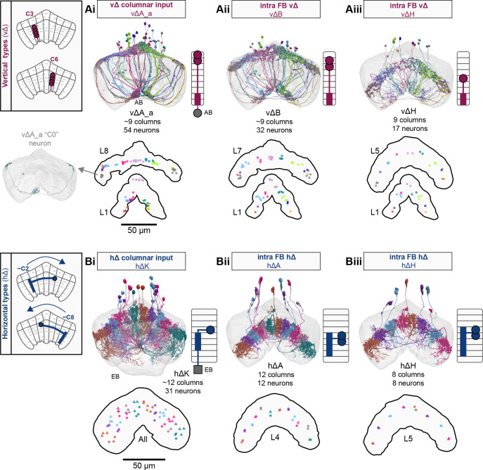
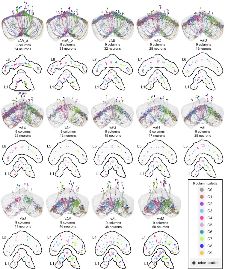
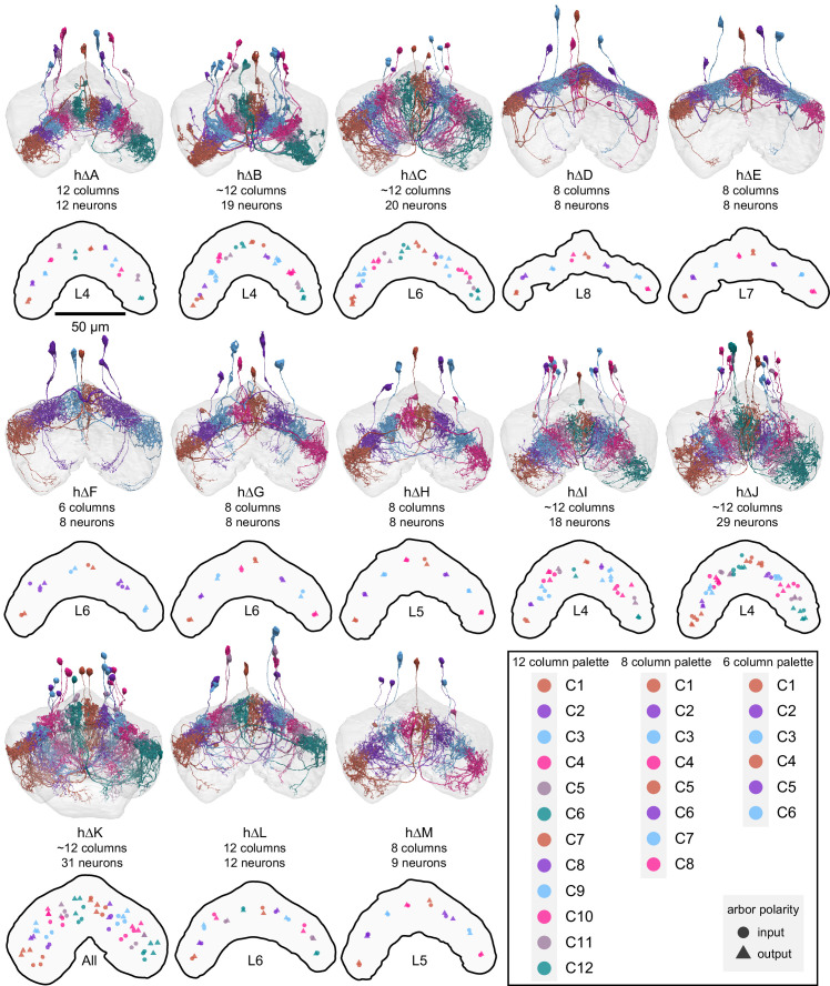
Figure 31. Overview of vΔ and hΔ columnar structure.
(A) Vertical columnar interneurons – the vΔ neuron types – have individual neurons with processes centered around one fan-shaped body (FB) column. Schematic on left shows two schematized neurons with arbors centered on C3 and C6. (Ai) Morphological rendering of the vΔA_a population, along with their schematized innervation pattern. Individual neurons are colored by FB column (from C1 to C9). In addition to innervating the FB, vΔA_a neurons (and some vΔA_b) are unique among vΔ neurons in that they innervate an extra-FB area, the asymmetric body (AB). Also notice the high degree of overlap of processes in the dorsal FB and the messy columnar structure of the population. Inset to the left shows a ‘C0’ neuron, which has arbors in both C1 and C9. (Aii) Same as in (Ai), but for the vΔB population. As with all other vΔ types, these neurons have processes restricted to the FB and receive most of their input in ventral layers while sending most of their output to more dorsal layers. (Aiii) Same as in (Ai), but for the vΔH population. (B) Horizontal columnar interneurons – the hΔ types – have individual neurons with processes centered on two distant FB columns, as shown in the illustration for two generic hΔ neurons. In particular, each hΔ neuron has a dendritic compartment that is ~180° away from its axonal compartment (i.e., separated by half the FB’s width). Half of the population has dendrites in right FB columns and project to left FB columns, while the other half of the population does the opposite. Individual hΔ neurons are assigned to columns based on the location of their dendritic compartment. (Bi) Morphological rendering of the hΔK population, along with their schematized innervation pattern. Individual neurons are colored according to FB column, with paired columns given matching colors. To achieve the ~180° phase shift, all hΔ types form an even number of columns. In this case, 12 columns (marked with six colors). In addition to innervating the FB, hΔK neurons are unique among hΔ neurons in that they innervate an extra-FB area, the EB. (Bii) Same as in (Bi) but for the hΔA population, which also forms 12 columns. Like most hΔ neurons, hΔA receives most of its input in ventral FB layers and provides most of its output to more dorsal FB layers. (Biii) Same as in (Bi) but the for the hΔH population, which forms eight columns instead of 12. Note the highly columnar structure of hΔ neuron types compared to the vΔ neuron types from (Ai) to (Aiii).


