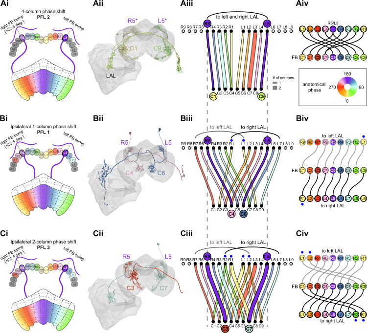
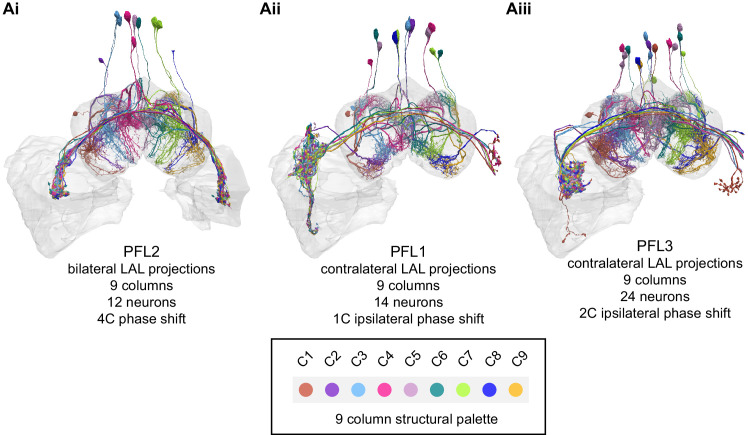
Figure 39. PFL neurons, a major fan-shaped body (FB) output, have type-specific phase shifts in protocerebral bridge-to-fan-shaped body (PB-to-FB) projections.
(A) PFL2 neurons have a four-column (~180°) PB-FB phase shift and bilateral LAL projections. (Ai) Schematic of a PB-to-FB projection pattern with a four-column phase shift, as shown for the R5 and L5 glomeruli. PB glomeruli and FB columns are colored according to anatomical phase, which indicates matching bump locations. (Aii) Morphological renderings of PFL2 neurons innervating R5 and L5. Neurons are colored according to their FB column. Notice that R5 and L5 neurons end up at C1 and C9, respectively. R5/L5 have been given asterisks because individual PFL neurons can innervate multiple PB glomeruli (in this case, R4/R5 and L4/R5). (Aiii) Graph showing the projection pattern from PB glomeruli to FB columns for all neurons in the PFL2 population. R5 and L5 projections have been highlighted as in (Ai), and edges are colored according to PB glomerulus. Unlike all other PB-FB-* neurons, the PFL2 population should only inherit one bump in the PB since the neurons sample from an ~360° region of PB space, split between left and right halves (R5–R1 and L1–L5). (Aiv) Functional graph showing the mapping between phases in the PB (top row) and phases in the FB (bottom row). Circles are colored by anatomical phase (legend). With one exception (R1), every PB glomerulus connects to a FB column with an ~180° phase shift. (B) PFL1 neurons have a one-column (~45°) ipsilateral phase shift and project to the contralateral LAL. (Bi) Same as in (Ai), but schematizing the one-column ipsilateral phase shift of PFL1 neurons. (Bii) Same as in (Aii), but for two PFL1 neurons. Notice that the R5 neuron projects to C4, and the L5 neuron projects to C6. (Biii) Similar to (Aiii), but for PFL1. Black and gray arched lines indicate groups of glomeruli that project to the right or left LAL, respectively. R1 and L1 are marked with blue dots because they project to the ipsilateral LAL, unlike the other neurons in the population. (Biv) Similar to (Aiv), but for PFL1. Here, instead of dividing glomeruli by their left vs. right PB innervation (as in Biii), glomeruli are grouped by whether they project to the left LAL (top row, gray outlined circles) or the right LAL (bottom row, black outlined circles), and sorted by anatomical phase. With the exception of R7 and L7, each glomerulus projects to FB columns with a one-column (~45°) ipsilateral phase shift. (C) PFL3 neurons have a two-column (~90°) ipsilateral phase shift and project to the contralateral LAL. (Ci) Same as in (Bi), but schematizing the two-column ipsilateral phase shift of PFL3 neurons. (Cii) Same as in (Bii), but for two PFL3 neurons. Notice that the R5 neuron projects to C3, and the L5 neuron projects to C7. (Ciii) Same as in (Biii), but for PFL3. R1/R2 and L1/L2 are marked with blue dots because these glomeruli contain neurons that either project to the contralateral LAL (like most of the population) or project to the ipsilateral LAL (unlike most of the population). (Civ) Same as in (Biv), but for PFL3. Notice that every neuron that projects to the left LAL (top row) and right LAL (bottom row) samples FB columns with a two-column (~90°) phase shift.


