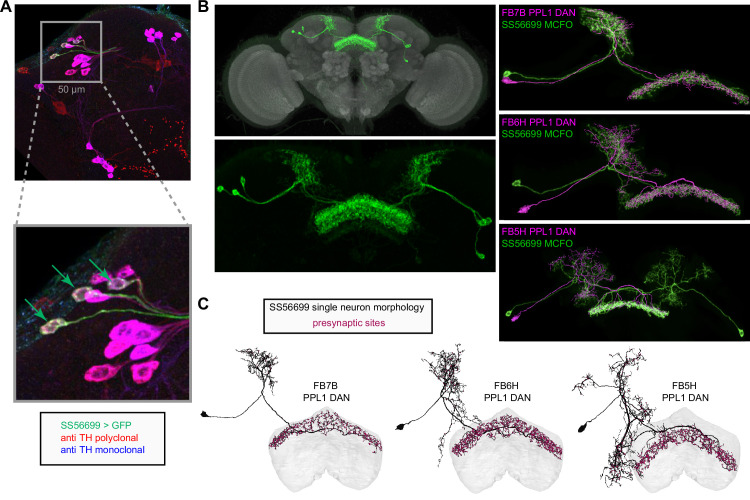
Figure 49. Identification of wake-promoting, PPL1 dopaminergic dorsal fan-shaped body (dFB) tangential neuron types.
(A) Confocal micrographs showing a portion of the expression pattern of a split-GAL4 line, SS56699 (green), focused on the cell bodies of the three neurons expressed in each brain hemisphere of this line along with immunohistochemical staining against TH using a polyclonal (red) and monoclonal (blue) antibody. Inset shows a zoomed-in view of the three SS56699 soma in the right hemisphere, marked by green arrows, which are all TH+. This result was consistent across 12 hemispheres from six brains. (B) Expression pattern of the SS56699 line with nc82 reference staining (top) and zoomed-in view of the expression pattern alone (bottom). One of hemidriver parents of this line uses an 11kb genomic segment of the TH tyrosine hydroxylase (TH) gene (see Aso et al., 2014a) to drive its expression. Morphological renderings comparing the three putative dFB dopaminergic neuron types (magenta)—FB7B (top panel), FB6H (middle panel), and FB5H (bottom panel)—to individual neurons from SS56699, generated by MCFO stochastic labeling (green; Nern et al., 2015). (C) Single neuron morphological renderings from each of the three identified PPL1 dopaminergic neuron types: FB7B, FB6H, FB5H. Magenta circles mark presynaptic sites. See Video 16 for 3D comparisons.

