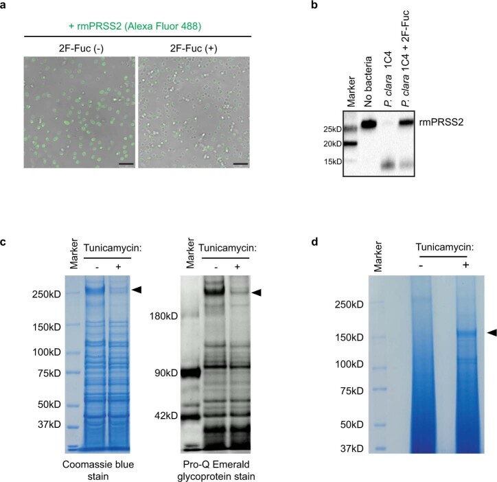
Extended Data Fig. 4. Shedding of Paraprevotella proteins into the supernatant following treatment with tunicamycin.
a, b, P. clara 1C4 was pre-treated with 2F-Fuc [2F-Fuc (+)] or vehicle control [2F-Fuc (−)] followed by incubation with rmPRSS2. Association of rmPRSS2 with P. clara 1C4 was examined by confocal microscopy (a) and degradation of rmPRSS2 was analysed by Western Blot with anti-His-tag antibody (b). Scale bar: 5 μm (a). c, P. clara 1C4 was treated with tunicamycin or vehicle control, and whole cell lysates were analysed for protein (left) and glycan (right) contents with Colloidal Coomassie Blue staining and Pro-Q Emerald 300 staining, respectively. d, Supernatant proteins from samples in (c) were analysed with Colloidal Coomassie Blue staining. Arrowheads indicate the bands that were decreased (c) or increased (d) after tunicamycin treatment. a–d, Representative images from two independent experiments with similar results are shown. See Supplementary Figure 1 for gel and blot source data.

