Abstract
The integration of large-scale two-dimensional (2D) materials onto semiconductor wafers is highly desirable for advanced electronic devices, but challenges such as transfer-related crack, contamination, wrinkle and doping remain. Here, we developed a generic method by gradient surface energy modulation, leading to a reliable adhesion and release of graphene onto target wafers. The as-obtained wafer-scale graphene exhibited a damage-free, clean, and ultra-flat surface with negligible doping, resulting in uniform sheet resistance with only ~6% deviation. The as-transferred graphene on SiO2/Si exhibited high carrier mobility reaching up ~10,000 cm2 V−1 s−1, with quantum Hall effect (QHE) observed at room temperature. Fractional quantum Hall effect (FQHE) appeared at 1.7 K after encapsulation by h-BN, yielding ultra-high mobility of ~280,000 cm2 V−1 s−1. Integrated wafer-scale graphene thermal emitters exhibited significant broadband emission in near-infrared (NIR) spectrum. Overall, the proposed methodology is promising for future integration of wafer-scale 2D materials in advanced electronics and optoelectronics.
Subject terms: Two-dimensional materials, Electronic properties and devices, Two-dimensional materials
Defect-free integration of 2D materials onto semiconductor wafers is desired to implement heterogeneous electronic devices. Here, the authors report a method to transfer high-quality graphene on target wafers via gradient surface energy modulation, leading to improved structural and electronic properties.
Introduction
The integration of two-dimensional (2D) materials into current silicon technology may embed high mobility, dangling band-free interface, atomic-scale channel size into practical electronic and optoelectronic devices1–4. Note that, an essential prerequisite is transferring 2D materials from their growth substrates to industrial wafers. Yet, a significant gap still exists in transfer method, which frustrates the recent progress in wafer-scale single-crystal growth of 2D materials5–10. Conventionally, wet transfer methods commonly use poly(methyl methacrylate) (PMMA) film as a transfer medium to support 2D materials when separated from the growth substrate and scooped from the liquid surface to a target substrate11,12. For graphene, the wet transfer methods may significantly degrade its properties due to introduced cracks, wrinkles, polymer contaminations, and water doping by water adsorptions on the graphene surface13–18. These factors act as extra scattering centers to limit carrier mobility and decrease device performance. To overcome these issues, previous attempts have shown that the optimization of PMMA and replacement of PMMA with small molecules or other polymers would facilitate clean graphene transfer19–27, conformal contact with the target substrate may reduce the formation of cracks and wrinkles28–31, and the development of dry transfer methods may diminish water doping by preventing the submersion of target substrate in liquids32–39. However, no method has so far entirely solved these issues, and most approaches are not compatible with high-volume semiconductor technology at the wafer level40,41.
Here, we designed a multi-functional tri-layer transfer medium with gradient surface energy distribution, according to the thin-film adhesion theory that transfer of thin film from one layer to another layer is mainly dominated by the difference in surface energy of each layer42,43. In this case, the higher surface energy of a target substrate, the better it serves as the thin film ‘acceptor’ due to the better wetting and larger adhesion strength at interface44. Thus, the surface energies of transfer medium and target substrates should be engineered to ensure reliable adhesion and release45, critical features for securing the wafer-scale 2D materials integration1,2,46. As a result, the gradient surface energy (GSE) modulation approach conduced to the integration of 4-inch single-crystal ultra-flat graphene onto silicon wafers. The transferred graphene wafer maintained its flatness, exhibiting intact and clean surfaces with negligible water doping. Consequently, the resulting wafer-scale graphene illustrated a uniform sheet resistance of only ~6% deviation over a 4-inch area. The as-transferred graphene on SiO2/Si exhibited outstanding electrical performances with smaller Dirac points and much higher carrier mobility (~10,000 cm2 V−1 s−1) at room temperature when compared to conventional wet transfer (~2000 cm2 V−1 s−1). The quantum Hall effect (QHE) was also observed at room temperature in graphene transferred on SiO2/Si, and fractional quantum Hall effect (FQHE) was recorded at 1.7 K in transferred graphene encapsulated by h-BN with high mobility reaching ~280,000 cm2 V−1 s−1. Furthermore, the integrated thermal emitter arrays fabricated on a 4-inch graphene/silicon wafer showed significant emission with a broad spectrum in NIR region.
Results
Design of wafer-scale graphene integration
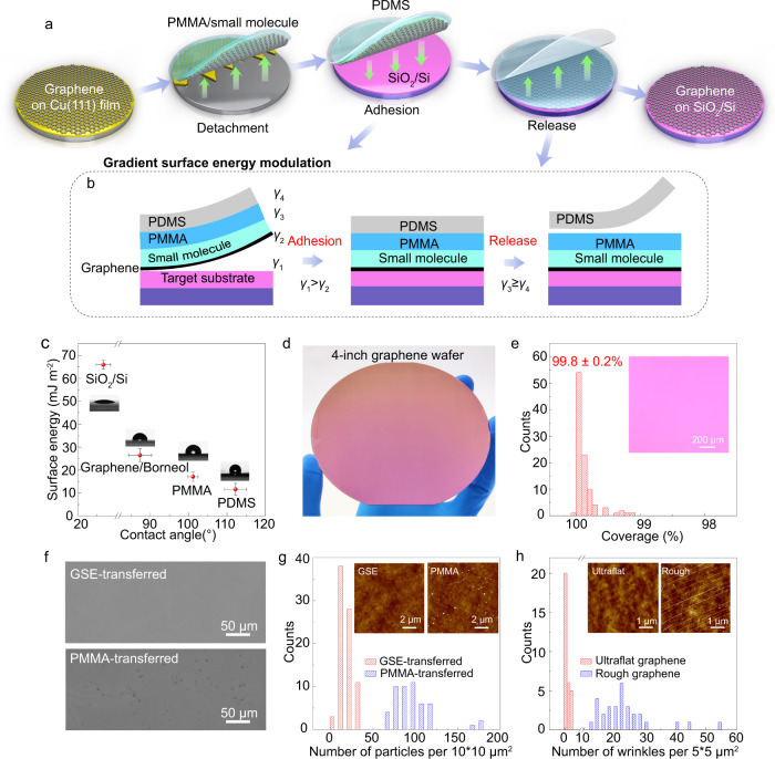
To minimize the adverse effects of grain boundaries and wrinkles on the charge carrier mobility, single-crystal ultra-flat graphene films were grown on 4-inch Cu(111)/sapphire wafers and the details can be found in Methods. A multi-functional tri-layer transfer medium was designed to support the wafer-scale graphene during transfer (Fig. 1a, b, Supplementary Figs. 1 and 2). The bottom layer of small molecules (borneol) was adsorbed on the graphene to reduce the surface energy of graphene, as well as working as a buffer layer to prevent direct contaminations caused by the upper PMMA layer (Fig. 1b). The PMMA layer ensured the integrity of graphene during transfer (Supplementary Fig. 3), and the topmost polydimethylsiloxane (PDMS) layer served as a self-supporting layer, allowing dry transfer of graphene and preventing water doping (Fig. 1a, b).
Fig. 1. Wafer-scale graphene integration by gradient surface energy modulation.
a Schematic illustration of wafer-scale ultra-flat graphene transfer via gradient surface energy modulation. b The structure of transfer medium, in which different layers with gradient surface energy are designed (γ1 > γ2, γ3 ≥ γ4). Left and right figures show the adhesion and release procedures in panel (a). Note that the surface energy of SiO2/Si (γ1) is larger than that of graphene/borneol (γ2), enabling reliable adhesion as the middle picture shows. Also, the surface energy of PDMS (γ4) is the lowest, leading to the intact release of graphene onto the target substrate. c The surface energies of different surfaces calculated from measured contact angles. Insets: images showing contact angles of water on different surfaces. Error bars indicate standard deviations of surface energies and contact angles for different surface. d Optical image of 4-inch transferred graphene on SiO2/Si wafer. e Histograms of coverage of transferred graphene. Inset: optical microscopy image of transferred graphene. f Scanning electron microscopy images of GSE-transferred graphene and PMMA-transferred graphene. g Histograms of particle number per 10 × 10 μm2 from 80 AFM images of GSE-transferred and 50 AFM images of PMMA-transferred graphene. Insets: Typical AFM images of GSE-transferred and PMMA-transferred graphene. h Histograms of wrinkle number per 5 × 5 μm2 issued from AFM images of transferred ultra-flat and rough graphene. Insets: AFM images of transferred ultra-flat and rough graphene.
Most importantly, the surface energy gradually decreased from the destination SiO2/Si wafer to the topmost PDMS layer (Fig. 1b, c), whose surface energy was calculated by measuring the contact angles based on Owen-Wendt and Young’s equations28,44 (Supplementary Table 1 and 2, Supplementary Fig. 4). The film with low surface energy tends to adsorb strongly on the substrate with high surface energy, according to the thin-film adhesion theory31. The driving force for wetting the interface is the spreading coefficient λAB:
| 1 |
where γB and γA are the surface energies of phase B (the adherend) and A (the adhesive), respectively; and γAB is the interface energy between phase A and B. The fracture strength σf of the interface is related to λAB by:
| 2 |
where Km is a function of the mechanical properties. Considering that γAB ≪ γA, the fracture strength σf is proportional to the surface energy ratio of the adherend to the adhesive (γB /γA).
For the wafer-scale graphene transfer, both the reliable adhesion and release of graphene film are critical, which determine the integrity of transferred wafer-scale graphene. Since the surface energy of SiO2/Si (γ1) was much larger than that of graphene/borneol (γ2), the complete wetting and reliable adhesion of graphene to the SiO2/Si wafer was facilitated (Fig. 1b, c). Moreover, the very small surface energy of PDMS (γ4) that is close to PMMA (γ3) ensured the damage-free release of wafer-scale graphene onto SiO2/Si wafer (Fig. 1b, c, Supplementary Fig. 5) due to the weak adhesion bond strength. By comparison, the use of thermal release tape with larger surface energy and sticky surface as a self-supporting layer led to uncontrolled release of the wafer-scale graphene with poor macroscopic and microscopic integrity (Supplementary Fig. 6), indicating that the gradient surface energy is the key to the successful adhesion and release of wafer-scale 2D materials during transfer.
The design of gradient surface energy (GSE) allowed successful integration of the 4-inch single-crystal graphene onto the SiO2/Si wafer with high intactness (99.8 ± 0.2%, Fig. 1d, e, Supplementary Figs. 7 and 8). The GSE-transferred graphene also exhibited a clean surface with significantly reduced polymer residues when compared to conventional PMMA-transferred graphene17,22 (Fig. 1f, g, Supplementary Figs. 9–12), owing to the much lower adsorption energy of borneol on graphene than that of PMMA (Supplementary Fig. 11). In general, the flatness of graphene was influenced by the density of particles and wrinkles on the surface. In addition to the negligible surface particles, the GSE-transferred graphene maintained its flat morphology with few wrinkles, benefiting from the ultra-flat nature of graphene/Cu(111)/sapphire with significantly inhibited graphene wrinkles and Cu step bunches (Fig. 1h, Supplementary Figs. 13–14). In this way, a graphene film with an intact, clean, and ultra-flat surface was obtained on wafer-scale SiO2/Si substrate.
The proposed GSE strategy can also be used for the graphene integration onto 4-inch industrial sapphire substrates (Supplementary Fig. 15). Meanwhile, wafer-scale graphene grown on Cu foil and h-BN could also be integrated onto SiO2/Si using the GSE strategy (Supplementary Figs. 16 and 17). Similar results were obtained by using rosin as small molecule buffer layer20, implying the versatility of the GSE method (Supplementary Fig. 18). To show more details of the GSE transfer method, we have included a step-by-step protocol within the Methods section, Supplementary Movie 1 and 2.
Uniform wafer-scale graphene
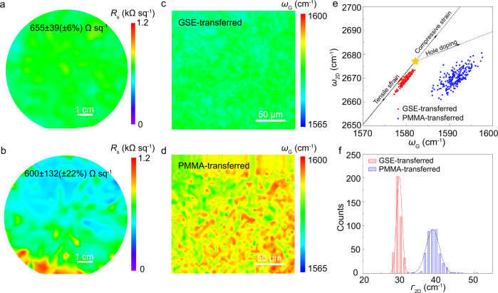
The uniformity of transferred graphene is vital for advanced wafer-scale integrated devices1–4,46. Owing to the intact and clean surface, the GSE-transferred graphene had a very uniform sheet resistance (655 ± 39 Ω sq-1), whose standard deviation was only ~6% over the 4-inch wafer (Fig. 2a). By contrast, the sheet resistance of PMMA-transferred looked inhomogeneous with a much higher standard deviation of ~22% (600 ± 132 Ω sq-1), resulting from the uneven distribution of cracks and contaminations introduced during the transfer20,21 (Fig. 2b). The uniformity of graphene at the microscopic level was further evaluated by Raman mapping. As shown in Supplementary Fig. 19, no D band peak was observed for GSE-transferred graphene. Also, the distribution of G-band position became remarkably narrower when compared to that of PMMA-transferred graphene (Fig. 2c, d). These observations further evidenced the advantages of the proposed GSE strategy for integrating wafer-scale damage-free and clean graphene with a uniform surface.
Fig. 2. Uniformity of transferred graphene.
a, b Spatial sheet resistance maps of GSE-transferred graphene (a) and PMMA-transferred graphene (b) on 4-inch SiO2/Si wafer. Note that the sheet resistance deviation of GSE-transferred graphene is ~6%. c, d Spatial G-peak’s position maps of GSE-transferred (c) and PMMA-transferred (d) graphene. The peak position of G-band of PMMA-transferred graphene is blue shifted. e Correlation map of the Raman G and 2D peak positions of GSE- and PMMA-transferred graphene. A total of 225 Raman spectra were taken from each type of transferred graphene. The yellow star represents the G and 2D peak positions of the pristine graphene with neither doping nor strain. f 2D peak’s full width at half maximum (Γ2D) of GSE- and PMMA-transferred graphene. The solid lines are fitting lines of distribution function for Γ2D.
To examine the doping and strain level of GSE-transferred graphene, the peak positions of G band (ωG) and 2D band (ω2D) were extracted from the Raman spectra, and the correlation maps were plotted in Fig. 2e. The yellow star reveals the location of intrinsic graphene (ωG = 1582 cm-1, ω2D = 2677 cm-1), corresponding to neither doping nor strain47. The PMMA-transferred graphene films are often deeply p-doped when using wet transfer methods due to water doping at the interface13,18,21, leading to changes in the Fermi level of graphene and declined carrier mobility. As shown in Fig. 2e, the PMMA-transferred graphene film on SiO2/Si wafer experienced deeply p-doping. By comparison, GSE-transferred graphene on SiO2/Si wafer nearly experienced no p-doping and strain, thereby close to the intrinsic graphene (Fig. 2e). The histograms of 2D peak’s full width at half maximum (Γ2D) of transferred graphene were gathered in Fig. 2f. The average Γ2D of GSE-transferred graphene (~30 cm-1) was smaller than that of PMMA-transferred graphene (~40 cm-1), indicating GSE-transferred graphene with little random strain fluctuation and potentially high charge carrier mobility33,48,49.
Electronic properties of GSE-transferred graphene
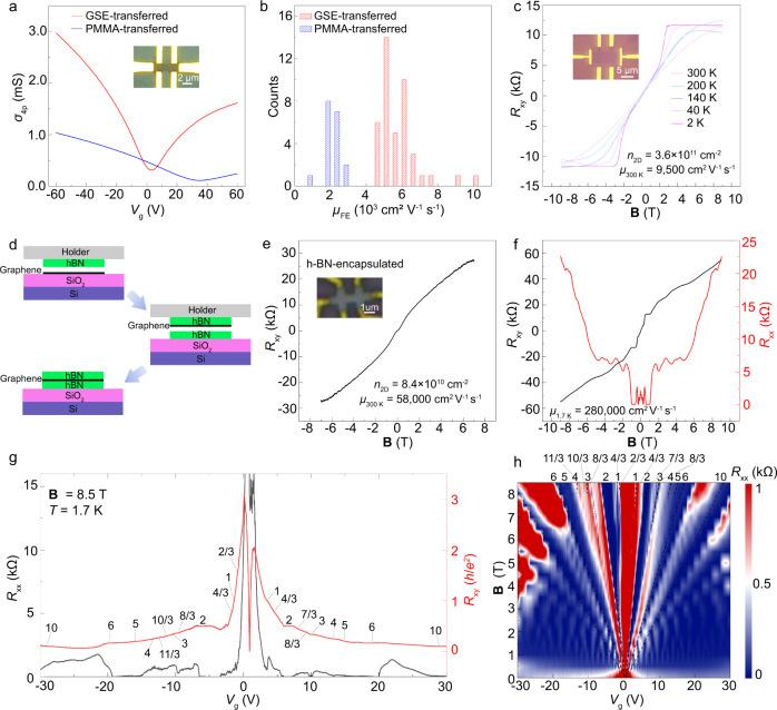
The electrical performances of devices fabricated with GSE-transferred graphene were investigated. Hall-bar devices were fabricated with standard electron beam lithography (EBL) to measure the field-effect carrier mobility of graphene on SiO2/Si. The typical transfer characteristics of 60 Hall-bar devices fabricated with GSE- and PMMA-transferred graphene are summarized in Fig. 3a. The Dirac point of GSE-transferred graphene was near zero, and the carrier concentration was about 3 × 1011 cm−2, revealing a very small doping level of graphene consistent with the Raman results. The extracted hole mobility reached up 10,000 cm2 V−1 s−1 (Fig. 3a), comparable to previously reported values of state-of-the-art CVD graphene5,16,21. By contrast, the Dirac point of deeply-doped PMMA-transferred graphene was close to 35 V, and the carrier concentration (~3 × 1012 cm−2) was an order of magnitude higher than that of GSE-transferred graphene, showing a relatively low μ of 1930 cm2 V−1 s−1. Accordingly, the average hole mobility of GSE-transferred graphene reached ~6000 cm2 V−1 s−1 (Fig. 3b), a value 3-fold higher than that of PMMA-transferred graphene (~2000 cm2 V−1 s−1). In addition, the mobility of wet-transferred graphene by only using PMMA/borneol as the transfer medium is ~3950 cm2 V−1 s−1, much lower than that of GSE-transferred graphene, which indicate the water-adsorption-induced doping will significantly degrade the electrical properties of graphene (Supplementary Fig. 20).
Fig. 3. Electrical properties of transferred graphene.
a Transfer characteristics comparison of two typical Hall-bar devices fabricated with PMMA- and GSE-transferred graphene. Inset: image of graphene Hall-bar device on SiO2/Si. b Histograms of FET mobility of GSE- and PMMA-transferred graphene. The average mobility of 42 GSE-transferred and 18 PMMA-transferred graphene devices are 6000 cm2 V−1 s−1 and 2000 cm2 V−1 s−1, respectively. c The change in Hall resistance as a function of magnetic field of GSE-transferred graphene on SiO2/Si at different temperature. Inset: image of graphene Hall-bar device on SiO2/Si. d The fabrication scheme of h-BN-encapsulated transferred graphene. e The change in Hall resistance as a function of magnetic field (B) of h-BN-encapsulated transferred graphene at 300 K. Inset: image of h-BN encapsulated graphene Hall-bar device. f The variation in Hall resistance (Rxy) (left axis) and longitudinal resistance (Rxx) (right axis) as a function of B at 1.7 K. g Rxx (left axis) and Rxy (right axis) as a function of the back gate (Vg) at 1.7 K and 8.5 T. h 2D contour plot of Rxx as a function of B and Vg. The dash black lines show LLs at filling factors ν = ±2, ±6, and ±10, as well as some new emerging fractional filling factors ν = 2/3, ±4/3, 7/3, ±8/3…, due to the degeneracy lifting of LLs.
As shown in Fig. 3c, the Hall mobility of the GSE-transferred graphene on SiO2/Si extracted from the Hall effect measurement was 9500 cm2 V−1 s−1 at room temperature. By measuring the Hall resistance (Rxy) and magnetoresistance (Rxx) at different temperatures (Fig. 3c, Supplementary Fig. 21a), we confirmed that the nonlinearity in the large magnetic field at room temperature was caused by the quantum Hall effect (QHE), further demonstrating the outstanding electrical performances and little scattering centers of GSE-transferred graphene50 (Supplementary Fig. 22). The Hall mobilities and carrier concentrations at different temperature was shown in Supplementary Fig. 21b, and Hall mobility can reach up 19,500 cm2 V−1 s−1 at 2 K.
To further confirm the intrinsic mobility of transferred graphene, hexagonal boron nitride (h-BN) flake was employed to pick up and encapsulate the transferred graphene from SiO2/Si substrate (Fig. 3d) to fabricate Hall-bar devices with 1D edge contact (inset of Fig. 3e). The Hall mobility at 300 K was calculated as ~58,000 cm2 V−1 s−1, and the carrier concentration was about 8.4 × 1010 cm−2, indicating excellent electrical properties of transferred graphene (Fig. 3e). According to the longitudinal magnetoresistance and Hall curve at 1.7 K (Fig. 3f), the extracted Hall mobility reached as high as 280,000 cm2 V−1 s−1, thus rivaling mechanically exfoliated graphene51,52. In the Longitudinal (Rxx) and Hall (Rxy) magnetoresistance measurements at 1.7 K with a fixed magnetic field (B = 8.5 T), quantized Hall platform and magnetoresistance zeros were observed at all possible integer fillings of n = 0 and n = 1 Landau Levels (LLs) (Fig. 3g). Furthermore, quantization of Rxy to (1/υ)h/e2 with minimum Rxx at fractional filling factors υ = 2/3 and 4/3 in n = 0 LL, as well as υ = 7/3, 8/3, 10/3, and 11/3 in n = 1 LL were observed, confirming the fractional quantum Hall effect (FQHE) feature. The back gate (Vg) dependent resistant measurements at the different magnetic fields on h-BN-encapsulated transferred graphene Hall-bar device were then performed to resolve the broken Landau level degeneracy and FQHE in the Landau fan diagram (Fig. 3h). The observation of FQHE further demonstrated the mobility of GSE-transferred graphene should be comparable to the high-quality exfoliated graphene53,54 with average mobilities exceeding 100,000 cm2 V−1 s−1. These data confirmed the ultrahigh quality of the as-transferred graphene.
Wafer-scale integrated graphene thermal emitter devices
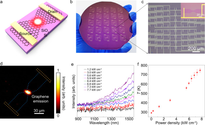
Graphene-based silicon-chip blackbody emitters in the near-infrared region, including telecommunication wavelength hold promise in applications in on-Si-chip, small footprint, and high-speed emitters of highly integrated optoelectronics and silicon photonics55. As shown in Fig. 4a, the passage of a current I through graphene thermal emitter device with narrow constrictions led to enhancements in the Joule heating, as well as localized light emission of graphene at the middle of the constriction56. As shown in Fig. 4b, the integrated graphene emitter device arrays with 4-inch GSE-transferred graphene wafer were successfully obtained. The representative device array was enlarged in Fig. 4c, showing an array of 8 × 8 graphene emitter devices with a graphene channel length of 120 μm and width of 10 μm at the center of graphene.
Fig. 4. Integration and radiation properties of wafer-scale graphene thermal emitters.
a A schematic diagram of graphene thermal emitter. b Wafer-scale graphene thermal emitter arrays on GSE-transferred graphene. c Optical microscopy image of 8 × 8 graphene thermal emitters. Inset: single graphene thermal emitter device. d Image of thermal emission at P = 3.0 kW cm−2 captured by IR camera with false-color treatment. The bright spot in the center represents the radiation emitted by graphene. The blue dashed line and yellow dashed line indicate the graphene and metal electrodes, respectively. e Emission spectra issued from a graphene emitter at P = 1.2–7.7 kW cm−2. f Graphene lattice temperatures obtained by the shift in 2D peak’s position. The temperatures varied approximately linearly with power density. Error bars indicate standard deviations of temperatures at different power density.
To protect the graphene channel of the thermal emitter device, a ~70-nm-thick Al2O3 layer was deposited on the graphene before voltage application (Supplementary Fig. 23a). Under continuous DC bias voltage, significant emission from Al2O3-capped graphene device between two electrodes was observed with an Infrared (IR) camera under vacuum at power density P = 3.0 kW cm−2 (Fig. 4d). Note that the blue and yellow dashed lines indicated the graphene and metal electrode, respectively. The emission from these devices induced a broad spectrum in NIR region, including telecommunication wavelength. In addition, the emission intensity increased with the applied voltage (Fig. 4e). The graphene lattice temperature obtained from the peak position shift of 2D band of biased graphene57 (Supplementary Fig. 23b) depicted a linear change with the applied power density due to the Joule heating effect58 that reached ~750 K under vacuum at the power density of 7.7 kW cm−2 (Fig. 4f). Thus, the graphene-based emitter is promising for high-density emitters on silicon chips, and the GSE-transferred graphene integration strategy will provide a key to the fabrication of wafer-scale graphene thermal emitter devices.
Discussion
A general method was successfully developed for the wafer-scale graphene integration onto silicon wafers, compatible with current semiconductor technologies. The physical adhesion model and data revealed the importance of the gradient surface energy in the transfer of wafer-scale graphene, enabling reliable adhesion and release during transfer. Accordingly, 4-inch damage-free graphene with preserved intrinsic properties was obtained, contributing to a uniform sheet resistance with ~6% deviation over a 4-inch area.
The transferred graphene enhanced the electrical performance due to the negligible doping level and much fewer scattering centers when compared to conventional PMMA-transferred graphene. The Hall-bar devices fabricated with graphene on SiO2/Si exhibited small Dirac points and high carrier mobilities (up to ~10,000 cm2 V−1 s−1), allowing the observation of quantum Hall effect (QHE) at room temperature. Fractional quantum Hall effect (FQHE) also appeared at 1.7 K in the transferred graphene encapsulated by h-BN, with mobility reaching ~280,000 cm2 V−1 s−1. Furthermore, integrated thermal emitter arrays with the 4-inch graphene/silicon wafer illustrated significant emission with a broad spectrum in NIR region. In sum, the proposed methodology can be used as a universal approach for the integration of other intrinsic 2D materials, such as h-BN and 2D MoS2 on the wafer level, paving the way for the development of integrated high-performance electronics and optoelectronics.
Furthermore, our GSE transfer method was successfully reproduced by an independent research group (as shown in the Peer Review file), demonstrating the robust reproducibility of GSE method in the transfer of large-area 2D materials.
Methods
Preparation of single-crystal Cu(111) film on sapphire
The Cu(111) film was deposited on a single-crystal sapphire (4 inch, c plane with misorientation <0.5°, 600 μm thickness, epi-ready with Ra < 0.2 nm) using a sputtering system (ULVAC QAM-4W). The deposition rate was 0.3 nm s-1 with a power of 300 W at 8 × 10⁻⁴ Torr, and a ~500-nm-thick Cu film was obtained on sapphire after 30 min. After that, the Cu/sapphire was then annealed at 1000 °C with 2000 sccm Ar and 10 sccm H2 for 2 h at atmospheric pressure in a homemade annealing furnace to obtain Cu(111) single crystal.
Growth of ultra-flat single-crystal graphene wafers
The 4-inch Cu(111)/sapphire was heated to 1000 °C with 2000 sccm Ar at atmospheric pressure, then 40 sccm CH4 (0.1% diluted in Ar) and 40 sccm H2 was introduced for graphene growth. Fully covered graphene was obtained after ~2 h, and CH4 gas flow was turned off while the sample cooled to room temperature.
Preparation of single-crystal Cu(111) foil
Commercially available polycrystalline Cu foils were placed in a homemade annealing furnace equipped with a 6-inch quartz tube. Three heating zones were asynchronously heated up from room temperature to target temperatures (1040 °C, 1020 °C, 1000 °C, respectively) in 40 min and kept for 1 h, which leads to the formation of a temperature gradient through the Cu foils (about 2 °C/cm) and promotes the anomalous Cu grain growth59. The heating and annealing processes were carried out under 500 sccm H2 and 100 sccm Ar.
Growth of rough graphene on single-crystal Cu foil
The rough graphene was grown on Cu(111) foil using a low-pressure CVD system. Firstly, The Cu(111) foil was heated to 1000 °C with 500 sccm Ar, followed by annealing with 500 sccm H2 for 30 min. Then the growth of graphene was initialed by the introduction of 1 sccm CH4. Fully covered graphene was produced after 1-h growth. Finally, the system was cooled down to room temperature.
Transfer of ultra-flat single-crystal graphene wafers with GSE strategy
A step-by-step protocol is available as a Supplementary Protocol in Supplementary Information. First, a layer of (-)-borneol (>97% purity, Alfa Aesar) dissolving in isopropyl alcohol (Crystal Clear Electronic Material Co., Ltd.) (25 wt%) and a layer of PMMA (950 K A4, Microchem Inc.) were sequentially spin-coated on graphene/Cu(111)/sapphire at 1000 rpm for 1 min, and baked at 130 °C for 3 min to form a composite support film. After that, PMMA/Borneol/graphene film was detached from growth substrate by etching the Cu film for 8~12 h in an aqueous solution of 1 mol/L (NH)4S2O8 (Rawhn, Shanghai Yien Chemical Technology Co., Ltd.). After washing with deionized water to remove residual etchant, the PMMA/Borneol/graphene film was attached to the Si substrates, and the PDMS (WF-40×40-0060-X4, Gel-Pak) was laminated on the surface of PMMA after the graphene was dried using the commercial laminator (LM-330ID, Rayson Co., Ltd.). The composite film of PDMS/PMMA/Borneol/Graphene was detached from Si substrate in water because the water will intercalate into the interface of graphene and Si substrate due to the hydrophilic surface of SiO2/Si (Supplementary Fig. 2, Supplementary Movie 1). The composite film is fully dried in atmosphere, followed by laminating onto SiO2/Si (sapphire), and the PDMS was exfoliated from the PMMA at 180 °C in 5 min. To further enhance the interaction of graphene and substrate, we baked the graphene at 180 °C for 3 h before removing PMMA and borneol with the vapor of hot acetone (UP, 99%, Crystal Clear Electronic Material Co., Ltd.), leaving the monolayer graphene on target substrate.
The detachment of graphene from Cu(111)/sapphire can also be achieved by electrochemical bubbling delamination when PDMS/PMMA/borneol was used as a composite support layer, which reduced the time of transfer of graphene and preserved the Cu(111)/sapphire wafer.
Transfer of ultra-flat and rough single-crystal graphene wafers with PMMA
First, a layer of PMMA was spin-coated on graphene/Cu(111)/sapphire or graphene/Cu(111) foil at 1000 rpm for 1 min, and baked at 170 °C for 3 min. After that, PMMA/graphene film was detached from growth substrate by etching the Cu film in an aqueous solution of 1 mol/L (NH)4S2O8. After washing with deionized water to remove residual etchant, the PMMA/graphene film was attached to the SiO2/Si substrates at the surface of water. To further enhance the interaction of graphene and SiO2/Si, the PMMA/graphene/SiO2/Si was baked at 180 °C for 3 h before removing PMMA with the vapor of hot acetone, leaving the monolayer graphene on SiO2/Si.
DFT calculation of adsorption energies of borneol and PMMA on graphene surface
To evaluate the adsorption energies of borneol and PMMA on graphene surface Ead, short chain of PMMA (C22H38O8) terminated with -CH3 without introducing additional polarity of the polymer was used in our first-principles calculations. Both borneol and PMMA short chain were placed on the surface of graphene with a ~3.3 A distance, measured from the center plane of molecule to the plane of graphene. The adsorption energy is defined as:
| 3 |
All the structures were optimized using the density functional theory (DFT) implemented in VASP. To keep consistency, all the periodic cells are of the same size with a = 24.56 Å, b = 24.56 Å, c = 21.70 Å. The 11 × 11 supercell of graphene was chosen for all the calculations. The PBE with the optB86b-vdW correction functional was used to determine the total energies for each structure, taking into account the Van der Waals interaction between polymer and graphene.
Fabrication of Hall-bar devices and electrical transport measurement
Hall-bar devices were fabricated on the graphene/SiO2/Si with marks for alignment. Electron-beam lithography and plasma etching with air (Diener Pico) were employed to pattern graphene into a Hall-bar geometry. After a PMMA mask (PMMA 950 K A4 @ 4000 rpm) was patterned by EBL, Pd/Au (5/40 nm) electrodes were deposited by thermal evaporation (ZHD300, Beijing Technol Science Co., Ltd), followed by a standard lift-off technique. Limited by the stage size of electron beam lithography, we fabricated 42 graphene Hall-bar devices on 10 slices from the same 4-inch GSE-transferred graphene wafer and 18 graphene Hall-bar devices on 5 slices from another 4-inch PMMA-transferred graphene wafer. The area of each slice is 1 × 1 cm2.
Electrical transport at room temperature was determined using a vacuum-probe station (Lakeshore CRX-VF) with a semiconductor characterization system (B1500A, KeySight). Electrical transport at low temperature and magneto-transport data were acquired using a low temperature and strong magnetic electronic measurement system (AttoDry2100, Attocube). Device resistance was measured using a lock-in amplifier (Stanford Research 830) with an AC driving current of 0.1–1 μA.
Fabrication of thermal emitters and thermal radiation measurement
The arrays of graphene thermal emitters were fabricated by UV lithography machine, and Al2O3 layer was grown by atomic layer deposition method. The Raman spectroscopy of graphene were measured by a confocal microRaman spectroscope (Renishaw inVia Qontor, UK) with a solid-state laser at 532 nm. The Raman signals were dispersed by a grating of 1800 lines/mm ensuring a high spectral resolution of ~1.0 cm−1. The spectrometer was calibrated by a quartz tungsten lamb at temperature of 3200 K before measurement. The thermal emission spectra were recorded using a spectrometer and a liquid nitrogen cooled Si CCD with a 50× objective lens. The radiation signal was acquired using an infrared camera. All measurements were carried out at room temperature and in vacuum.
Characterization
Optical measurement
Optical microscopy was conducted on a Nikon Olympus LV100ND. Raman spectra of transferred graphene were collected on a Horiba LabRAM HR Evolution Raman system using a 532 nm laser with a laser spot size 1 μm, and a 100x objective and 600 lines/mm grating were used to collect the Raman signal.
Contact angle measurement
The contact angle images of tested liquids on different surfaces were obtained using Biolin THETA optical tensiometer, the volume of the droplet on surface was controlled at about 4 μL.
SEM and TEM measurement
SEM images were obtained on an FEI Quattro S field-emission scanning electron microscope using a 5 kV acceleration voltage. The aberration-corrected STEM images of graphene were performed using a Nion U-HERMS200 microscope at 60 kV.
AFM measurement
The AFM morphology images were collected on a Brucker Dimension Icon using the ScanAsyst mode.
Sheet resistance measurement
The sheet resistances of transferred graphene on 4-inch wafers were collected by CDE ResMap 178 four-probe resistance tester.
Supplementary information
Acknowledgements
This work was supported by the National Natural Science Foundation of China (Nos. 52021006, T2188101, 22105009, 12174444, 52072043), Beijing National Laboratory for Molecular Sciences (BNLMS-CXTD-202001), the Tencent Foundation (XPLORER PRIZE), the National Key R&D Program of China (2018YFA0306900), the Natural Science Foundation of Hunan Province (Nos. 2020JJ3039, 2020RC3032) and the Beijing Nova Program of Science and Technology (Z191100001119067). We acknowledge Molecular Materials and Nanofabrication Laboratory (MMNL) in the College of Chemistry at Peking University for the use of instruments.
Source data
Author contributions
H.P., X.G. and L.Z. conceived the original idea for the project. Y.C., J.T., Y.L., L.S. and Z.L. carried out the synthesis of graphene. Y.W. carried out the synthesis of h-BN. X.G., L.Z., M.Y. and G.G. carried out the transfer and characterization of graphene. L.L. carried out the DFT calculation of adsorption energy. W.W. and L.L. carried out the fabrication of h-BN-encapsulated transferred graphene. J.Q., J.W., X.G., Q.W., W.W., J.T., C.T. and J.Y. carried out the fabrication and electrical transport measurement of graphene Hall-bar devices and data analysis. F.L., M.Z. and S.Q. carried out the fabrication and measurement of graphene thermal emitters. The manuscript was written by H.P., L.Z., and X.G. with the input from other authors. The whole work was supervised by H.P. All authors contributed to the scientific planning and discussions.
Peer review
Peer review information
Nature Communications thanks Peter Bøggild, Po-Wen Chiu and the other, anonymous, reviewer(s) for their contribution to the peer review of this work. Peer reviewer reports are available.
Data availability
The data that support the findings of this study are available within the article and its Supplementary Information files. The source data of Fig. 1c, e, g, h, 2a–f, 3a–c, e–h, 4e, f, and Supplementary Figs. 8b, 10a, 12c, 14c, 15b, d, 16b, 18c, 19a, b, 20b, c, 21a, b, and 23b are provided as “Source Data File”. All raw data generated during the current study are available from the corresponding authors upon request. Source data are provided with this paper.
Competing interests
The authors declare no competing interests.
Footnotes
Publisher’s note Springer Nature remains neutral with regard to jurisdictional claims in published maps and institutional affiliations.
These authors contributed equally: Xin Gao, Liming Zheng, Fang Luo, Jun Qian.
Contributor Information
Mengjian Zhu, Email: zhumengjian11@nudt.edu.cn.
Shiqiao Qin, Email: sqqin8@nudt.edu.cn.
Hailin Peng, Email: hlpeng@pku.edu.cn.
Supplementary information
The online version contains supplementary material available at 10.1038/s41467-022-33135-w.
References
- 1.Akinwande D, et al. Graphene and two-dimensional materials for silicon technology. Nature. 2019;573:507–518. doi: 10.1038/s41586-019-1573-9. [DOI] [PubMed] [Google Scholar]
- 2.Liu Y, et al. Promises and prospects of two-dimensional transistors. Nature. 2021;591:43–53. doi: 10.1038/s41586-021-03339-z. [DOI] [PubMed] [Google Scholar]
- 3.Das S, et al. Transistors based on two-dimensional materials for future integrated circuits. Nat. Electron. 2021;4:786–799. doi: 10.1038/s41928-021-00670-1. [DOI] [Google Scholar]
- 4.Zhu K, et al. The development of integrated circuits based on two-dimensional materials. Nat. Electron. 2021;4:775–785. doi: 10.1038/s41928-021-00672-z. [DOI] [Google Scholar]
- 5.Deng B, et al. Wrinkle-free single-crystal graphene wafer grown on strain-engineered substrates. ACS Nano. 2017;11:12337–12345. doi: 10.1021/acsnano.7b06196. [DOI] [PubMed] [Google Scholar]
- 6.Yuan G, et al. Proton-assisted growth of ultra-flat graphene films. Nature. 2020;577:204–208. doi: 10.1038/s41586-019-1870-3. [DOI] [PubMed] [Google Scholar]
- 7.Chen T-A, et al. Wafer-scale single-crystal hexagonal boron nitride monolayers on Cu (111) Nature. 2020;579:219–223. doi: 10.1038/s41586-020-2009-2. [DOI] [PubMed] [Google Scholar]
- 8.Li T, et al. Epitaxial growth of wafer-scale molybdenum disulfide semiconductor single crystals on sapphire. Nat. Nanotechnol. 2021;16:1201–1207. doi: 10.1038/s41565-021-00963-8. [DOI] [PubMed] [Google Scholar]
- 9.Chang C, et al. Recent progress on two-dimensional materials. Acta Phys. Chim. Sin. 2021;37:2108017. doi: 10.3866/PKU.WHXB202108017. [DOI] [Google Scholar]
- 10.Wang M, et al. Single-crystal, large-area, fold-free monolayer graphene. Nature. 2021;596:519–524. doi: 10.1038/s41586-021-03753-3. [DOI] [PubMed] [Google Scholar]
- 11.Reina A, et al. Transferring and identification of single- and few-layer graphene on arbitrary substrates. J. Phys. Chem. C. 2008;112:17741–17744. doi: 10.1021/jp807380s. [DOI] [Google Scholar]
- 12.Li X, et al. Large-area synthesis of high-quality and uniform graphene films on copper foils. Science. 2009;324:1312–1314. doi: 10.1126/science.1171245. [DOI] [PubMed] [Google Scholar]
- 13.Pirkle A, et al. The effect of chemical residues on the physical and electrical properties of chemical vapor deposited graphene transferred to SiO2. Appl. Phys. Lett. 2011;99:122108. doi: 10.1063/1.3643444. [DOI] [Google Scholar]
- 14.Huang PY, et al. Grains and grain boundaries in single-layer graphene atomic patchwork quilts. Nature. 2011;469:389–392. doi: 10.1038/nature09718. [DOI] [PubMed] [Google Scholar]
- 15.Zhu W, et al. Structure and electronic transport in graphene wrinkles. Nano Lett. 2012;12:3431–3436. doi: 10.1021/nl300563h. [DOI] [PubMed] [Google Scholar]
- 16.Lin L, et al. Towards super-clean graphene. Nat. Commun. 2019;10:1912. doi: 10.1038/s41467-019-09565-4. [DOI] [PMC free article] [PubMed] [Google Scholar]
- 17.Lin YC, et al. Graphene annealing: how clean can it be? Nano Lett. 2012;12:414–419. doi: 10.1021/nl203733r. [DOI] [PubMed] [Google Scholar]
- 18.Peng Z, et al. Influence of O2, H2O and airborne hydrocarbons on the properties of selected 2D materials. RSC Adv. 2017;7:27048–27057. doi: 10.1039/C7RA02130E. [DOI] [Google Scholar]
- 19.Kim HH, et al. Clean transfer of wafer-scale graphene via liquid phase removal of polycyclic aromatic hydrocarbons. ACS Nano. 2015;9:4726–4733. doi: 10.1021/nn5066556. [DOI] [PubMed] [Google Scholar]
- 20.Zhang Z, et al. Rosin-enabled ultraclean and damage-free transfer of graphene for large-area flexible organic light-emitting diodes. Nat. Commun. 2017;8:14560. doi: 10.1038/ncomms14560. [DOI] [PMC free article] [PubMed] [Google Scholar]
- 21.Leong WS, et al. Paraffin-enabled graphene transfer. Nat. Commun. 2019;10:867. doi: 10.1038/s41467-019-08813-x. [DOI] [PMC free article] [PubMed] [Google Scholar]
- 22.Lin YC, et al. Clean transfer of graphene for isolation and suspension. ACS Nano. 2011;5:2362–2368. doi: 10.1021/nn200105j. [DOI] [PubMed] [Google Scholar]
- 23.Wood JD, et al. Annealing free, clean graphene transfer using alternative polymer scaffolds. Nanotechnology. 2015;26:055302. doi: 10.1088/0957-4484/26/5/055302. [DOI] [PubMed] [Google Scholar]
- 24.Chen M, et al. Large-scale cellulose-assisted transfer of graphene toward industrial applications. Carbon. 2016;110:286–291. doi: 10.1016/j.carbon.2016.09.029. [DOI] [Google Scholar]
- 25.Li X, et al. Transfer of large-area graphene films for high-performance transparent conductive electrodes. Nano Lett. 2009;9:4359–4363. doi: 10.1021/nl902623y. [DOI] [PubMed] [Google Scholar]
- 26.Liang X, et al. Toward clean and crackless transfer of graphene. ACS Nano. 2011;5:9144–9153. doi: 10.1021/nn203377t. [DOI] [PubMed] [Google Scholar]
- 27.Van Ngoc H, et al. PMMA-etching-free transfer of wafer-scale chemical vapor deposition two-dimensional atomic crystal by a water soluble polyvinyl alcohol polymer method. Sci. Rep. 2016;6:33096. doi: 10.1038/srep33096. [DOI] [PMC free article] [PubMed] [Google Scholar]
- 28.Kim SJ, et al. Ultraclean patterned transfer of single-layer graphene by recyclable pressure sensitive adhesive films. Nano Lett. 2015;15:3236–3240. doi: 10.1021/acs.nanolett.5b00440. [DOI] [PubMed] [Google Scholar]
- 29.Hong J-Y, et al. A rational strategy for graphene transfer on substrates with rough features. Adv. Mater. 2016;28:2382–2392. doi: 10.1002/adma.201505527. [DOI] [PubMed] [Google Scholar]
- 30.Jung W, et al. Ultraconformal contact transfer of monolayer graphene on metal to various substrates. Adv. Mater. 2014;26:6394–6400. doi: 10.1002/adma.201400773. [DOI] [PubMed] [Google Scholar]
- 31.Seo Y-M, et al. Defect-free mechanical graphene transfer using n-doping adhesive gel buffer. ACS Nano. 2021;15:11276–11284. doi: 10.1021/acsnano.0c10798. [DOI] [PubMed] [Google Scholar]
- 32.Bae S, et al. Roll-to-roll production of 30-inch graphene films for transparent electrodes. Nat. Nanotechnol. 2010;5:574–578. doi: 10.1038/nnano.2010.132. [DOI] [PubMed] [Google Scholar]
- 33.Quellmalz A, et al. Large-area integration of two-dimensional materials and their heterostructures by wafer bonding. Nat. Commun. 2021;12:917. doi: 10.1038/s41467-021-21136-0. [DOI] [PMC free article] [PubMed] [Google Scholar]
- 34.Kim J, et al. Layer-resolved graphene transfer via engineered strain layers. Science. 2013;342:833–836. doi: 10.1126/science.1242988. [DOI] [PubMed] [Google Scholar]
- 35.Moon, J. Y. et al. Layer-engineered large-area exfoliation of graphene. Sci. Adv. 6, 40 (2020). [DOI] [PMC free article] [PubMed]
- 36.Kim KS, et al. Large-scale pattern growth of graphene films for stretchable transparent electrodes. Nature. 2009;457:706–710. doi: 10.1038/nature07719. [DOI] [PubMed] [Google Scholar]
- 37.Banszerus L, et al. Ultrahigh-mobility graphene devices from chemical vapor deposition on reusable copper. Sci. Adv. 2015;1:e1500222. doi: 10.1126/sciadv.1500222. [DOI] [PMC free article] [PubMed] [Google Scholar]
- 38.Luo D, et al. Role of graphene in water-assisted oxidation of copper in relation to dry transfer of graphene. Chem. Mater. 2017;29:4546–4556. doi: 10.1021/acs.chemmater.7b01276. [DOI] [Google Scholar]
- 39.Kang J, et al. Efficient transfer of large-area graphene films onto rigid substrates by hot pressing. ACS Nano. 2012;6:5360–5365. doi: 10.1021/nn301207d. [DOI] [PubMed] [Google Scholar]
- 40.Kang J, et al. Graphene transfer: key for applications. Nanoscale. 2012;4:5527–5537. doi: 10.1039/c2nr31317k. [DOI] [PubMed] [Google Scholar]
- 41.Langston X, et al. Graphene transfer: a physical perspective. Nanomaterials. 2021;11:2837. doi: 10.3390/nano11112837. [DOI] [PMC free article] [PubMed] [Google Scholar]
- 42.Wu, S. Polymer Interface and Adhesion 1st edn (Routledge, 1982).
- 43.Kim H, et al. Micropatterning of thin P3HT films via plasma enhanced polymer transfer printing. J. Mater. Chem. 2008;18:3489–3495. doi: 10.1039/b807285j. [DOI] [Google Scholar]
- 44.Kotsidi M, et al. Preventing colour fading in artworks with graphene veils. Nat. Nanotechnol. 2021;16:1004–1010. doi: 10.1038/s41565-021-00934-z. [DOI] [PubMed] [Google Scholar]
- 45.Mannix AJ, et al. Robotic four-dimensional pixel assembly of van der Waals solids. Nat. Nanotechnol. 2022;17:361–366. doi: 10.1038/s41565-021-01061-5. [DOI] [PubMed] [Google Scholar]
- 46.Giambra MA, et al. Wafer-scale integration of graphene-based photonic devices. ACS nano. 2021;15:3171–3187. doi: 10.1021/acsnano.0c09758. [DOI] [PMC free article] [PubMed] [Google Scholar]
- 47.Lee JE, et al. Optical separation of mechanical strain from charge doping in graphene. Nat. Commun. 2012;3:1024. doi: 10.1038/ncomms2022. [DOI] [PubMed] [Google Scholar]
- 48.Couto NJG, et al. Random strain fluctuations as dominant disorder source for high-quality on-substrate graphene devices. Phys. Rev. X. 2014;4:041019. [Google Scholar]
- 49.Neumann C, et al. Raman spectroscopy as probe of nanometre-scale strain variations in graphene. Nat. Commun. 2015;6:8429. doi: 10.1038/ncomms9429. [DOI] [PMC free article] [PubMed] [Google Scholar]
- 50.Novoselov KS, et al. Room-temperature quantum Hall effect in graphene. Science. 2007;315:1379–1379. doi: 10.1126/science.1137201. [DOI] [PubMed] [Google Scholar]
- 51.Dean CR, et al. Boron nitride substrates for high-quality graphene electronics. Nat. Nanotechnol. 2010;5:722–726. doi: 10.1038/nnano.2010.172. [DOI] [PubMed] [Google Scholar]
- 52.Wang L, et al. One-dimensional electrical contact to a two-dimensional material. Science. 2013;342:614–617. doi: 10.1126/science.1244358. [DOI] [PubMed] [Google Scholar]
- 53.Schmitz M, et al. Fractional quantum Hall effect in CVD-grown graphene. 2D Mater. 2020;7:041007. doi: 10.1088/2053-1583/abae7b. [DOI] [Google Scholar]
- 54.Pezzini S, et al. High-quality electrical transport using scalable CVD graphene. 2D Mater. 2020;7:041003. doi: 10.1088/2053-1583/aba645. [DOI] [Google Scholar]
- 55.Miyoshi Y, et al. High-speed and on-chip graphene blackbody emitters for optical communications by remote heat transfer. Nat. Commun. 2018;9:1279. doi: 10.1038/s41467-018-03695-x. [DOI] [PMC free article] [PubMed] [Google Scholar]
- 56.Luo F, et al. Graphene thermal emitter with enhanced joule heating and localized light emission in air. ACS Photonics. 2019;6:2117–2125. doi: 10.1021/acsphotonics.9b00667. [DOI] [Google Scholar]
- 57.Calizo I, et al. Variable temperature Raman microscopy as a nanometrology tool for graphene layers and graphene-based devices. Appl. Phys. Lett. 2007;91:071913. doi: 10.1063/1.2771379. [DOI] [Google Scholar]
- 58.Freitag M, et al. Thermal infrared emission from biased graphene. Nat. Nanotechnol. 2010;5:497–501. doi: 10.1038/nnano.2010.90. [DOI] [PubMed] [Google Scholar]
- 59.Sun L, et al. Toward epitaxial growth of misorientation-free graphene on Cu(111) foils. ACS Nano. 2021;16:285–294. doi: 10.1021/acsnano.1c06285. [DOI] [PubMed] [Google Scholar]
Associated Data
This section collects any data citations, data availability statements, or supplementary materials included in this article.
Supplementary Materials
Data Availability Statement
The data that support the findings of this study are available within the article and its Supplementary Information files. The source data of Fig. 1c, e, g, h, 2a–f, 3a–c, e–h, 4e, f, and Supplementary Figs. 8b, 10a, 12c, 14c, 15b, d, 16b, 18c, 19a, b, 20b, c, 21a, b, and 23b are provided as “Source Data File”. All raw data generated during the current study are available from the corresponding authors upon request. Source data are provided with this paper.


