Abstract
N7-methylguanosine (m7G) modification on internal RNA positions plays a vital role in several biological processes. Recent research shows m7G modification is associated with multiple cancers. However, in hepatocellular carcinoma (HCC), its implications remain to be determined. In this place, we need to interrogate the mRNA patterns for 29 key regulators of m7G RNA modification and assess their prognostic value in HCC. Initial, the details from The Cancer Genome Atlas (TCGA) database concerning transcribed gene data and clinical information of HCC patients were inspected systematically. Second, according to the mRNA profiles of 29 m7G RNA methylation regulators, two clusters (named 1 and 2, respectively) were identified by consensus clustering. Furthermore, robust risk signature for seven m7G RNA modification regulators was constructed. Last, we used the Gene Expression Omnibus (GEO) dataset to validate the prognostic associations of the seven-gene risk signature. We figured out that 24/29 key regulators of m7G RNA modification varied remarkably in their grades of expression between the HCC and the adjacent tumor control tissues. Cluster one compared with cluster two had a substandard prognosis and was also positively correlated with T classification (T), pathological stage, and vital status (fustat) significantly. Consensus clustering results suggested the expression pattern of m7G RNA modification regulators was correlated with the malignancy of HCC strongly. In addition, cluster one was extensively enriched in metabolic-related pathways. Seven optimal genes (METTL1, WDR4, NSUN2, EIF4E, EIF4E2, NCBP1, and NCBP2) were selected to establish the risk model for HCC. Indicating by further analyses and validation, the prognostic model has fine anticipating command and this probability signature might be a self supporting presage factor for HCC. Finally, a new prognostic nomogram based on age, gender, pathological stage, histological grade, and prospects were established to forecast the prognosis of HCC patients accurately. In essence, we detected association of HCC severity and expression levels of m7G RNA modification regulators, and developed a risk score model for predicting prognosis of HCC patients’ progression.
Keywords: m7G, hepatocellular carcinoma, bioinformatics, prognosis, risk signature
Introduction
In 2020, the sixth most commonly diagnosed cancer is primary liver cancer (∼906,000 new cases) and the third global lethal cancer (∼830,000 deaths) (Sung et al., 2021). HCC comprised 75–85% of primary liver cancer cases (Sung et al., 2021). Epidemiological studies have shown that chronic infection with hepatitis B or C viruses, ingestion of aflatoxin-contaminated foods, excessive drinking, overweight, type II diabetes, and smoking are the main threats for HCC (Thomas London et al., 2018). At early stages, most diagnosed patients of HCC are firstly identified in an advanced stage because of symptomless nature of HCC which results in limited curative options and poor prediction for one or both intrahepatic and extrahepatic metastasis (Llovet et al., 2018). For advanced-stage cases of HCC, only 1/3 patients can benefit from chemotherapy by using one or multiple kinase inhibitors, and the regimen brings evident drug resistance or toxicity with long-term therapeusis, suggesting no effective treatment can effectively improve the outcome of HCC (Wang et al., 2021). Presently, TNM staging system (Tumor T, Node N, and metastases M) remains the most extensively used predictive index to monitor HCC progression. Whereas, HCC is most diversified; thus, the same TNM stage patients tended to show significant differences in survival outcomes and responses of treatment. Hence, a finer comprehension of HCC lying molecular mechanisms can enhance dissatisfying outcomes in patients and identify new and reliable molecular signatures for predicting prognosis.
N7-methylguanosine (m7G) methylation is a very frequent RNA modification, which recently has shown an important role in balancing gene expression in both prokaryotes and eukaryotes (Wang et al., 2012; Létoquart et al., 2014; Haag et al., 2015). The m7G modification is widespread in cells for engaging in different intracellular biological function, such as transcription elongation, pre-mRNA splicing, nuclear export and mRNA translation etc. m7G modification has comprehensive effects on tRNA and mRNA (in the cytoplasm, an m7G methyltransferase complex, METLL1-WDR4, mediated m7G modification of tRNAs and mRNAs, regulating pre-mRNA splicing, RNA export, stability, and translation), and rRNA (in nucleus, 18S rRNA m7G modification mediated by WBSCR22, thus, the evolution of rRNAs and biosynthesis of 40S ribosomal subunit were regulated) molecules posttranscriptionally, and are associated with many biological processes, including the occurrence and development of human diseases (Jonkhout et al., 2017; Enroth et al., 2019; Rong et al., 2021). In mammals, deficiency in m7G tRNA modification is associated with developmental diseases (Shaheen et al., 2015; Braun et al., 2018). In addition, m7G mRNA modification participate in the pathogenesis of multiple human cancers (Rong et al., 2021). METTL1, a m7G methyltransferase, is upregulated in tumor-bearing patients compared to healthy controls (Liu et al., 2019; Tian et al., 2019). m7G exhibits the potential to become a biomarker for some types of cancer (Rong et al., 2021). These results indicate m7G modification plays conclusive roles in many diseases, involving tumors. However, the relationship between m7G modulators and tumors’ progression remains unclear, and requires a great attention and in-depth research. Therefore, further understanding of the roles of m7G regulators in cancer development might provide an attractive perspective for cancer therapy (Rong et al., 2021).
The cause of poor prognosis of cancer is its special invasive biological characteristics, such as epithelial-mesenchymal transition (EMT), signaling transduction, cancer stem cell formation, tumor angiogenesis and cancer metabolism, etc. (Zhang et al., 2020) Researches, recently, have revealed that abnormal m7G RNA modifications play crucial part in cancer progression. For example, the hepatocarcinogenesis in vitro and hydrodynamic transfection HCC mouse models could be promoted by METTL1-dependent m7G tRNA modification (Chen Z. et al., 2021). m7G tRNA modification and METTL1 are raised in HCC and correlated with worsening prognosis (Chen Z. et al., 2021). Overexpression of METTL1 suppresses colon cancer (CC) cells proliferation, invasion, migration, and induces cell apoptosis in a m7G dependent manner (Liu et al., 2020). The overexpression of METTL1 increases chemosensitivity of CC cells to cisplatin, because within the carcinoma treating process, the chemoresistance regulating process was crucially affected by METTL1-mediated m7G (Liu et al., 2019). In vitro and in vivo, the EGFR pathway genes and the genes involved in cell-cycle were scrupulously regulated by METTL1-mediated m7G tRNA modification and thus promoted intrahepatic cholangiocarcinoma (ICC) progression (Dai et al., 2021). The elevation of METTL1 and WDR4 were reported to be related with worse prognosis of human lung cancer and intrahepatic cholangiocarcinoma negatively associated with patient (Ma et al., 2021; Orellana et al., 2021). Two kinds of m7G tRNA modifying enzymes, METTL1 and NSUN2, determine 5-Fluorouracil (a drug used in the treatment of cancer) sensitivity in human cancer cells (Okamoto et al., 2014). In summary, the malignancy of cancer can significantly be advanced by the deregulation of m7G modification. This describes clearly that m7G RNA modification has prognostic effects in HCC patients. According to a review for m7G (Tomikawa, 2018), 29 related genes were downloaded from the Gene Set Enrichment Analysis (GSEA) database for subsequent studies.
In this exploration, the transcriptomic data of HCC from The Cancer Genome Atlas (TCGA) dataset is used to evaluate the expression profiles of the 29 key regulators of m7G RNA modification. Furthermore, based on the expression patterns of m7G RNA modification regulators, HCC cases were classified into 2 clusters via consensus clustering, and these 2 clusters displayed outstandingly contrasting clinical consequences. Moreover, based on risk signature, a prognostic prediction model was established, which had approving value of prediction for HCC patients. In addition, the prognostic correlation of this risk indication was successfully verified in the Gene Expression Omnibus (GEO) database. Our work helps to elucidate the important roles of m7G modification regulators in tumors’ progression and signifies the prospect of the genetic expression signature of m7G regulators in predicting the prognosis of HCC, which will contribute to the providing of personalized prognosis of clinical outcomes and pointing out a new orientation for targeting drugs discovery in HCC patients.
Materials and methods
Data collection
The transcriptome data of RNA-seq and the relevant clinical information of HCC patients were acquired from TCGA (website: https://portal.gdc.cancer.gov/; until 10 February 2022). For further analysis, 374 HCC tissues and 50 normal adjacent tumor tissues were involved altogether.
Currently, 29 known genes, including METTL1, WDR4, NSUN2, DCP2, DCPS, NUDT10, NUDT11, NUDT16, NUDT3, NUDT4, NUDT4B, AGO2, CYFIP1, EIF4E, EIF4E1B, EIF4E2, EIF4E3, GEMIN5, LARP1, NCBP1, NCBP2, NCBP3, EIF3D, EIF4A1, EIF4G3, IFIT5, LSM1, NCBP2L, and SNUPN are recognized as the regulators of m 7G methylation. For subsequent analyses, the transcribed profiles of the 29 key regulators were recovered from the HCC cohort of the TCGA database. An irrelevant cohort (GSE54236) derived from the GEO (http://www.ncbi.nlm.nih.gov/geo) database and containing 78 HCC cases with relevant mRNA expression data and the survival information was used for external verification.
Bioinformatics analysis
Between the RNA composition of HCC samples and normal tissues, we screened the differentially expressed genes (DEGs) which encoded the m7G RNA methylation regulators at different levels via the use of the Wilcoxon test method in R software (version 4.1.0). False discovery rate (FDR), a more liberal criterion, was adopted together with absolute log2 fold change (FC) to determine the significance criteria (FDR <0.05, and FC > 1). Thereafter, we used a vioplot to show the 29 m7G-associated genes’ expression in 374 HCC and 50 control tissues. In order to survey the correlation between any two regulators of m7G RNA methylation, the Spearman correlation analyses were performed via R software.
To evaluate the association between the expression of m7G RNA methylation regulators and HCC prognosis, we used the “ConsensusClusterPlus” R package to cluster the HCC cohort into two distinct subgroups. Then, the principal component analysis (PCA) was performed via the “limma” and “ggplot2” packages to confirm the classification outcomes. A survival curve was constructed to contrast survival between clusters on the basis of the Kaplan-Meier analysis log-rank test. We applied the Chi-square test to determine the differences in clinical parameters between the 2 clusters. To functionally annotate the genes differentially expressed in the two subgroups, the Gene Ontology (GO) and the Kyoto Encyclopedia of Genes and Genomes (KEGG) analyses were performed.
We used Univariate Cox regression analyses to evaluate the relationship between m7G-associated genes’ expression and overall survival (OS). Afterward, to keep away overflow, the least absolute shrinkage and selection operator (LASSO) Cox regression was performed to remove the high relevant genes by using the “glment” package. Ultimately, a risk signature of a seven-m7G-regulatory-gene was determined. In order to yield risk scores, we multiplied the gene expression and its coefficient acquired from the LASSO Cox regression. HCC cases were then allocated to the low-risk and high-risk groups by using median risk scores. By employing the “survival” package, the Kaplan-Meier analysis was executed to generate and analyze survival-time data. Receiver operating characteristic (ROC) curves were applied to inspect the prognostic accuracy of the predicted results derived from the model. The differences of clinicopathological variables between the low- and the high-risk groups were visualized by a heatmap and examined by Chi-square test. Moreover, as effective means, univariate and multivariate Cox regression analyses were performed to assess whether the risk scores were independent and appropriate prognostic indicators. To verify the prognostic value of th e seven-m7G-regulatory-gene risk signature, the GSE54236 dataset was applied as a validated cohort (Villa et al., 2016). Risk score of each patient was computed based on the formula described earlier. By applying the same cutoff criteria, all HCC patients were classified into low- and high-risk groups. Thereafter, the Kaplan-Meier analysis and the ROC curve analysis were respectively executed for evaluating the prognostic values.
Ultimately, a prognostic nomogram was established using clinical information (age, gender, histologic grade, and pathological stage) and risk scores to forecast 1-, 3-, and 5-years survival in HCC patients by the “rms” package.
From http://www.bioconductor.org, we acquired all of the aforementioned R packages.
The pipeline of the research was shown in Supplementary Figure S1.
Statistical analysis
For all statistical analyses, R software (version 4.1.0) was utilized. The significance of the threshold was p < 0.05.
Results
Recognition of m7G RNA modification regulators expressed differentially in HCC
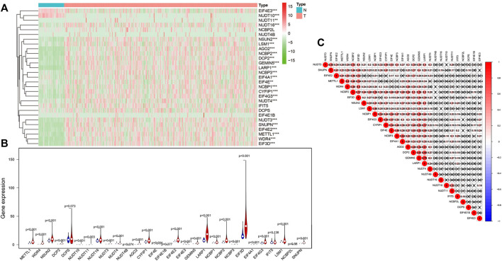
An analysis of 29 differential expressed m7G regulatory genes was performed in HCC (n = 374) and normal tissues (n = 50). The majority of m7G-associated genes (24/29) were expressed between HCC and adjacent tissues differentially showed by heatmap clearly (Figure 1A). Particularly, the levels of expressions for NUDT16, NSUN2, LSM1, AGO2, NCBP2, DCP2, GEMIN5, LARP1, NCBP3, EIF4A1, NCBP1, CYFIP1, EIF4G3, NUDT3, SNUPN, EIF4E2, METTL1, WDR4, and EIF3D in cancer tissues were significantly higher (all p < 0.001) than those in normal tissues. However, the higher expression levels of EIF4E3 and NUDT10 (both p < 0.001) were remarkably in normal tissues than those in tumor samples. No significant difference was found in DCPS, NUDT4B, EIF4E1B, IFIT5 and NCBP2L (all p > 0.05) (Figure 1B). Furthermore, we performed an association analysis to characterize further inherent connection among 29 m7G RNA modification regulators. As indicated in Figure 1C, the heatmap analysis showed that the connection between GEMIN5 and LARP1 was the most remarkable (r = 0.79). The most likely positive correlated expression level of GEMIN5 was with LARP1.
FIGURE 1.
The levels of m7G modification regulators in hepatocellular carcinoma (HCC). (A)The heatmap shows the mRNA level of m7G RNA modification regulators in each sample. “N” represents normal sample, and “T” represents cancer sample. Green stands for low expressions, and red stands for high expressions. (B) The vioplot shows the differential regulators of m7G RNA modification in HCC. Blue stands for normal samples, and red stands for HCC samples. White spots indicate the median value of the expression. (C) Spearman correlation analyses of 29 m7G RNA modification regulators in HCC. ***p < 0.001.
Identify two clusters of HCC patients with different clinical outcomes by using consensus clustering based on m7G RNA modification regulators
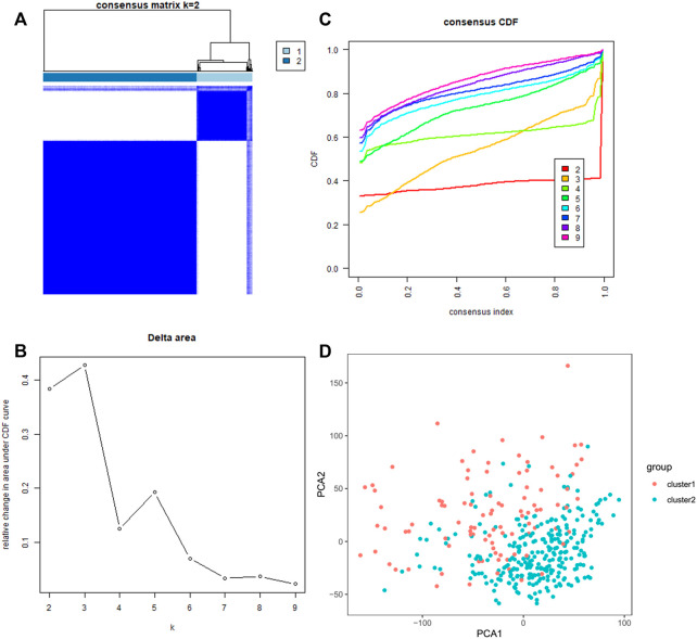
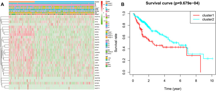
We classified the patients into clusters based on the patterns of mRNA expression to explore additionally the HCC clinical importance of 29 m7G RNA modification regulators. k = 2 grant the best clustering as well as the HCC cohorts could be divided into two definite and nonoverlapping clusters according to the analogy of the m7G RNA modification regulators (Figures 2A–C). We further performed the clustering analysis using the principal component analysis (PCA) to corroborate clustered results. As can be seen from Figure 2D, the PCA plot unveiled distinct differences between two clusters. Afterward, significant differences between the two clusters in the OS and the clinical parameters were evaluated. Hence, the OS of cluster two was more preferable than that of cluster one in significant level (p < 0.01) (Figure 3B). In addition, most m7G RNA modification regulators in cluster one exhibited higher expression levels than that in cluster 2 (Figure 3A). In contrast to cluster 2, cluster one was associated with T classification (T), later pathological stage, and vital status (fustat) significantly (all p < 0.001). There were no significant differences in the degree of Nodal involvement, Metastasis, tumor grading, age, and gender (Figure 3A). Therefore, the final outcomes of consensus clustering proposed that the mRNA profile pattern of m7G RNA modification regulators is strongly related to the malignancy of HCC.
FIGURE 2.
Consistent clustering analyses of the HCC. (A) The correlations between subgroups when the number of clusters is k = 2. (B) Cumulative distribution function (CDF) for k = 2–9 is displayed. (C) The relative variation of the area under the CDF curve of k = 2–9. (D) Principal component analysis of the RNA-seq data. Red dots stand for cluster 1, and cyan dots stand for cluster 2.
FIGURE 3.
Differences between cluster one and cluster two in clinicopathological features and overall survival. (A) Heatmap and clinicopathological features of the two clusters. Green stands for low expressions, and red stands for high expressions. (B) Compare the overall survival (OS) distribution of cluster one and cluster 2. *p < 0.05, ***p < 0.001.
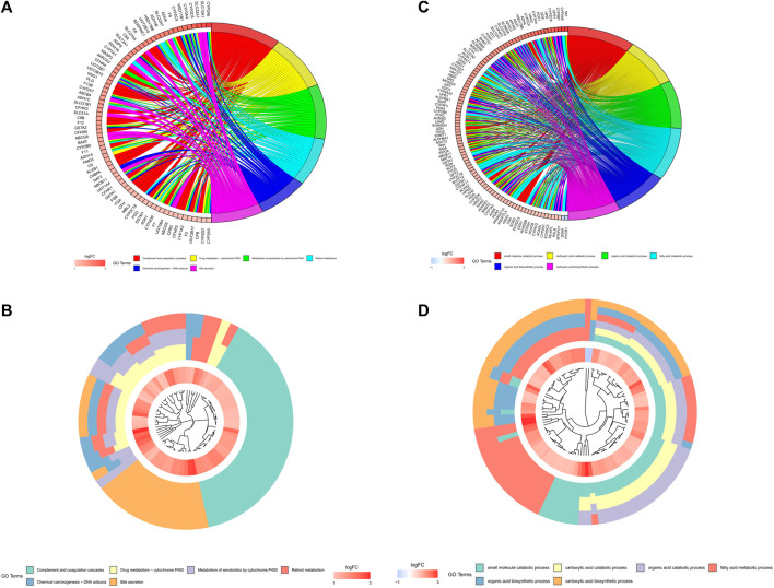
On the basis of biological processes, GO and KEGG analyses of the DEGs between two clusters were performed to explicate the clustered results. The outcomes of the GO analyses proposed that the up-regulated genes were enhancing metabolic-related biological processes, such as “small molecule catabolic process”, “carboxylic acid catabolic process”, “fatty acid metabolic process”, “organic acid catabolic process”, “organic acid biosynthetic process” and “carboxylic acid biosynthetic process” (Figures 4A,B). The KEGG analyses revealed that these up-regulated genes were adding to “Complement and coagulation cascades”, “Drug metabolism - cytochrome P450”, “Metabolism of xenobiotics by cytochrome P450”, “Retinol metabolism”, “Chemical carcinogenesis - DNA adducts” and “Bile secretion” pathways (Figures 4C,D).
FIGURE 4.
Analyses of the differentially expressed genes between the two clusters by the Kyoto Encyclopedia of Genes and Genomes (KEEG) and Gene Ontology (GO). The highly expressed genes in cluster one were functionally annotated applying the GO terms (A,B) and KEGG pathway (C,D).
A prognostic risk model established on the basis of the mRNA level of m7G regulator genes
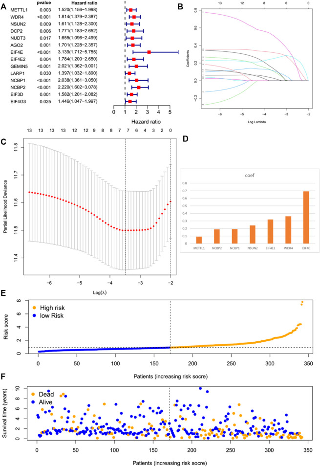
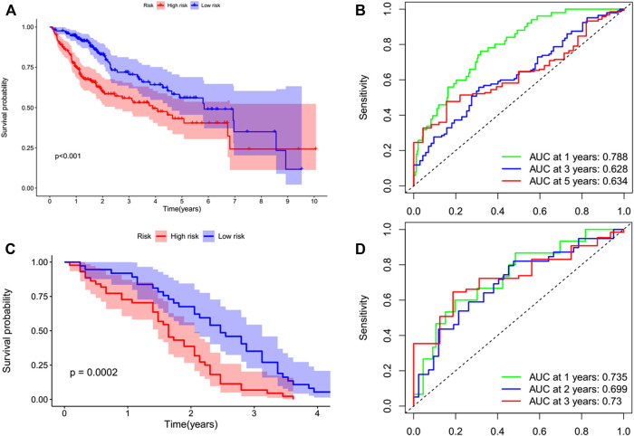
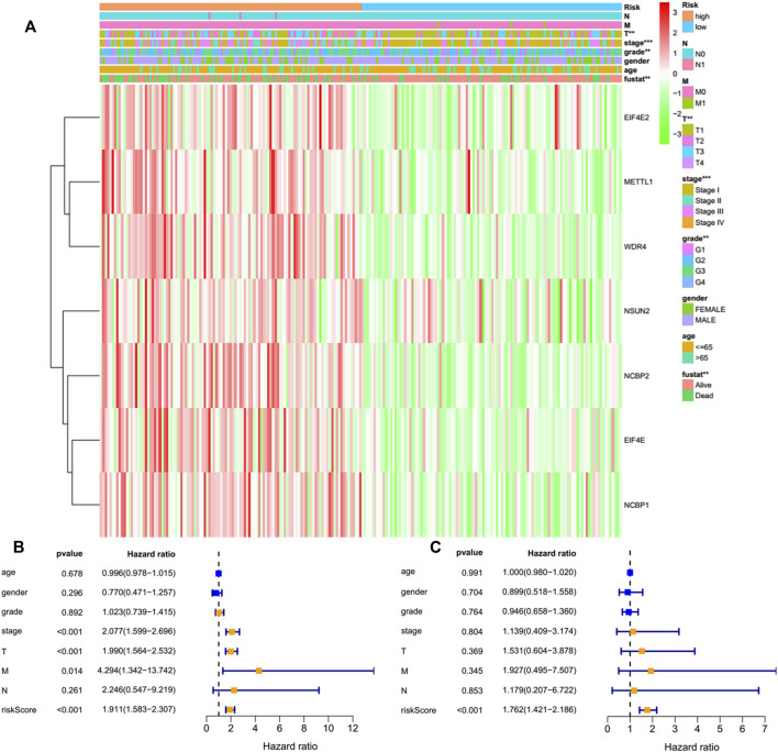
By performing a univariate Cox regression analysis on the mRNA levels of 29 key regulators, we specified a secure correlation between m7G RNA methylation regulators and the prognosis in HCC invalids. The results displayed those 14/29 regulators were outstandingly associated with OS (p < 0.05) (Figure 5A). The 14 regulators are all considered as risky genes with HR > 1. Thereafter, LASSO Cox regression analysis was applied to recognize the m7G RNA modification regulators possessing the best prognostic power (Figures 5B,C). Finally, seven effective genes (METTL1, WDR4, NSUN2, EIF4E, EIF4E2, NCBP1, and NCBP2) were selected to establish the risk model for HCC and also the corresponding coefficients calculated from the LASSO algorithm (Figure 5D). The risk score was calculated thus: RISK SCORE = (0.092 ∗ EXP value of METTL1) + (0.189 ∗ EXP value of NCBP2) + (0.193 ∗ EXP value of NCBP1) + (0.242 ∗ EXP value of NSUN2) + (0.320 ∗ EXP value of EIF4E2) + (0.362 ∗ EXP value of WDR4) + (0.693 ∗ EXP value of EIF4E). A ccording to the median risk score, HCC invalids were grouped as low-risk and high-risk to survey the prognostic function of the seven-gene signature model. Survival analysis showed that OS was worse in invalids with high-risk scores compared with low-risk scores (Figure 6A, p < 0.001). The 5-years OS rates of high-risk and low-risk group were 44.6% and 55.4%, respectively. Furthermore, ROC curve analyses were executed to estimate the area under the curve (AUC) of 0.788, 0.628, and 0.634 for the 1-, 3-, and 5-years OS, respectively. This showed eminent prognosticative power for survival results (Figure 6B). Besides, the distribution of risk scores for HCC invalids were also constructed (Figure 5E). For manifesting the survival status of each invalid, a dot pot was plotted (Figure 5F). The heatmap showed the mRNA levels of seven prognosticative genes in the groups of high-risk and low-risk (Figure 7A). Simultaneously, correlated clinical information was plotted above the heatmap. While contrasting the clinical parameters of the low-risk and high-risk groups, terms of T, stage, grade and vital status (fustat) were all significant at the 0.01 level.
FIGURE 5.
The prognostic risk model established on basis of m7G RNA modification regulator genes. (A) Univariate Cox regression analyses of m7G RNA methylation regulators. (B–D) The process of constructing signatures applying least absolute shrinkage and selection operator (LASSO) Cox regression. (E) Distribution of risk scores of the patients in the prognostic model. (F) Distributions of survival status of the patients in the prognostic model.
FIGURE 6.
Kaplan–Meier survival analysis of the prognostic model. Invalids in both datasets were divided into high-risk (red) and low-risk (blue) groups, applying the median risk score as the threshold. (A,B) In the TCGA cohort, the survival probability of the low-risk group was higher than that of the high-risk group (p < 0.001). The AUCs at 1-, 3-, and 5-years were 0.788, 0.628, and 0.634, respectively. (C,D) This prognostic model was verified in the GEO cohort. The survival probability of low-risk group was higher than that of high-risk group (p = 0.0002). The AUCs at 1 year, 2 years, and 3 years were 0.735, 0.699, and 0.73, respectively.
FIGURE 7.
Effects of the risk score and the clinicopathological characteristics on the prognosis of invalids with HCC. (A) Heatmap manifests the distribution of clinicopathological characteristics and the expressions of seven m7G RNA modification regulators in high- and low-risk groups. (B) Univariate Cox regression analyses of the clinicopathological parameters and OS. (C) Multivariate Cox regression analyses of the clinicopathological parameters and OS. **p < 0.01, ***p < 0.001.
Verifying the prognostic signatures employing GEO database
The prognostic value of the signature of seven genes for the survival predictions can be assessed by validating them using the GEO microarray data (GSE54236) (Villa et al., 2016). In the GSE54236 cohort, a total of 78 patients with HCC were grouped in high- (n = 42) and low-risk (n = 36) based on the cutoff values of the TCGA cohort. Similarly, it was also showed by the survival analysis that the OS of HCC invalids in the low-risk group was significantly finer than in high-risk invalids (Figure 6C, p = 0.0002). The AUCs for the OS of 1-, 2-, and 3-years were 0.735, 0.699, and 0.73, respectively, stating that this prognostic model was able to better predict OS in HCC invalids clearly (Figure 6D). As no patients survived for more than 5 years, therefore, no 5-years ROC curve was constructed.
The seven-gene risk signature independently forecasts the prognosis of HCC invalids
Only 226 patients were acceptable to perform the Cox regression analysis after removing deficient clinical cases. Univariate analysis disclosed that the seven-gene risk score, T, and stage were notably correlated with the OS of HCC cases (Figure 7B, all p < 0.001). We also performed a multivariate Cox regression analysis, revealing that the risk score was independently linked with OS in HCC invalids (Figure 7C, p < 0.001). All such outcomes manifested the independent prognosis of the risk signature of the seven genes for gender, age, histological grade, and pathological stage. Thus, indicating that the risk signature of these seven genes was able to serve as an independent prognosis for the patients of HCC.
Prognostic nomogram established for HCC
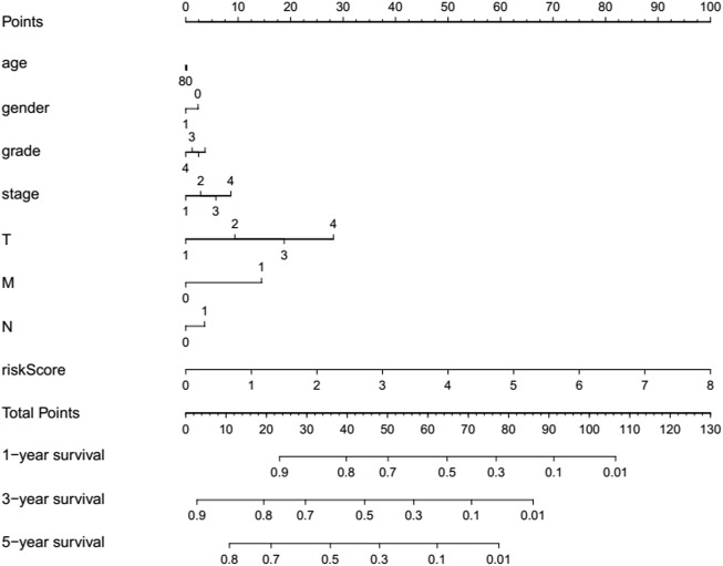
For providing a quantitative method to estimate the survival rate, a refreshing prognostic nomogram (Figure 8) based on age, gender, risk score, histological grade, and pathological stage was established. The results stated clearly the nomogram was able to systematically forecast the 1, 3, and 5-years OS in HCC invalids.
FIGURE 8.
Prognostic nomogram established via the combination with the clinicopathologic features and risk score.
Discussion
Globally, HCC is one of the most common cancers. Although great progress has been made in the diagnosis, treatment, and prognosis of HCC, which still retains the third place in cancer mortality (Sung et al., 2021). At present, there are no productive diagnostic as well as therapeutic targets for HCC. There is unsatisfactory overall survival and challenging treatment such as difficulty in predicting the prognosis, difficulties in clinical decision-making, and clinical management of patients with HCC. Hence, the molecular mechanisms implicit in contributing to tumorigenesis of HCC are very crucial and necessary to clarify. Over the years, RNA m7G modification has attracted great interest as a novel dimensionality of transcription regulation. Nevertheless, the research of m7G modification in the tumor area is yet in its infancy. As a result of the extensive appliance of RNA-seq and microarray technologies, risk scoring system in the light of multiple gene signatures is progressively used to forecast prognosis for cancers oftentimes (Chen et al., 2007; Zhang et al., 2013; Wang et al., 2019). In current study, an effective prognostic signature model through the use of seven m7G RNA modification regulators was established. It is encouraging that the risk score calculated from the seven-gene model could forecast the prognostic value of HCC invalids independently. Consequently, the risk signature reported in this paper can help clinicians make precise personalized survival prediction.
The prognostic model indicated all the seven m7G-related genes (METTL1, NCBP2, NCBP1, NSUN2, EIF4E2, WDR4, and EIF4E) might exert a stimulative effect on HCC. Tian et al. reported that METTL1 is upregulated in HCC and overexpressed METTL1 is linked to a poor prognosis of HCC (Tian et al., 2019). Overexpressed METTL1 induced oncogenic cell transformation and cancer (Orellana et al., 2021). METTL1-dependent m7G tRNA modification could promote hepatocarcinogenesis in vitro and hydrodynamic transfected in HCC mouse model, and METTL1 is raised in HCC and linked with worse prognosis (Chen Z. et al., 2021). METTL1-mediated m7G tRNA modification advances the progression of ICC (Dai et al., 2021). In lung cancer cases, METTL1 and WDR4 were both remarkably raised and had a negative correlation with invalids’ prognosis (Ma et al., 2021). The overexpression of METTL1 increases the chemotherapy sensitivity of CC cells to cisplatin, which is a common chemotherapy drug (Liu et al., 2019). METTL1 and NSUN2, determine 5-Fluorouracil sensitivity in human cancer cells (Okamoto et al., 2014). In function, METTL1 has not been shown to be associated with tumorigenesis, but it is in overexpression repeatedly (Orellana et al., 2021). In contrast, METTL1 is considered a latent tumor suppressor of colon cancer (Liu et al., 2020). Usually, METTL1 is considered as an oncogene. It plays an important role in cancerous cells’ growth and proliferation, invasion, and malignant phenotype transformation. Promisingly, METTL1 is a biomarker for the diagnosis and prognosis of HCC (Zhu L. R. et al., 2021). Despite being one of the most indispensable m7G-related genes, the overall relevance of METTL1 in cancer remains mostly unclear.
NCBP1 and NCBP2, both are the components of the nuclear cap-binding protein complex, and are essential for the processing and subcellular localization of capped RNAs. Silencing of NCBP1 reportedly results in reduced cell growth in HeLa cells (Gebhardt et al., 2015). A significant overexpression of NCBP1 was discovered in lung cancer tissues and NCBP1 promoted cancerous cells’ growth, wound healing capacity, migration, and epithelial-mesenchymal transition (Zhang et al., 2019). In the report of Zhu et al. (Zhu W. et al., 2021), an mRNA signature for prognosis prediction of liver cancer including seven mRNA was demonstrated, and most of all, like our model, NCBP2 was also included. Moreover, in their study (Zhu W. et al., 2021), the mRNA of NCBP2 in liver cancer tissues was also in significant upregulation (p < 0.0001) while in the comparison to normal tissues. In a meta-analysis of the molecular mechanisms of miR-193a-5p, it was found that NCBP2 was one key miR-193a-5p target gene, both of its mRNA and protein expression were significantly upregulated in lung cancer (Xie et al., 2018). In screening out the biomarkers related to chemoresistance of ovarian carcinomas, NCBP2 was selected as a potential key gene through the protein-protein interaction network analysis (Wei et al., 2015). For NCBP2 is one of the genes that is closely related to the prognosis of acute myeloid leukemia (AML), the risk score and nomogram results on pediatric AML research suggested that NCBP2 may be a risk factor for the malignant tumor (Zhang et al., 2022). These studies suggest that NCBP1 and NCBP2 are oncogenic proteins that significantly overexpressed or upregulated in cancerous tissues, as the same with our research, both of them were screened out as key genes.
WDR4, a member of the WD-repeat protein family, had been reported to play an important role in various kinds of malignant tumors (Zeng et al., 2021). In various malignant cancers, abnormal expression of WDR4 had been observed and was significantly implicated on overall survival outcomes. It is also strongly associated with tumor immunity. Furthermore, expression level of WDR4 could also be applied as one of the prognostic biomarkers of value for certain kinds of tumors (Zeng et al., 2021). Research showed the elevated WRD4 levels in cancer of liver could enhance m7G methylation levels remarkably and was also linked with the worse prognosis of HCC invalids (Xia et al., 2021). WDR4 is a necessary cofactor of METTL1, and coordinates with METTL1 transferring methyl groups to nucleic acids, proteins, lipids, etc., by forming a heterodimeric complex (Campeanu et al., 2021), also catalyzes m7G modification in tRNAs or rRNAs in eukaryotes (Jonkhout et al., 2017; Enroth et al., 2019; Rong et al., 2021). Reasonably, in our prognostic model, both METTL1 and WDR4 are included in seven key m7G-related genes. Mechanistically, WDR4 was identified as one kind of oncogenic protein, which regulates promyelocytic leukemia through ubiquitination negatively and promotes pulmonary tumor advance via cultivating a metastatic and immunological suppressive neoplastic microenvironment (Wang et al., 2017). The above studies testified that in the advancement of cancers, WDR4 acts as an oncoprotein, and can also be deemed as a treated target in prospect for live cancer. Therefore, as an important RNA m7G methyltransferase, WDR4 should be selected as a key gene in our prognostic model.
NSUN2 is not only the member of the m7G RNA modification system but also the m5C RNA member of an RNA methyltransferase, that has been proved to participate in tumorigenesis and progression of multiple cancers in an m5C-dependent manner (Chellamuthu and Gray, 2020; Hu et al., 2021). It has been revealed that NSUN2 is significantly involved in multiple biological functions, including cellular differentiation (Sajini et al., 2019), cellular proliferation (Xing et al., 2015), and cellular migration (Sun et al., 2019). It has been confirmed that the expression of NSUN2 in cancerous cells is higher than that in normal cells. Besides, NSUN2 promoted the proliferation and metastasis of tumor cells and the progression of gastric cancer (Hu et al., 2021) and breast cancer (Yi et al., 2017). Recently, Yan et al. reported that NSUN2 is an important intermediate element that was recruited to FOXC2 mRNA by FOXC2-AS1 (one kind of long noncoding RNA) to increase the m5C level of the mRNA and repress its degradation, thereby, elevating the expression of FOXC2-AS1 and advance TNM stage and shorten overall survival in gastric cancer patients (Yan et al., 2021). These studies showed that as one kind of RNA methyltransferase, NSUN2 could function through a variety of signaling pathways. However, the functional roles, overexpression mechanisms of NSUN2 and its relation between clinicopathological characteristics of tumor are still obscure. More importantly, the detailed biological functions and regulatory mechanisms of NSUN2 as a member of the m7G RNA modification system are still unclear.
EIF4E contributes to translation initiation, thus is the part to the eukaryotic translation initiation factor 4F (EIF4F) complex. Overexpression of eIF4E in invalids with liver cancer is associated with worse prognosis and high risk of recurrence (Jiang et al., 2016). Yang et al. reported it is through decreasing expression of EIF4E in HCC cells, that the up regulation of miR-503 prevented the proliferation of live cancer cells and increased chemosensitivity to a certain degree (Yang et al., 2017). Similarly, Zhang et al. reported it is via targeting EIF4E in HCC cells, that the inhibited miR-15a-5p promoted chemoresistance to pirarubicin (Zhang et al., 2021). Hence, targeting eIF4E may be an alternative treatment strategy for live cancer (Tan et al., 2018). It was shown that eIF4E level in colorectal cancer cell lines was higher than that in controls. Studies have also shown that high expression of eIF4E in invalids with colorectal cancer predict high risk of hepatic metastases and their related mechanism may be partly by the regulation of the levels of VEGF, cyclin D1, MMP-2, and MMP-9 (Xu et al., 2016). Li et al. discovered that the eIF4E levels in BRCA were higher than in controls, and invalids having elevated eIF4E level had lower survival and upper additive recurrence, hence they proved eIF4E level had prognosis significance for BRCA invalids (Li et al., 2021). In short, as an oncogene of several tumors, eIF4E can promote the transformation, carcinogenesis, metastasis, and chemoresistance (Pelletier et al., 2015).
EIF4E2 is an important paralog of EIF4E, suggesting that EIF4E2 acts by competing with EIF4E and block assembly of EIF4F at the cap, therefore, EIF4E2 was first characterized as a translation inhibitor. However, the pathway in which EIF4E2 participated was sensitized via hypoxia, which is a microenvironmental feature existing in numerous solid carcinomas (Uniacke et al., 2012). Furthermore, EIF4E2 is required for tumor progression, such as tumor growth in mouse xenografts (Uniacke et al., 2014). Among a variety of solid carcinomas, EIF4E2 is considered to be a valuable biomarker for distinguishing metastatic cancer from primary tumors (Ramaswamy et al., 2003). In the paper of Melanson et al., they considered eIF4E2 is like some drugs that prohibit standard translation thus, eIF4E might have the therapeutic potential in the treatment of hypoxic solid tumors (Melanson et al., 2017). Kelly et al. found it is via regulating EIF4E2 that the elevated cadherin-22 promotes the migration and invasion of cancerous cells (Kelly et al., 2018). To deepen our understanding of cancer progression, Evagelou et al. supported DDX28 as a tumor suppressor and prognostic marker through mechanistic insights into a negatively regulated hypoxic translation mediated by HIF-2α and eIF4E2 (Evagelou et al., 2020). The findings of Yang et al. showed the elevated EIF4E2 level was a distinct risk factor for prognosis of invalids with uveal melanoma, where EIF4E2 played a significant role in hypoxia related signaling pathway in the advancement of uveal melanoma (Yang et al., 2021). It was demonstrated that the phenotypic expressions of cancer genomes need to be translated through the hypoxia protein synthesis mechanism guided by eIF4E2 (Uniacke et al., 2014). These studies suggest that eIF4E2 can be related to the advancement of carcinoma and that may be an oncogenic protein that significantly overexpressed or upregulated in cancerous tissues, as the same with our research, was screened out as a key biomarker.
The results of this paper may suggest the dysregulation of m7G may play a significant role in the development of HCC. In the present study, heatmap (Figure 1A) exposed that many of such m7G associated genes (24/29) had differential expressions between HCC and controls. It should be noted that 22 of 29 m7G RNA methylation regulators were up-regulated in cancer tissues. On the contrary, only the levels of EIF4E3 and NUDT10 were down-regulated in HCC tissues. Osborne et al. demonstrated that EIF4E3 relies on the atypical mode of m7G cap recognition to act as a tumor suppressor (Osborne et al., 2013). The reduction of eIF4E3 in tumors of elevated eIF4E levels proposed that eIF4E3 identifies a related repressive mechanism in clinical disappearing into a few malignant tumors (Osborne et al., 2013). In addition, eIF4E3 level was significantly decreased in AML samples, while it was missing in HNSCC. Nevertheless, eIF4E1 expressions were considered to be extremely promoted in the two cases (Volpon et al., 2013). Considering the latent roles of eIF4E3 in inhibiting eIF4E1 functions, these carcinomas seem to be motivated through the increase of eIF4E1 carcinogenic activities and the deletion of eIF4E3 inhibitory activities (Volpon et al., 2013). Research by Kamdar et al. has shown that reduced level of NUDT10 may improve promoter methylation in prostate cancer by showing tumoral suppressive characteristics (Kamdar et al., 2019). Similar to our study, a recent study has been concluded that the expressions of NUDT10 mRNA and protein in gastric cancer samples were remarkably reduced than those in controls (Chen D. et al., 2021).
However, we even so admit that some restrictions of this research are worth mentioning. Initially, our data being obtained from TCGA and GEO data banks, and experimental evidences are also needed for the verification of all findings. Secondly, due to the deficiency of detected proteomic information, the differences of the protein expressions of m7G regulators had not been brought into bioinformatical analyses. Thirdly, the sample size differed distinctively between the cancer group and the control group, which affected the dependability of the outcomes. Fourth, certain clinical parameters related to HCC, such as alcohol consumption and, hepatitis virus DNA levels, were not considered. Ultimately, selection bias inevitably occurred, since the main patients studied are Americans and Italians. Consequently, our findings may not apply to every population.
In conclusion, we signified the prospect of the genetic expression signature of m7G modification regulators in predicting the prognosis of HCC. Our work has provided additional evidences for more investigation of m7G RNA modification in HCC. Meanwhile, METTL1, NCBP2, NCBP1, NSUN2, EIF4E2, WDR4, and EIF4E might be potential predictors and might have a prognostic value for HCC. What we found can provide personalized prognosis of clinical outcomes and point out a new orientation for targeting drugs discovery in HCC invalids. However, additional biochemical researches and functional experiments will be necessary to demonstrate our findings in the future.
Data availability statement
The original contributions presented in the study are included in the article/Supplementary Material, further inquiries can be directed to the corresponding author.
Author contributions
LL and JC designed the study. SY, YW and JY collected the data. JC, SY, and YW performed the bioinformatics analysis. JC and ZS searched the literature and wrote the manuscript. LL, YC, CY and HX were responsible for the supervision of the work. All authors contributed to the article and approved the submitted version.
Funding
This work was supported by the Open Fund of Key Laboratory of Hepatosplenic Surgery, Ministry of Education, Harbin, China (Grant Nos. GPKF202006); Natural Science Foundation of Inner Mongolia Autonomous Region (Grant Nos. 2020MS03074), and First-class disciplines construction project of Chifeng University (Grant Nos. CFXYYLXKB202102), the postdoctoral scientific research developmental fund from Heilongjiang province (No:LBH-Q19158 to LL).
Conflict of interest
The authors declare that the research was conducted in the absence of any commercial or financial relationships that could be construed as a potential conflict of interest.
Publisher’s note
All claims expressed in this article are solely those of the authors and do not necessarily represent those of their affiliated organizations, or those of the publisher, the editors and the reviewers. Any product that may be evaluated in this article, or claim that may be made by its manufacturer, is not guaranteed or endorsed by the publisher.
Supplementary material
The Supplementary Material for this article can be found online at: https://www.frontiersin.org/articles/10.3389/fgene.2022.894325/full#supplementary-material
References
- Braun D. A., Shril S., Sinha A., Schneider R., Tan W., Ashraf S., et al. (2018). Mutations in WDR4 as a new cause of Galloway-Mowat syndrome. Am. J. Med. Genet. A 176 (11), 2460–2465. 10.1002/ajmg.a.40489 [DOI] [PMC free article] [PubMed] [Google Scholar]
- Campeanu I. J., Jiang Y., Liu L., Pilecki M., Najor A., Cobani E., et al. (2021). Multi-omics integration of methyltransferase-like protein family reveals clinical outcomes and functional signatures in human cancer. Sci. Rep. 11 (1), 14784. 10.1038/s41598-021-94019-5 [DOI] [PMC free article] [PubMed] [Google Scholar]
- Chellamuthu A., Gray S. G. (2020). The RNA methyltransferase NSUN2 and its potential roles in cancer. Cells 9 (8), 1758. 10.3390/cells9081758 [DOI] [PMC free article] [PubMed] [Google Scholar]
- Chen H. Y., Yu S. L., Chen C. H., Chang G. C., Chen C. Y., Yuan A., et al. (2007). A five-gene signature and clinical outcome in non-small-cell lung cancer. N. Engl. J. Med. 356 (1), 11–20. 10.1056/NEJMoa060096 [DOI] [PubMed] [Google Scholar]
- Chen Z., Zhu W., Zhu S., Sun K., Liao J., Liu H., et al. (2021). METTL1 promotes hepatocarcinogenesis via m7 G tRNA modification-dependent translation control. Clin. Transl. Med. 11 (12), e661. 10.1002/ctm2.661 [DOI] [PMC free article] [PubMed] [Google Scholar]
- Chen D., Zhang R., Xie A., Yuan J., Zhang J., Huang Y., et al. (2021). Clinical correlations and prognostic value of Nudix hydroxylase 10 in patients with gastric cancer. Bioengineered 12 (2), 9779–9789. 10.1080/21655979.2021.1995104 [DOI] [PMC free article] [PubMed] [Google Scholar]
- Dai Z., Liu H., Liao J., Huang C., Ren X., Zhu W., et al. (2021). N7-Methylguanosine tRNA modification enhances oncogenic mRNA translation and promotes intrahepatic cholangiocarcinoma progression. Mol. Cell 81 (16), 3339–3355. e8. 10.1016/j.molcel.2021.07.003 [DOI] [PubMed] [Google Scholar]
- Enroth C., Poulsen L. D., Iversen S., Kirpekar F., Albrechtsen A., Vinther J. (2019). Detection of internal N7-methylguanosine (m7G) RNA modifications by mutational profiling sequencing. Nucleic Acids Res. 47 (20), e126. 10.1093/nar/gkz736 [DOI] [PMC free article] [PubMed] [Google Scholar]
- Evagelou S. L., Bebenek O., Specker E. J., Uniacke J. (2020). DEAD box protein family member DDX28 is a negative regulator of hypoxia-inducible factor 2α- and eukaryotic initiation factor 4E2-directed hypoxic translation. Mol. Cell. Biol. 40 (6), e00610–19. 10.1128/MCB.00610-19 [DOI] [PMC free article] [PubMed] [Google Scholar]
- Gebhardt A., Habjan M., Benda C., Meiler A., Haas D. A., Hein M. Y., et al. (2015). mRNA export through an additional cap-binding complex consisting of NCBP1 and NCBP3. Nat. Commun. 6, 8192. 10.1038/ncomms9192 [DOI] [PMC free article] [PubMed] [Google Scholar]
- Haag S., Kretschmer J., Bohnsack M. T. (2015). WBSCR22/Merm1 is required for late nuclear pre-ribosomal RNA processing and mediates N7-methylation of G1639 in human 18S rRNA. RNA 21 (2), 180–187. 10.1261/rna.047910.114 [DOI] [PMC free article] [PubMed] [Google Scholar]
- Hu Y., Chen C., Tong X., Chen S., Hu X., Pan B., et al. (2021). NSUN2 modified by SUMO-2/3 promotes gastric cancer progression and regulates mRNA m5C methylation. Cell Death Dis. 12 (9), 842. 10.1038/s41419-021-04127-3 [DOI] [PMC free article] [PubMed] [Google Scholar]
- Jiang X. M., Yu X. N., Huang R. Z., Zhu H. R., Chen X. P., Xiong J., et al. (2016). Prognostic significance of eukaryotic initiation factor 4E in hepatocellular carcinoma. J. Cancer Res. Clin. Oncol. 142 (11), 2309–2317. 10.1007/s00432-016-2232-2 [DOI] [PubMed] [Google Scholar]
- Jonkhout N., Tran J., Smith M. A., Schonrock N., Mattick J. S., Novoa E. M. (2017). The RNA modification landscape in human disease. RNA 23 (12), 1754–1769. 10.1261/rna.063503.117 [DOI] [PMC free article] [PubMed] [Google Scholar]
- Kamdar S., Isserlin R., Van der Kwast T., Zlotta A. R., Bader G. D., Fleshner N. E., et al. (2019). Exploring targets of TET2-mediated methylation reprogramming as potential discriminators of prostate cancer progression. Clin. Epigenetics 11 (1), 54. 10.1186/s13148-019-0651-z [DOI] [PMC free article] [PubMed] [Google Scholar]
- Kelly N. J., Varga J. F. A., Specker E. J., Romeo C. M., Coomber B. L., Uniacke J. (2018). Hypoxia activates cadherin-22 synthesis via eIF4E2 to drive cancer cell migration, invasion and adhesion. Oncogene 37 (5), 651–662. 10.1038/onc.2017.372 [DOI] [PMC free article] [PubMed] [Google Scholar]
- Létoquart J., Huvelle E., Wacheul L., Bourgeois G., Zorbas C., Graille M., et al. (2014). Structural and functional studies of Bud23-Trm112 reveal 18S rRNA N7-G1575 methylation occurs on late 40S precursor ribosomes. Proc. Natl. Acad. Sci. U. S. A. 111 (51), E5518–E5526. 10.1073/pnas.1413089111 [DOI] [PMC free article] [PubMed] [Google Scholar]
- Li F., Sun H., Li Y., Bai X., Dong X., Zhao N., et al. (2021). High expression of eIF4E is associated with tumor macrophage infiltration and leads to poor prognosis in breast cancer. BMC Cancer 21 (1), 1305. 10.1186/s12885-021-09010-0 [DOI] [PMC free article] [PubMed] [Google Scholar]
- Liu Y., Yang C., Zhao Y., Chi Q., Wang Z., Sun B. (2019). Overexpressed methyltransferase-like 1 (METTL1) increased chemosensitivity of colon cancer cells to cisplatin by regulating miR-149-3p/S100A4/p53 axis. Aging (Albany NY) 11 (24), 12328–12344. 10.18632/aging.102575 [DOI] [PMC free article] [PubMed] [Google Scholar]
- Liu Y., Zhang Y., Chi Q., Wang Z., Sun B. (2020). Methyltransferase-like 1 (METTL1) served as a tumor suppressor in colon cancer by activating 7-methyguanosine (m7G) regulated let-7e miRNA/HMGA2 axis. Life Sci. 249, 117480. 10.1016/j.lfs.2020.117480 [DOI] [PubMed] [Google Scholar]
- Llovet J. M., Montal R., Sia D., Finn R. S. (2018). Molecular therapies and precision medicine for hepatocellular carcinoma. Nat. Rev. Clin. Oncol. 15, 599–616. 10.1038/s41571-018-0073-4 [DOI] [PubMed] [Google Scholar]
- Ma J., Han H., Huang Y., Yang C., Zheng S., Cai T., et al. (2021). METTL1/WDR4-mediated m7G tRNA modifications and m7G codon usage promote mRNA translation and lung cancer progression. Mol. Ther. 29 (12), 3422–3435. 10.1016/j.ymthe.2021.08.005 [DOI] [PMC free article] [PubMed] [Google Scholar]
- Melanson G., Timpano S., Uniacke J. (2017). The eIF4E2-directed hypoxic cap-dependent translation machinery reveals novel therapeutic potential for cancer treatment. Oxid. Med. Cell. Longev. 2017, 6098107. 10.1155/2017/6098107 [DOI] [PMC free article] [PubMed] [Google Scholar]
- Okamoto M., Fujiwara M., Hori M., Okada K., Yazama F., Konishi H., et al. (2014). tRNA modifying enzymes, NSUN2 and METTL1, determine sensitivity to 5-fluorouracil in HeLa cells. PLoS Genet. 10 (9), e1004639. 10.1371/journal.pgen.1004639 [DOI] [PMC free article] [PubMed] [Google Scholar]
- Orellana E. A., Liu Q., Yankova E., Pirouz M., De Braekeleer E., Zhang W., et al. (2021). METTL1-mediated m7G modification of Arg-TCT tRNA drives oncogenic transformation. Mol. Cell 81 (16), 3323–3338. e14. 10.1016/j.molcel.2021.06.031 [DOI] [PMC free article] [PubMed] [Google Scholar]
- Osborne M. J., Volpon L., Kornblatt J. A., Culjkovic-Kraljacic B., Baguet A., Borden K. L. (2013). eIF4E3 acts as a tumor suppressor by utilizing an atypical mode of methyl-7-guanosine cap recognition. Proc. Natl. Acad. Sci. U. S. A. 110 (10), 3877–3882. 10.1073/pnas.1216862110 [DOI] [PMC free article] [PubMed] [Google Scholar]
- Pelletier J., Graff J., Ruggero D., Sonenberg N. (2015). Targeting the eIF4F translation initiation complex: a critical nexus for cancer development. Cancer Res. 75 (2), 250–263. 10.1158/0008-5472.CAN-14-2789 [DOI] [PMC free article] [PubMed] [Google Scholar]
- Ramaswamy S., Ross K. N., Lander E. S., Golub T. R. (2003). A molecular signature of metastasis in primary solid tumors. Nat. Genet. 33 (1), 49–54. 10.1038/ng1060 [DOI] [PubMed] [Google Scholar]
- Rong D., Sun G., Wu F., Cheng Y., Sun G., Jiang W., et al. (2021). Epigenetics: roles and therapeutic implications of non-coding RNA modifications in human cancers. Mol. Ther. Nucleic Acids 25, 67–82. 10.1016/j.omtn.2021.04.021 [DOI] [PMC free article] [PubMed] [Google Scholar]
- Sajini A. A., Choudhury N. R., Wagner R. E., Bornelöv S., Selmi T., Spanos C., et al. (2019). Loss of 5-methylcytosine alters the biogenesis of vault-derived small RNAs to coordinate epidermal differentiation. Nat. Commun. 10 (1), 2550. 10.1038/s41467-019-10020-7 [DOI] [PMC free article] [PubMed] [Google Scholar]
- Shaheen R., Abdel-Salam G. M., Guy M. P., Alomar R., Abdel-Hamid M. S., Afifi H. H., et al. (2015). Mutation in WDR4 impairs tRNA m(7)G46 methylation and causes a distinct form of microcephalic primordial dwarfism. Genome Biol. 16, 210. 10.1186/s13059-015-0779-x [DOI] [PMC free article] [PubMed] [Google Scholar]
- Sun Z., Xue S., Xu H., Hu X., Chen S., Yang Z., et al. (2019). Effects of NSUN2 deficiency on the mRNA 5-methylcytosine modification and gene expression profile in HEK293 cells. Epigenomics 11 (4), 439–453. 10.2217/epi-2018-0169 [DOI] [PubMed] [Google Scholar]
- Sung H., Ferlay J., Siegel R. L., Laversanne M., Soerjomataram I., Jemal A., et al. (2021). Global cancer statistics 2020: GLOBOCAN estimates of incidence and mortality worldwide for 36 cancers in 185 countries. Ca. Cancer J. Clin. 71 (3), 209–249. 10.3322/caac.21660 [DOI] [PubMed] [Google Scholar]
- Tan J., Ye J., Song M., Zhou M., Hu Y. (2018). Ribavirin augments doxorubicin's efficacy in human hepatocellular carcinoma through inhibiting doxorubicin-induced eIF4E activation. J. Biochem. Mol. Toxicol. 32 (1), e22007. 10.1002/jbt.22007 [DOI] [PubMed] [Google Scholar]
- Thomas London W., Petrick J. L., McGlynn K. A. (2018). “Liver cancer,” in Cancer epidemiology and prevention. Editors Thun M., Linet M. S., Cerhan J. R., Haiman C. A., Schottenfeld D.. 4th ed. (New York: Oxford University Press; ), 635–660. 10.1093/oso/9780190238667.003.0033 [DOI] [Google Scholar]
- Tian Q. H., Zhang M. F., Zeng J. S., Luo R. G., Wen Y., Chen J., et al. (2019). METTL1 overexpression is correlated with poor prognosis and promotes hepatocellular carcinoma via PTEN. J. Mol. Med. 97 (11), 1535–1545. 10.1007/s00109-019-01830-9 [DOI] [PubMed] [Google Scholar]
- Tomikawa C. (2018). 7-Methylguanosine modifications in transfer RNA (tRNA). Int. J. Mol. Sci. 19 (12), 4080. 10.3390/ijms19124080 [DOI] [PMC free article] [PubMed] [Google Scholar]
- Uniacke J., Holterman C. E., Lachance G., Franovic A., Jacob M. D., Fabian M. R., et al. (2012). An oxygen-regulated switch in the protein synthesis machinery. Nature 486 (7401), 126–129. 10.1038/nature11055 [DOI] [PMC free article] [PubMed] [Google Scholar]
- Uniacke J., Perera J. K., Lachance G., Francisco C. B., Lee S. (2014). Cancer cells exploit eIF4E2-directed synthesis of hypoxia response proteins to drive tumor progression. Cancer Res. 74 (5), 1379–1389. 10.1158/0008-5472.CAN-13-2278 [DOI] [PubMed] [Google Scholar]
- Villa E., Critelli R., Lei B., Marzocchi G., Cammà C., Giannelli G., et al. (2016). Neoangiogenesis-related genes are hallmarks of fast-growing hepatocellular carcinomas and worst survival. Results from a prospective study. Gut 65 (5), 861–869. 10.1136/gutjnl-2014-308483 [DOI] [PubMed] [Google Scholar]
- Volpon L., Osborne M. J., Culjkovic-Kraljacic B., Borden K. L. (2013). eIF4E3, a new actor in mRNA metabolism and tumor suppression. Cell Cycle 12 (8), 1159–1160. 10.4161/cc.24566 [DOI] [PMC free article] [PubMed] [Google Scholar]
- Wang K. T., Desmolaize B., Nan J., Zhang X. W., Li L. F., Douthwaite S., et al. (2012). Structure of the bifunctional methyltransferase YcbY (RlmKL) that adds the m7G2069 and m2G2445 modifications in Escherichia coli 23S rRNA. Nucleic Acids Res. 40 (11), 5138–5148. 10.1093/nar/gks160 [DOI] [PMC free article] [PubMed] [Google Scholar]
- Wang Y. T., Chen J., Chang C. W., Jen J., Huang T. Y., Chen C. M., et al. (2017). Ubiquitination of tumor suppressor PML regulates prometastatic and immunosuppressive tumor microenvironment. J. Clin. Invest. 127 (8), 2982–2997. 10.1172/JCI89957 [DOI] [PMC free article] [PubMed] [Google Scholar]
- Wang Y., Zhang Q., Gao Z., Xin S., Zhao Y., Zhang K., et al. (2019). A novel 4-gene signature for overall survival prediction in lung adenocarcinoma patients with lymph node metastasis. Cancer Cell Int. 19, 100. 10.1186/s12935-019-0822-1 [DOI] [PMC free article] [PubMed] [Google Scholar]
- Wang H., Ellipilli S., Lee W. J., Li X., Vieweger M., Ho Y. S., et al. (2021). Multivalent rubber-like RNA nanoparticles for targeted co-delivery of paclitaxel and MiRNA to silence the drug efflux transporter and liver cancer drug resistance. J. Control. Release 330, 173–184. 10.1016/j.jconrel.2020.12.007 [DOI] [PMC free article] [PubMed] [Google Scholar]
- Wei S. Y., Wang Y. F., Xu H., Kuang Y. (2015). Screening of potential biomarkers for chemoresistant ovarian carcinoma with miRNA expression profiling data by bioinformatics approach. Oncol. Lett. 10 (4), 2427–2431. 10.3892/ol.2015.3610 [DOI] [PMC free article] [PubMed] [Google Scholar]
- Xia P., Zhang H., Xu K., Jiang X., Gao M., Wang G., et al. (2021). MYC-targeted WDR4 promotes proliferation, metastasis, and sorafenib resistance by inducing CCNB1 translation in hepatocellular carcinoma. Cell Death Dis. 12 (7), 691. 10.1038/s41419-021-03973-5 [DOI] [PMC free article] [PubMed] [Google Scholar]
- Xie Z. C., Tang R. X., Gao X., Xie Q. N., Lin J. Y., Chen G., et al. (2018). A meta-analysis and bioinformatics exploration of the diagnostic value and molecular mechanism of miR-193a-5p in lung cancer. Oncol. Lett. 16 (4), 4114–4128. 10.3892/ol.2018.9174 [DOI] [PMC free article] [PubMed] [Google Scholar]
- Xing J., Yi J., Cai X., Tang H., Liu Z., Zhang X., et al. (2015). NSun2 promotes cell growth via elevating cyclin-dependent kinase 1 translation. Mol. Cell. Biol. 35 (23), 4043–4052. 10.1128/MCB.00742-15 [DOI] [PMC free article] [PubMed] [Google Scholar]
- Xu T., Zong Y., Peng L., Kong S., Zhou M., Zou J., et al. (2016). Overexpression of eIF4E in colorectal cancer patients is associated with liver metastasis. Onco. Targets. Ther. 9, 815–822. 10.2147/OTT.S98330 [DOI] [PMC free article] [PubMed] [Google Scholar]
- Yan J., Liu J., Huang Z., Huang W., Lv J. (2021). FOXC2-AS1 stabilizes FOXC2 mRNA via association with NSUN2 in gastric cancer cells. Hum. Cell 34 (6), 1755–1764. 10.1007/s13577-021-00583-3 [DOI] [PubMed] [Google Scholar]
- Yang X., Zang J., Pan X., Yin J., Xiang Q., Yu J., et al. (2017). miR-503 inhibits proliferation making human hepatocellular carcinoma cells susceptible to 5-fluorouracil by targeting EIF4E. Oncol. Rep. 37 (1), 563–570. 10.3892/or.2016.5220 [DOI] [PubMed] [Google Scholar]
- Yang B., Gu A., Wu Y. (2021). High EIF4E2 expression is an independent prognostic risk factor for poor overall survival and recurrence-free survival in uveal melanoma. Exp. Eye Res. 206, 108558. 10.1016/j.exer.2021.108558 [DOI] [PubMed] [Google Scholar]
- Yi J., Gao R., Chen Y., Yang Z., Han P., Zhang H., et al. (2017). Overexpression of NSUN2 by DNA hypomethylation is associated with metastatic progression in human breast cancer. Oncotarget 8 (13), 20751–20765. 10.18632/oncotarget.10612 [DOI] [PMC free article] [PubMed] [Google Scholar]
- Zeng H., Xu S., Xia E., Hirachan S., Bhandari A., Shen Y. (2021). Aberrant expression of WDR4 affects the clinical significance of cancer immunity in pan-cancer. Aging (Albany NY) 13 (14), 18360–18375. 10.18632/aging.203284 [DOI] [PMC free article] [PubMed] [Google Scholar]
- Zhang J. X., Song W., Chen Z. H., Wei J. H., Liao Y. J., Lei J., et al. (2013). Prognostic and predictive value of a microRNA signature in stage II colon cancer: a microRNA expression analysis. Lancet. Oncol. 14 (13), 1295–1306. 10.1016/S1470-2045(13)70491-1 [DOI] [PubMed] [Google Scholar]
- Zhang H., Wang A., Tan Y., Wang S., Ma Q., Chen X., et al. (2019). NCBP1 promotes the development of lung adenocarcinoma through up-regulation of CUL4B. J. Cell. Mol. Med. 23 (10), 6965–6977. 10.1111/jcmm.14581 [DOI] [PMC free article] [PubMed] [Google Scholar]
- Zhang L., Qiao Y., Huang J., Wan D., Zhou L., Lin S., et al. (2020). Expression pattern and prognostic value of key regulators for m6A RNA modification in hepatocellular carcinoma. Front. Med. 7, 556. 10.3389/fmed.2020.00556 [DOI] [PMC free article] [PubMed] [Google Scholar]
- Zhang Y., Tie Q., Bao Z., Shao Z., Zhang L. (2021). Inhibition of miR-15a-5p promotes the chemoresistance to pirarubicin in hepatocellular carcinoma via targeting eIF4E. Comput. Math. Methods Med. 2021, 6468405. 10.1155/2021/6468405 [DOI] [PMC free article] [PubMed] [Google Scholar] [Retracted]
- Zhang H., Cheng L., Liu C. (2022). Regulatory networks of prognostic mRNAs in pediatric acute myeloid leukemia. J. Healthc. Eng. 2022, 2691997. 10.1155/2022/2691997 [DOI] [PMC free article] [PubMed] [Google Scholar] [Retracted]
- Zhu L. R., Ni W. J., Cai M., Dai W. T., Zhou H. (2021). Advances in RNA epigenetic modifications in hepatocellular carcinoma and potential targeted intervention strategies. Front. Cell Dev. Biol. 9, 777007. 10.3389/fcell.2021.777007 [DOI] [PMC free article] [PubMed] [Google Scholar]
- Zhu W., Zhang Q., Liu M., Yan M., Chu X., Li Y. (2021). Identification of DNA repair-related genes predicting pathogenesis and prognosis for liver cancer. Cancer Cell Int. 21 (1), 81. 10.1186/s12935-021-01779-1 [DOI] [PMC free article] [PubMed] [Google Scholar]
Associated Data
This section collects any data citations, data availability statements, or supplementary materials included in this article.
Supplementary Materials
Data Availability Statement
The original contributions presented in the study are included in the article/Supplementary Material, further inquiries can be directed to the corresponding author.


