Abstract
Purpose
To compare ultrasound measurements of diaphragmatic mobility, diaphragm thickness, and diaphragm thickening fraction with one another and also with lung function parameters in patients with chronic obstructive pulmonary disease (COPD).
Patients and Methods
We conducted a prospective, observational study from 2015 to 2018. A total of 140 patients were randomly selected for this study. Diaphragmatic thickness was measured at deep expiration and deep inspiration with a linear 10-MHz ultrasound probe. Diaphragm thickening fraction was calculated as the ratio between diaphragm thickness at deep inspiration and end expiratory diaphragm thickness. Diaphragmatic mobility was measured with a 3.5-MHz curved probe. Forced expiratory volume in one second (FEV1), residual lung volume, Pimax, and P0.1max were also measured. Sonographic results were compared to FEV1 and other lung function parameters.
Results
There was a significant positive correlation between diaphragmatic mobility and the following measurements: FEV1 (P < 0.01), diaphragm thickening fraction (P = 0.013), and lung function parameters reflecting ventilatory muscle strength such as Pimax (P < 0.017) and P0.1/Pimax (P < 0.01). There was a significant negative correlation between diaphragmatic mobility and both residual volume (P < 0.01) and diaphragmatic thickness (P = 0.022). In contrast, there was no correlation between diaphragmatic thickness and FEV1, Pimax, and P0.1/Pimax. Diaphragm thickening fraction had a significant correlation with FEV1 (P = 0.041).
Conclusion
In patients with COPD, diaphragm mobility measured sonographically correlates with different lung function parameters and also with sonographically measured diaphragm thickness (negative correlation) and diaphragm thickening fraction (positive correlation).
Keywords: COPD, ultrasound, diaphragmatic thickness, diaphragmatic thickening, diaphragmatic mobility
Introduction
Impairment of diaphragmatic function in patients with chronic obstructive pulmonary disease (COPD) is caused by a decrease in both diaphragmatic muscle strength and diaphragmatic mobility. A decrease in diaphragm muscle strength is caused by various factors including remodeling of the muscle,1 oxidative stress,2 reduction of myosin filaments due to reduced protein production, and increased apoptosis of muscle cells.3,4
A decrease in diaphragmatic mobility in patients with COPD is secondary to overinflation of the lungs, which creates a mechanical disadvantage by decreasing the space available for diaphragm movement.4–8
Ultrasound can assess both diaphragm muscle strength and diaphragm mobility. Diaphragm thickness and diaphragm thickening fraction are presumed to reflect muscle strength, whereas diaphragm mobility reflects diaphragm movement.
Diaphragm thickness and diaphragm thickening fraction are measured in the zone of apposition of the diaphragm.9 There are various methods of assessing diaphragm mobility; however, they all measure the distance of diaphragm or lung movement from deep expiration to deep inspiration. Our preferred method of assessing diaphragm mobility is the measurement of the distance of the upward and downward movement of the lungs. We previously demonstrated that this correlates well with forced expiratory volume in one second (FEV1) in patients with COPD.10
In this study, we compared diaphragmatic thickness, diaphragmatic thickening fraction, and diaphragmatic mobility. We also compared these different ultrasound measurements with the lung function parameters FEV1, residual volume (RV), muscle strength (Pimax), and respiratory capacity (P0.1/Pimax).
Materials and Methods
Study Population and Clinical Measurements
This was a prospective observational study conducted from 2015 to 2018. A total of 140 patients who fulfilled the inclusion criteria were randomly selected for this study. Ultrasonographic measurements of diaphragmatic mobility, diaphragmatic thickness, and diaphragm thickening fraction were obtained for all patients. Lung function tests for FEV1, residual volume, and Pimax were performed.
Ethical Approval of the Study Protocol
The protocol of this prospective study was approved by the local Ethics Committee of Marienkrankenhaus Kassel, Germany (MKH 04Ba/2015). Guidelines outlined through Declaration of Helsinki were followed. All patients gave written informed consent for the scientific use of their data acquired during hospitalization.
Inclusion/Exclusion Criteria
The inclusion criteria were the following: age >40 years; diagnosis of COPD; smoking history of at least 20 pack-years; and able to cooperate. Patients were excluded from the study if they had pulmonary edema, pleural effusion >100 mL, interstitial lung disease or any other restrictive thoracic disease, obesity (body mass index >40), or if a doctor was not available to perform the ultrasound.
Pimax, and P0.1/Pimax
Pimax is a lung function surrogate for maximal muscle strength, and P0.1/Pimax is an index to assess respiratory capacity.
P0.1/Pimax expresses the relation of the respiratory muscle load of a normal breath (P0.1) to the maximal strength of the respiratory muscles (Pimax). It expresses the normal utilization rate of the respiratory muscles. The index P0.1/PImax is an attempt to link the ventilatory demands and muscle ventilatory reserve.11
Diaphragm Thickness and Diaphragm Thickening Fraction
Ultrasound measurements of diaphragm thickness were performed using a linear 10-MHz ultrasound probe with the Ascendus™ ultrasound system (Hitachi, Tokyo, Japan). The probe was positioned close to the costophrenic sinus at the mid-axillary line. In this position, the pleura and peritoneum are visible as two echogenic lines; between them are two non-echogenic muscle layers, which represent the diaphragm. The diaphragm thickness is measured at deep expiration and deep inspiration, and the diaphragm thickening fraction is the ratio between the two.
Diaphragmatic Mobility
Sonographic measurements of diaphragm mobility were performed using a 3.5-MHz curved probe with the Ascendus™ ultrasound system (Hitachi, Tokyo, Japan). The transducer was placed on the lowest part of the lung silhouette at the scapular line. We followed a previously described technique using the upward and downward movement of the lung silhouette at the scapular line on both sides with the patient in a sitting position.9 The patient was instructed to exhale as deeply as possible and then to inhale as deeply as possible; meanwhile, the maneuver was filmed. Afterwards, the distance between the highest and lowest point of the lung silhouette was measured and the mean value between both sides was calculated.
Statistical Analyses
In order to examine the association of diaphragm thickness, diaphragm thickening fraction and diaphragm mobility with lung function parameters, Pearson correlations were calculated. The correlations are classified according to Cohen: r = 0.1 is interpreted as a small effect, r = 0.3 a medium effect, and r = 0.5 as a large effect. The correlations are visualized by means of scatterplots with the respective regression lines included in the diagrams.
Results
A total of 140 hospitalized patients were included in the study, of which 70 were male. The mean age of the study participants was 68 years (range, 50–92 yr).
Correlation Between Diaphragm Parameters and FEV1
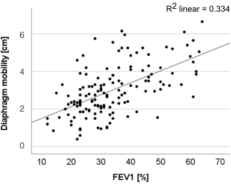
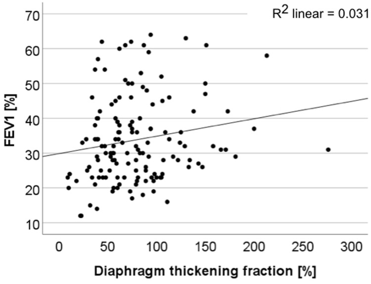
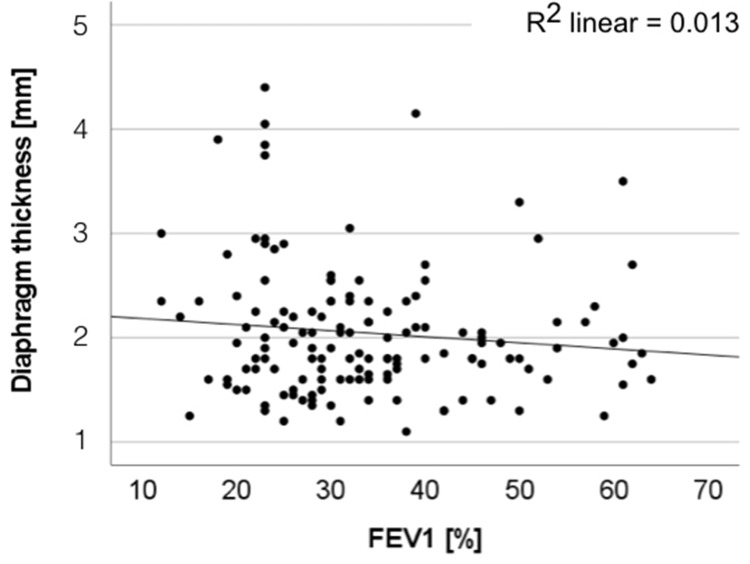
The mean FEV1 was 33.8%. Out of 140 total patients, 59 patients had an FEV1 <30%, 62 patients had an FEV1 30% to <50%, and 19 patients had an FEV1 50% to <80%. There was a significant correlation between FEV1 and diaphragm mobility (P < 0.01; Figure 1), as well as between FEV1 and diaphragm thickening fraction (P = 0.041; Figure 2) In contrast, no significant correlation was found between FEV1 and diaphragm thickness (Figure 3).
Figure 1.
FEV1 and diaphragm mobility (measured sonographically) have a statistically significant and clinically relevant correlation (p<0.01).
Figure 2.
There is a significant correlation between diaphragm thickening fraction and FEV1 (P = 0.041).
Figure 3.
There is no correlation between diaphragm thickness and FEV1.
Correlation Between Diaphragm Mobility and Other Diaphragm Parameters
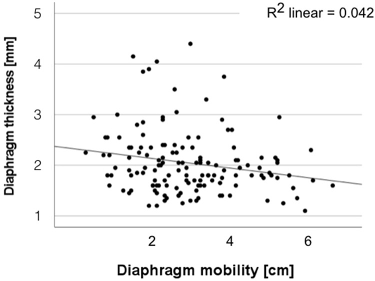
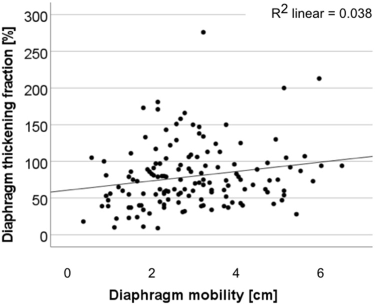
There was a significant negative correlation between diaphragm mobility and diaphragm thickness (P = 0.022; Figure 4). The higher the diaphragm mobility the lower the diaphragm thickness. In contrast, diaphragmatic mobility was significantly positively correlated with diaphragm thickening fraction (P = 0.013; Figure 5).
Figure 4.
There is a negative correlation between diaphragm mobility and diaphragm thickness. The higher the diaphragm mobility the lower the diaphragm thickness (P = 0.022).
Figure 5.
There is a significant positive correlation between diaphragm mobility and diaphragm thickening fraction (P = 0.013).
Correlation Between Diaphragm Mobility and Lung Function Parameters
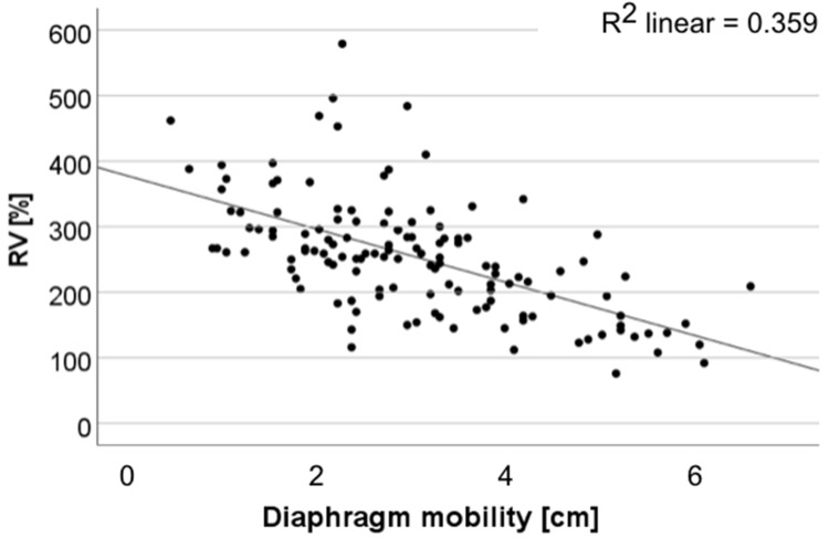
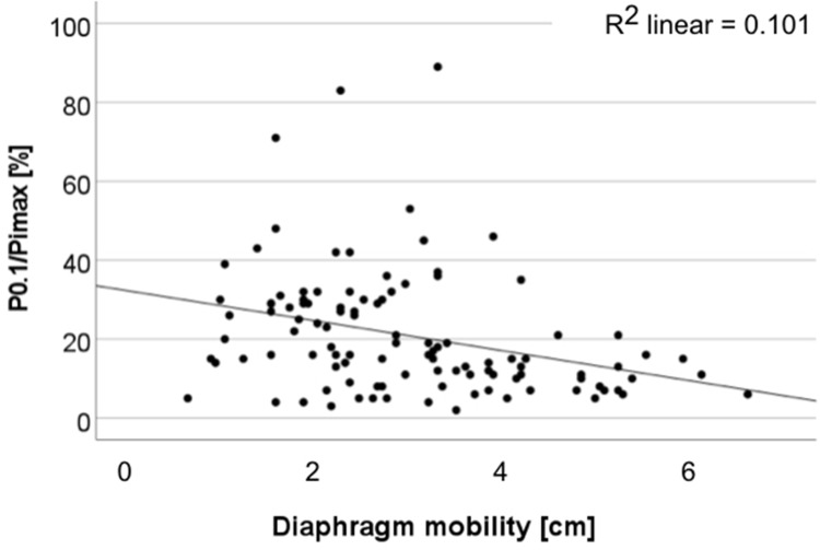
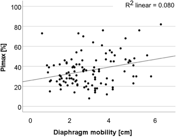
There was a significant negative correlation between diaphragm mobility and both residual volume (P < 0.01, r = −0.62; Figure 6) and respiratory capacity P0.1/Pimax (P < 0.01, r = −0.4; Figure 7). However, there was a significant and relevant positive correlation between diaphragm mobility and diaphragm maximal muscle strength (Pimax) (P < 0.017, r = 0.23; Figure 8).
Figure 6.
There is a statistically significant and highly clinically relevant negative correlation between diaphragm mobility and residual volume (P < 0.01, r = −0.62). A high residual volume reduces diaphragm mobility.
Figure 7.
There is a significant and relevant negative correlation between diaphragm mobility and respiratory capacity (P0.1/Pimax) (P < 0.01, r = −0.4).
Figure 8.
There is a statistically significant and relevant positive correlation between diaphragm mobility and diaphragm maximal muscle strength (Pimax) (P < 0.017, r = 0.23).
Correlation Between Diaphragm Thickness or Diaphragm Thickening Fraction and Lung Function
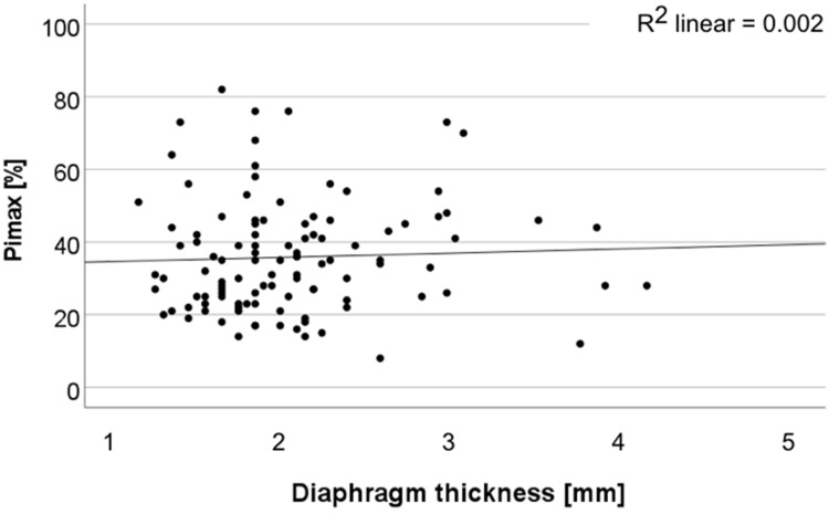
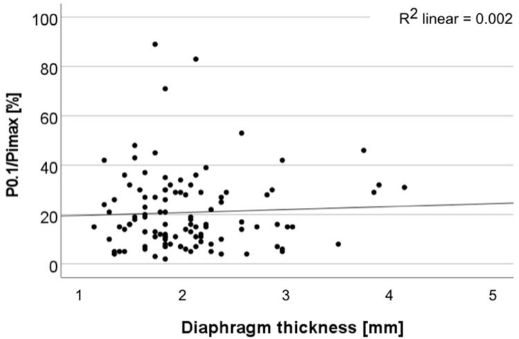
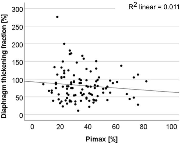
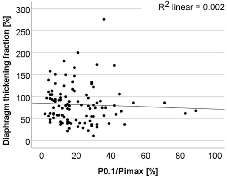
There was no correlation between diaphragm thickness and muscle strength (Pimax) or between diaphragm thickness and muscle (respiratory) capacity (P0.1/Pimax) (Figures 9 and 10). Similarly, there was no significant correlation between diaphragm thickening fraction and Pimax or P0.1/Pimax (Figures 11 and 12).
Figure 9.
Diaphragm thickness does not correlate with diaphragm maximal muscle strength (Pimax).
Figure 10.
Diaphragm thickness does not correlate with respiratory capacity (P0.1/Pimax).
Figure 11.
Correlation between diaphragm thickening fraction and diaphragm maximal muscle strength (Pimax) is not significant.
Figure 12.
Correlation between diaphragm thickening fraction and respiratory capacity (P0.1/Pimax) is not significant.
Discussion
Our results confirm the close correlation between sonographically measured diaphragm mobility and FEV1 in patients with COPD that we had already found in a prior study.10
In contrast to what we had expected, our current results found no correlation between diaphragm thickness and FEV1. This suggests that the dimension of the most important ventilatory muscle, as represented by diaphragm thickness, is not important in terms of FEV1, which is the most relevant lung function parameter for COPD. This observation corresponds with computed tomography (CT) data12 which show that the volume of the diaphragm does not correlate with COPD severity but the density of the diaphragm does. A lower diaphragm density correlates with a lower FEV1; the lower density reflects the substitution of muscle fibers by adipose tissue.12 Another small study also found no correlation between diaphragm thickness and severity of COPD.13 These results may be explained by the decrease in size of cross-sectional fibers as they are replaced by edema and adipose tissue as COPD increases in severity; this finding has been demonstrated not only via CT but also histopathologically.3,4 A case report has also addressed this observation; a patient admitted to the hospital with an exacerbation of COPD had a normal diaphragm muscle thickness and diaphragm thickening fraction but severely decreased diaphragm mobility as measured by ultrasound.14 In this case, the authors` attributed their findings to the high degree of lung hyperinflation present during an acute exacerbation of COPD, which decreases the available space for diaphragm movement, even with a well-preserved diaphragm strength.
COPD is defined by irreversible bronchial obstruction and is frequently associated with lung emphysema, which reduces the elastic recoil forces of the lung. The resulting limitation of expiratory flow leads to air trapping that increases during exacerbations and during exercising when flow limitation increases because of an increase in breathing frequency and reduced expiration time. This dynamic hyperinflation causes a flattening of the diaphragm resulting in a downward positioned diaphragmatic dome. The diaphragm of patients with severe emphysema is flattened even without additional exercise or exacerbations; therefore, the space for further downward movement of the diaphragm during inspiration is reduced even if the diaphragm muscle strength is fully maintained. If this is correct, the degree of hyperinflation as represented by the residual volume (RV) should correlate with a decrease in diaphragm mobility. We have shown this correlation in this study, where we found a significant and highly negative correlation between diaphragm mobility and residual volume; the higher the residual volume the lower the diaphragm mobility. We had demonstrated similar results in a study with a different patient cohort where we had shown that endobronchial valve implantation and the consequent atelectasis of the targeted lobe resulted in improvement not only of FEV1 and RV but also of diaphragm mobility.15 This positive outcome correlated with the achievement of atelectasis and therefore an increase in the space available for diaphragmatic downward movement.
The diaphragm of patients with COPD has a high workload because the work of breathing is increased. Therefore, we expected an increased diaphragmatic thickness to be an indicator of increased muscle strength in patients with severe COPD. It seemed logical that the higher workload would result in an increase of muscle mass and diaphragmatic diameter as occurs in other striated muscles secondary to a training effect, at least up to a certain point when the muscle becomes exhausted from overuse. If the training effect occurred in the diaphragm muscle, then maximal muscle strength measurement in lung function (Pimax) should correlate with an increased diaphragm thickness measured by ultrasound. However, we did not find a correlation between diaphragm thickness and Pimax, just as we also did not find a correlation between diaphragm thickness and FEV1. The above-mentioned replacement of diaphragm muscle tissue by edema and adipose tissue in COPD might be the explanation.
In COPD, not only the absolute strength of the diaphragm is interesting but also its capacity, reflected by the part of the maximal force of the diaphragm, which is needed for a normal breath (P0.1/Pimax). A low P0.1/Pimax indicates a well-preserved ventilatory capacity. In patients with severe COPD, the muscle strength required for normal (tidal) breathing, reflected by P0.1 is higher, and the maximum force reflected by Pimax is almost always diminished. The result is an increase in P0.1/Pimax. Higher P0.1/Pimax values reflect lower ventilatory capacity, meaning ventilatory exhaustion. A low P0.1/Pimax should correlate with the diaphragm thickening fraction; thickening of a muscle should reflect its strength. We could not find a correlation between diaphragm thickening fraction and P0.1/Pimax; therefore, a well-preserved ventilatory capacity is not necessarily associated with a high diaphragm thickening fraction. On the other hand, a well-preserved ventilatory capacity, a low P0.1/Pimax in lung function, correlated significantly with diaphragm mobility. The lower the P0.1/Pimax, the higher the diaphragm mobility. Both measurements, the P0.1/Pimax in lung function and the sonographically measured diaphragm mobility, are measurements of muscle strength.
In patients suffering from intensive care unit-acquired weakness, diaphragm thickness decreases day by day during controlled mechanical ventilation.16 This is the expected consequence of a muscle without the training effect. On the other hand, an increase in muscle strength when complete ventilatory support can be reduced and a patient can breath by her/himself again is also to be expected. A recent study demonstrated that diaphragm thickness significantly decreased during controlled mechanical ventilation (1.84 ± 0.44 to 1.49 ± 0.37 mm; P < 0.001) and was partially restored during assisted ventilation (1.49 ± 0.37 to 1.75 ± 0.43 mm; P < 0.001).17 This theory of an increase in diaphragm thickness during work challenge or training does not always apply. After pulmonary rehabilitation, many outcome parameters including diaphragm mobility improved but diaphragm thickness did not.18 This again might be because the diaphragm muscle is already permanently working at a high workload, comparable to strong training, in all patients with COPD that are in need of rehabilitation; therefore, a further increase in muscle thickness is difficult to achieve.
Conclusion
To achieve normal healthy breathing, diaphragm muscle strength is necessary as well as space for the diaphragm to move downward. Diaphragm muscle strength and diaphragm mobility can be measured sonographically. Our results suggest that in patients with COPD the space of the lung to move downward reflected by measurement of diaphragm mobility seems to be more important at least regarding FEV1 as a surrogate than diaphragm strength reflected by diaphragm thickness and diaphragm thickening fraction.
Funding Statement
There was no financial support for this study.
Data Sharing Statement
All data relevant to the study can be acquired from the corresponding author by email.
Author Contributions
All authors made a significant contribution to the work reported, whether that is in the conception, study design, execution, acquisition of data, analysis and interpretation, or in all these areas; took part in drafting, revising or critically reviewing the article; gave final approval of the version to be published; have agreed on the journal to which the article has been submitted; and agree to be accountable for all aspects of the work.
Disclosure
None of the authors has competing interests or financial interests in the publication of this study.
References
- 1.Levine S, Nguyen T, Kaiser LR, et al. Human diaphragm remodeling associated with chronic obstructive pulmonary disease: clinical implications. Am J Respir Crit Care Med. 2003;168(6):706–713. doi: 10.1164/rccm.200209-1070OC [DOI] [PubMed] [Google Scholar]
- 2.Barreiro E, de la Puente B, Minguella J, et al. Oxidative stress and respiratory muscle dysfunction in severe chronic obstructive pulmonary disease. Am J Respir Crit Care Med. 2005;171(10):1116–1124. doi: 10.1164/rccm.200407-887OC [DOI] [PubMed] [Google Scholar]
- 3.Ottenheijm CA, Heunks LM, Sieck GC, et al. Diaphragm dysfunction in chronic obstructive pulmonary disease. Am J Respir Crit Care Med. 2005;172(2):200–205. doi: 10.1164/rccm.200502-262OC [DOI] [PMC free article] [PubMed] [Google Scholar]
- 4.Ottenheijm CA, Heunks LM, Li YP, et al. Activation of the ubiquitin- proteasome pathway in the diaphragm in chronic obstructive pulmonary disease. Am J Respir Crit Care Med. 2006;174(9):997–1002. doi: 10.1164/rccm.200605-721OC [DOI] [PMC free article] [PubMed] [Google Scholar]
- 5.Similowski T, Yan S, Gauthier AP, Macklem PT, Bellemare F. Contractile properties of the human diaphragm during chronic hyperinflation. N Engl J Med. 1991;325(13):917–923. doi: 10.1056/NEJM199109263251304 [DOI] [PubMed] [Google Scholar]
- 6.Laghi F, Tobin MJ. Disorders of the respiratory muscles. Am J Respir Crit Care Med. 2003;168(1):10–48. doi: 10.1164/rccm.2206020 [DOI] [PubMed] [Google Scholar]
- 7.Decramer M. Effects of hyperinflation on the respiratory muscles. Eur Respir J. 1989;2(4):299–302. [PubMed] [Google Scholar]
- 8.Tzelepis G, McCool FD, Leith DE, Hoppin FG. Increased lung volume limits endurance of inspiratory muscles. J Appl Physiol. 1988;64(5):1796–1802. doi: 10.1152/jappl.1988.64.5.1796 [DOI] [PubMed] [Google Scholar]
- 9.Vivier E, Mekontso Dessap A, Dimassi S, et al. Diaphragm ultrasonography to estimate the work of breathing during non-invasive ventilation. Intensive Care Med. 2012;38(5):796–803. doi: 10.1007/s00134-012-2547-7 [DOI] [PubMed] [Google Scholar]
- 10.Scheibe N, Sosnowski N, Pinkhasik A, Vonderbank S, Bastian A. Sonographic evaluation of diaphragmatic dysfunction in COPD patients. Int J Chron Obstruct Pulmon Dis. 2015;10:1925–1930. doi: 10.2147/COPD.S85659 [DOI] [PMC free article] [PubMed] [Google Scholar]
- 11.Fernfindez R, Cabrera J, Calaf N, Benito S. P O.1/PIMax: an index for assessing respiratory capacity in acute respiratory failure. Intensiv Care Med. 1990;16:175–179. doi: 10.1007/BF01724798 [DOI] [PubMed] [Google Scholar]
- 12.Donovan AA, Johnston G, Moore M, et al. Diaphragm morphology assessed by computed tomography in chronic obstructive pulmonary disease. Ann Am Thorac Soc. 2020. doi: 10.1513/AnnalsATS.202007-865OC [DOI] [PubMed] [Google Scholar]
- 13.Ogan N, Aydemir Y, EVrin T, et al. Diaphragmatic thickness in chronic obstructive lung disease and relationship with clinical severity parameters. Turkish J Med Sci. 2019;49(4):1073–1078. doi: 10.3906/sag-1901-164 [DOI] [PMC free article] [PubMed] [Google Scholar]
- 14.Kracht J, Ogna A, Fayssoil A. Dissociation between reduced diaphragm inspiratory motion and normal diaphragm thickening in acute chronic pulmonary obstructive disease exacerbation: a case report. Medicine. 2020;99(10):e19390. doi: 10.1097/MD.0000000000019390 [DOI] [PMC free article] [PubMed] [Google Scholar]
- 15.Boyko M, Vonderbank S, Gürleyen H, et al. Endoscopic lung volume reduction results in improvement of diaphragm mobility as measured by sonography. Int J Chron Obstruct Pulmon Dis. 2020;15:1465–1470. doi: 10.2147/COPD.S247526 [DOI] [PMC free article] [PubMed] [Google Scholar]
- 16.Jaber S, Petrof BJ, Jung B, et al. Rapidly progressive diaphragmatic weakness and injury during mechanical ventilation in humans. Am J Respir Crit Care Med. 2011;183(3):364–371. doi: 10.1164/rccm.201004-0670OC [DOI] [PubMed] [Google Scholar]
- 17.Grassi A, Ferlicca D, Lupieri E, et al. Assisted mechanical ventilation promotes recovery of diaphragmatic thickness in critically ill patients: a prospective observational study. Crit Care. 2020;24:85. doi: 10.1186/s13054-020-2761-6 [DOI] [PMC free article] [PubMed] [Google Scholar]
- 18.Crimi C, Heffler E, Augelletti T, et al. Utility of ultrasound assessment of diaphragmatic function before and after pulmonary rehabilitation in COPD patients. Int J Chron Obstruct Pulmon Dis. 2018;13:3131–3139. doi: 10.2147/COPD.S171134 [DOI] [PMC free article] [PubMed] [Google Scholar]


