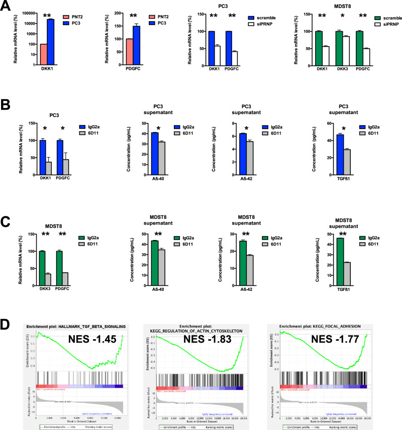
Fig. 3. Identification of potential Aβ-PrPC targets and impact of antibody-mediated blockade of the Aβ-PrPC interaction.
A qRT-PCR analysis of the relative expression of DKK1 and PDGFC in PC3 prostate cancer cells versus PNT2 normal prostate cells (left panels) as well as in PRNP-silenced versus control PC3 cells (middle panel) and qRT-PCR analysis of the relative expression of DKK1, DKK3 and PDGFC in PRNP-silenced versus control MDST8 cells (right panel). B DKK1 and PDGFC mRNA levels and extracellular Aβ40, Aβ42 and TGFβ levels were measured in cell extracts and supernatants of PC3 prostate cancer cells exposed to 6D11 antibodies versus control isotype antibodies. C DKK3 and PDGFC mRNA levels and extracellular Aβ40, Aβ42 and TGFβ levels were measured in cell extracts and supernatants of MDST8 colon cancer cells exposed to 6D11 antibodies versus control isotype antibodies. Results are expressed as means of n = 2 independent triplicates of cell preparations (except for A and B n = 2 independent duplicates of cell preparations) ± s.e.m. *p < 0.05, ***p < 0.001 vs. control (Mann-Whitney test). D GSEA analysis showing the downregulation of the TGFβ signalling, regulation of actin cytoskeleton and focal adhesion signatures in 6D11-treated versus control MDST8 cells. NES normalized enrichment score.

