Abstract
Backgrounds
Manual compression (MC) and vascular closure device (VCD) are two methods of vascular access site hemostasis after cardiac interventional procedures. However, there is still controversial over the use of them and a lack of comprehensive and systematic meta-analysis on this issue.
Methods
Original articles comparing VCD and MC in cardiac interventional procedures were searched in PubMed, EMbase, Cochrane Library, and Web of Science through April 2022. Efficacy, safety, patient satisfaction, and other parameters were assessed between two groups. Heterogeneity among studies was evaluated by I2 index and the Cochran Q test, respectively. Publication bias was assessed using the funnel plot and Egger's test.
Results
A total of 32 studies were included after screening with inclusion and exclusion criteria (33481 patients). This meta-analysis found that VCD resulted in shorter time to hemostasis, ambulation, and discharge (p < 0.00001). In terms of vascular complication risks, VCD group might be associated with a lower risk of major complications (p = 0.0001), but the analysis limited to randomized controlled trials did not support this result (p = 0.68). There was no significant difference in total complication rates (p = 0.08) and bleeding-related complication rates (p = 0.05) between the two groups. Patient satisfaction was higher in VCD group (p = 0.002). Meta-regression analysis revealed no specific covariate as an influencing factor for above results (p > 0.05).
Conclusions
Compared with MC, the use of VCDs significantly shortens the time of hemostasis and allows earlier ambulation and discharge, meanwhile without increase in vascular complications. In addition, use of VCDs achieves higher patient satisfaction and leads cost savings for patients and institutions.
1. Introduction
Invasive cardiac examinations and interventional procedures have become the important diagnostic and therapeutic means of cardiovascular diseases [1, 2]. More than 7 million invasive cardiac procedures are performed worldwide each year [3], and with a growing trend year by year. The modified Seldinger technique has become the standard technique to vascular puncture and sheath insertion in cardiac interventional procedures [4], but postoperative hemostasis, prolonged bed rest, and vascular-related complications remain clinical problems to be improved [5–8]. The radial approach is the preferred way of percutaneous coronary intervention (PCI) recommended by guidelines [9], which improves postoperative discomfort and complications to a certain extent. However, there are still a large number of interventional procedures requiring femoral approach, including structural cardiac intervention, catheter ablation (CA), and some PCIs under special circumstances. Effective and safe hemostasis techniques are essential to reduce the patient discomfort and the burden of complications.
Manual compression (MC) remains the current gold standard to achieve closure of percutaneous angiotomy site. However, it can be time-consuming and requires intensive compression by operator; even prolonged bed rest upon completion is required [10]. For patients, the most uncomfortable process is often not the procedure itself but the long bed rest afterwards. Therefore, vascular closure devices (VCDs) were created more than 20 years ago as an alternative to MC and have been increasingly utilized for angiotomy site closure and postoperative hemostasis. On the one side, VCDs have been reported to significantly shorten the time to hemostasis (TTH) and enable patients to ambulate at an early stage [11–13]. On the flip side, published studies have conflicting results on placement success rate and vascular complications of VCDs [14–17].
A variety of VCDs are currently available in clinical practice and can be categorized into two main groups based on closure mechanism: passive approximators, which deploy a plug, sealant, or procoagulant gel to the angiotomy site without physically occluding the angiotomy (e.g., AngioSeal, FemoSeal, Vascade, ExoSeal, SiteSeal, Celt ACD, and MynxGrip) and active approximators that physically close the angiotomy site with a suture, staple or clip (e.g., Perclose ProGlide, ProStar, and Starclose) [18, 19], indicating that the technology has changed dramatically over the past 20 years. Meta-analysis of VCDs was available as decade ago [14], but current techniques and materials have changed, and it is necessary to reevaluate the advantage of VCD and MC in clinical practice. We conducted a new systematic review and meta-analysis to analyze this issue comprehensively from multiaspect including efficacy, safety, success rates, patient satisfaction, and economic benefits.
2. Methods
2.1. Data Sources and Search Strategies
This systematic review and meta-analysis was performed referring to established methods [20]. Databases including PubMed, EMbase, Cochrane Library, and Web of Science were independently searched by two reviewers (N.P and J.G) through April 2022. Predefined search terms included “vascular closure device,” “manual compression,” “cardiovascular interventional procedure,” “cardiac intervention,” “invasive cardiac procedure,” and “cardiac catheterization” with no language restriction. Additional studies were searched from reviewing review articles and references of relevant researches manually. Any discrepancies were arbitrated by the third reviewer (R.W).
2.2. Inclusion and Exclusion Criteria
Inclusion criteria were applied as follows: (a) randomized-controlled trials (RCTs), observational studies, and propensity-score matched studies were included; (b) compared VCD with MC in cardiac interventional procedures; (c) contained hemostasis time parameters (efficacy) or vascular complications (safety) such as TTH, time to ambulation (TTA), access site related bleeding, hematoma, pseudoaneurysm, arteriovenous fistula, etc.; and (d) had complete and accurate outcome data. Review, case report, editorial, letter, animal study, and single cohort study were excluded. Studies were not restricted by race, sex, age, or country where the studies were conducted.
2.3. Data Extraction and Quality Assessment
Relevant information was obtained from the original articles and raw data files of all eligible studies and entered into a predetermined spreadsheet as follows: (a) study information (first author's name, publication year, country where the study was conducted, type of study design, operation type, sample size, VCD type, and vascular access site); (b) participant characteristics (mean age, male gender, race, and underlying disease); and (c) outcome indicators: efficacy and safety of hemostasis (TTH, TTA, time to discharge (TTD), time to discharge eligibility (TTDE), same-day discharges, hemostasis success rates, vascular complications, and patient-reported outcomes). The Cochrane Collaboration recommending tool was used for quality assessments of RCTs [21]. Non-RCTs were assessed using the Newcastle-Ottawa Scale (NOS), with scores varying from 0 to 9 depending on the quality of studies, and papers were considered high quality if they scored 7 or higher. Two reviewers preformed data extraction and quality assessment independently (N.P and J.G). Any disagreements were adjudicated by the third reviewer (R.W).
2.4. Statistical Analysis
Review Manager (RevMan, version 5.3) and Stata (version 12.0) were used for statistical calculations in this meta-analysis. Data of RCT studies and non-RCT were merged and analyzed separately. Statistical significance was set as p value of less than 0.05. Data of continuous variables represented by median and interquartile range (or max-and-min) were converted to mean and standard deviation to perform statistical analysis and data synthesis [22, 23]. Heterogeneity was assessed by calculating I2 and Cochran Q test, with I2 value more than 50% or p value of the Q test less than 0.1 was considered evidence of significant inconsistency [24, 25]. If heterogeneity was present, sensitivity analysis was conducted to inspect the effect of a single study on the overall risk estimate by omitting one study at a time. Meta-regression analysis was also performed to examine the sources of differences among studies. If a particular covariate had a significant effect on heterogeneity, further subgroup analysis was performed. We generated funnel plot to assess potential publication bias, and the asymmetry of the plot was evaluated by Egger's test, with p value of less than 0.05 indicating apparent asymmetry. Trim-and-fill analysis was used to estimate the effect of publication bias on the interpretation of the results [26].
2.5. Related Terms and Definitions
Due to the large number of included studies, some outcome indicators had different names or vague expressions, so we redefined the terms of important indicators and classified them consistently. TTH was defined as the time from the onset of VCD deployment or compression to complete cessation of bleeding. TTA was defined as the time from the end of procedure or leaving the cardiac catheterization laboratory to mobilization. TTD was defined as the time from the beginning of TTA to hospital discharge. Major vascular complication was defined as adverse event related to vascular puncture and closure that may cause serious consequence, require therapy, or prolong hospitalization, including large groin hematoma (usually larger than 5 cm), major bleeding that compromises hemodynamics or requiring blood transfusion, access site-related infection requiring intravenous antibiotics, retroperitoneal bleeding, and pseudoaneurysm requiring surgical repair. Minor vascular complication was defined as adverse event related to puncture and closure blood vessel that may resolve spontaneously or require no human intervention, such as small hematoma, persistent pain at vascular access site, slight bleeding of access site requiring no recompression, transient access site-related nerve injury, and pseudoaneurysm requiring no therapy. Bleeding-related complication was defined as access site bleeding, groin hematoma, and retroperitoneal bleeding. Injury-related complication was defined as tissue damage around the vascular access site, including pseudoaneurysm, arteriovenous fistula, infection, nerve injury, and pain. Related terms were used according to the definitions of previous clinical trials [27].
3. Results
3.1. Search Results
A total of 1175 studies were initially identified through database search (1169 records) and additional manual search (6 records). After removing 462 duplicate studies, step by step screening was performed based on inclusion and exclusion criteria. Eventually, 32 studies comprising 12 RCTs [28–39], 17 observational studies [40–55], and 3 propensity-score matched studies [56–58] were included in this meta-analysis. Figure 1 shows the flowchart of inclusions and exclusions.
Figure 1.
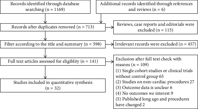
Flow diagram for study identification and inclusion.
3.2. Study Characteristics
The included studies comprised 34381 patients and were conducted in centers across the United States, Germany, China, Denmark, France, Canada, Italy, and India. The mean age of the entire cohort was 64.6 years, and participants were predominantly male (63.0%). Regarding the type of procedure, most studies were coronary angiography (CAG) and PCIs; the rest were structural cardiac procedures, CA, cardiac catheterization, etc. Regarding vascular access site, 26 studies performed procedures via femoral arteries, 4 studies via femoral veins, and 2 studies via brachial arteries. There were passive and active approximators involving 11 product types about the VCD types. The detailed characteristics of all included studies are showed in Table 1.
Table 1.
Summary of included studies.
| Study | Publication year | Research country | Study type | Operation type | Access site | VCD type | Sample size | Age (mean ± SD) | Male gender n (%) |
|---|---|---|---|---|---|---|---|---|---|
| Ben-Dor [28] | 2018 | USA | RCT | PCI/CAG | Femoral vein | MynxGrip | 208 | 72.5 ± 14.2 | 117 (56.3) |
| Ben-Dor [40] | 2011 | USA | Retrospective study | BAV | Femoral artery | AngioSeal/Perclose/Prostar | 333 | 81.8 ± 9.3 | 146 (43.8) |
| Bhat [41] | 2021 | India | Retrospective study | PCI | Femoral artery | Perclose | 1743 | 52.1 ± 11.2 | 1097 (62.9) |
| Christ [42] | 2015 | Germany | Retrospective study | PCI/CAG | Femoral artery | AngioSeal | 76 | 64.2 ± 12.8 | 46 (60.5) |
| De Poli [43] | 2014 | France | Retrospective study | PCI/CAG | Femoral artery | FemoSeal | 211 | 63.2 ± 12.2 | 76 (76.0) |
| De Poli past [43] | 2014 | France | Retrospective study | PCI/CAG | Femoral artery | Unknown | 3826 | Unknown | Unknown |
| Hermiller [29] | 2005 | USA | RCT | CAG | Femoral artery | Starclose | 208 | 61.7 ± 11.8 | 139 (66.8) |
| Hermiller [30] | 2006 | USA | RCT | PCI | Femoral artery | Starclose | 275 | 62.8 ± 9.9 | 221 (80.4) |
| Hermiller [31] | 2015 | USA | RCT | CC | Femoral artery | Vascade | 420 | 62.0 ± 10.9 | 298 (71.0) |
| Holm [32] | 2014 | Denmark | RCT | CAG | Femoral artery | FemoSeal | 1001 | 64.8 ± 11.0 | 621 (62.0) |
| Iqtidar [56] | 2011 | USA | Propensity match | PCI | Femoral artery | AngioSeal/Starclose/Perclose | 4221 | 65.4 ± 12.5 | 2076 (64.1) |
| Jakobsen [33] | 2022 | Denmark | RCT | CAG | Femoral artery | MynxGrip | 865 | 66.0 ± 11.0 | 570 (65.9) |
| Junquera [57] | 2021 | Canada | Propensity match | TAVR | Femoral artery | AngioSeal/Perclose | 4031 | 80.8 ± 7.8 | 1921 (47.7) |
| Kuno [58] | 2021 | USA | Propensity match | PCI | Femoral artery | AngioSeal/Perclose | 694 | 66.7 ± 9.7 | 529 (76.2) |
| Leclercq [44] | 2015 | France | Prospective study | BAV | Femoral artery | AngioSeal | 180 | 83.8 ± 6.8 | 84 (46.7) |
| Lupi [45] | 2012 | Italy | Retrospective study | PCI/CAG | Femoral artery | AngioSeal | 1913 | Unknown | Unknown |
| Mirza [46] | 2014 | USA | Retrospective study | CC | Brachial artery | Starclose | 148 | 69.5 ± 8.6 | 79 (53.4) |
| Mohammed [47] | 2021 | USA | Prospective study | CA | Femoral vein | Perclose | 231 | 64.9 ± 10.7 | 145 (62.8) |
| Mohanty [48] | 2019 | USA | Retrospective study | CA/LAAC | Femoral vein | Vascade | 803 | 66.1 ± 10.2 | 538 (70.0) |
| Natale [34] | 2020 | USA | RCT | CA | Femoral vein | Vascade | 204 | 62.5 ± 11.3 | 131 (64.2) |
| O'Neill [49] | 2013 | USA | Retrospective study | BAV | Femoral artery | Perclose | 428 | 83.7 ± 8.9 | 194 (45.3) |
| Owens [50] | 2017 | USA | Retrospective study | CC | Femoral artery | Cardiva catalyst II | 1470 | 63.9 ± 9.7 | 1419 (96.5) |
| Pieper [51] | 2016 | Germany | Prospective study | CC | Femoral artery | ExoSeal | 48 | 62.5 ± 12.6 | 29 (60.4) |
| Schulz-Schüpke [ 35] | 2014 | Germany | RCT | CAG | Femoral artery | FemoSeal/ExoSeal | 4524 | 67.0 ± 11.8 | 3129 (69.2) |
| Sekhar [52] | 2016 | USA | Prospective study | CC | Femoral artery | Perclose | 170 | 59.5 ± 11.0 | 149 (87.6) |
| Sharma [36] | 2020 | USA | RCT | CC | Femoral artery | SiteSeal | 39 | 60.5 ± 9.5 | 23 (59.0) |
| Stegemann [53] | 2011 | Germany | Retrospective study | PCI/CAG | Femoral artery | AngioSeal | 4653 | 65.0 ± 11.6 | 3233 (69.5) |
| Su [54] | 2018 | China | Retrospective study | PCI | Femoral artery | AngioSeal | 73 | 66.8 ± 12.1 | 52 (71.2) |
| Wei [55] | 2020 | China | Retrospective study | TBAD | Brachial artery | ExoSeal | 157 | 57.8 ± 13.1 | 124 (79.0) |
| Wong [37] | 2017 | USA | RCT | PCI | Femoral artery | Celt ACD | 207 | 67.0 ± 11.0 | 159 (76.8) |
| Wong [38] | 2009 | USA | RCT | PCI/CAG | Femoral artery | ExoSeal | 401 | 62.7 ± 10.9 | 265 (66.1) |
| Yeni [39] | 2016 | Germany | RCT | PCI | Femoral artery | AngioSeal/Starclose | 620 | 65.7 ± 11.1 | 444 (71.6) |
VCD = vascular closure device; USA = the United States of America; RCT = randomized controlled trial; PCI = percutaneous coronary intervention; CAG = coronary angiography; BAV = balloon aortic valvuloplasty; CC = cardiac catheterization; TAVR = transcatheter aortic valve replacement; CA = catheter ablation; LAAC = left atrial appendage closure; TBAD = type B aortic dissection.
3.3. Quality Assessment
All included studies were classified as high quality according to the Cochrane Collaboration criteria or NOS. Figure 2 and Supplementary Figure 1 show the details of quality assessment for RCTs, and results of assessment for non-RCTs is shown in Table 2.
Figure 2.
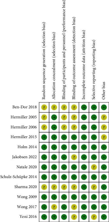
Risk of bias summary of included RCTs in the meta-analysis.
Table 2.
Quality assessment of non-RCTs.
| Study | Publication year | NOS score |
|---|---|---|
| Ben-Dor [40] | 2021 | 8 |
| Bhat [41] | 2021 | 9 |
| Christ [42] | 2015 | 8 |
| De Poli [43] | 2014 | 9 |
| De Poli past [43] | 2014 | 7 |
| Iqtidar [56] | 2011 | 8 |
| Junquera [57] | 2021 | 7 |
| Kuno [58] | 2021 | 9 |
| Leclercq [44] | 2015 | 8 |
| Lupi [45] | 2012 | 7 |
| Mirza [46] | 2014 | 8 |
| Mohammed [47] | 2021 | 9 |
| Mohanty [48] | 2019 | 7 |
| O'Neill [49] | 2013 | 8 |
| Owens [50] | 2017 | 8 |
| Pieper [51] | 2016 | 8 |
| Sekhar [52] | 2016 | 8 |
| Stegemann [53] | 2011 | 7 |
| Su [54] | 2018 | 8 |
| Wei [55] | 2020 | 8 |
RCT = randomized controlled trial; NOS = Newcastle-Ottawa scale.
3.4. Hemostasis Time Parameters
The main included clinical outcomes of hemostasis time parameters contained TTH, TTA, and TTD, and there were obvious differences in results between two groups and among studies. Notably, in terms of TTD, due to some confounding factors (e.g., delayed discharge formalities, additional examination, or consultation due to other indisposition) in included studies, TTD might not accurately reflect the efficacy of hemostasis. Therefore, the concept of time to discharge eligibility (TTDE) was introduced to reduce the error and incorporated in the subsequent quantitative synthesis on TTD.
15 studies reported the TTH, which in VCD group was significantly shorter than that in MC group (SMD: − 4.44, random-effect model, 95% CI, − 5.67 to − 3.21, p < 0.00001; Figure 3(a)) with high heterogeneity across studies (I2 = 100%, p < 0.00001 of Q test). 9 studies reported parameters of TTA. Similar to TTH, the result of pooled analysis suggested that use of VCD had a shorter TTA than MC (SMD: − 2.93, random-effect model, 95% CI, − 3.79 to − 2.06, p < 0.00001; Figure 3(b)) with high heterogeneity (I2 = 99%, p < 0.00001 of Q test). 9 studies provided related data of TTD. Data synthesis showed that VCD group had a significantly shorter length of stay (SMD: − 1.47, random-effect model, 95% CI, − 1.99 to − 0.95, p < 0.00001; Figure 3(c)) with high heterogeneity (I2 = 99%, p < 0.00001 of Q test). Results of RCT subgroup and non-RCT subgroup were consistent on statistical significance.
Figure 3.
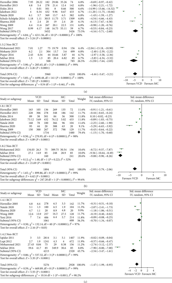
Forest plots comparing (a) TTH, (b) TTA, and (c) TTD between the VCD group and MC group.
Sensitivity analysis excluding one study at a time did not find any single study significantly affecting above results and overall heterogeneity. Heterogeneity was further explored in subsequent meta-regression analysis, as described in 3.9.
No significant publication biases of TTH and TTD were observed in funnel plots and Egger's tests (Figures 4(a) and 4(c)). However, significant publication bias of TTA was revealed by funnel plot and Egger's test (p = 0.003, Figure 4(b)). The trim-and-fill computation was further performed to estimate the effect of publication bias on the interpretation of results. After two iterations of linear estimation and incorporating possible missing studies into the meta-analysis, the results showed no trimming was required, indicating that the impact of publication bias on the results was within an acceptable range and the result of pooled analysis was robust (Supplementary Figure 2).
Figure 4.
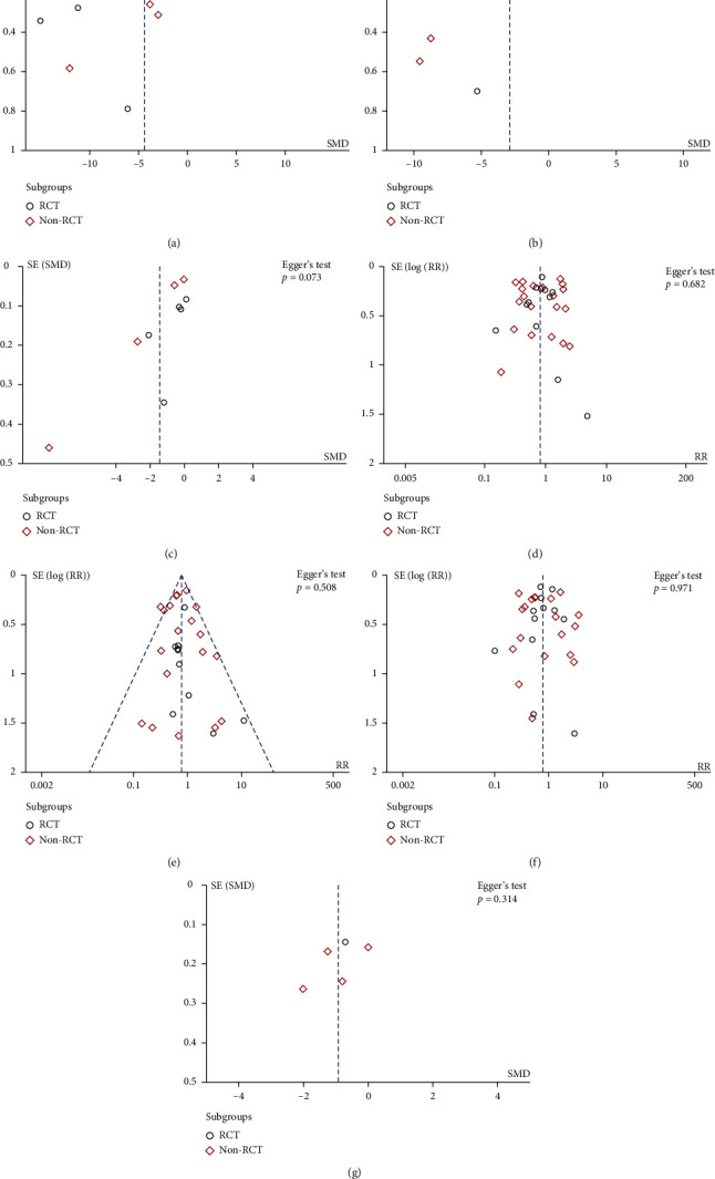
Funnel plots and Egger's test were used to assess publication bias of (a) TTH, (b) TTA, (c) TTD, (d) total vascular complication rate, (e) major vascular complication rate, (f) bleeding-related complication rate, and (g) patient-reported outcome.
3.5. Vascular-Related Complications
3.5.1. Total Complications
All 32 studies reported vascular-related complications of cardiac interventional procedures. Of these, 13 studies favored MC, whereas 19 studies suggested that VCD could reduce complication rates. The results of quantitative synthesis showed similar total complication risks between the two methods (5.5% in VCD group and 6.0% in MC group), with no statistical significance (RR: 0.81, random-effect model, 95% CI, 0.63 to 1.02, p = 0.08; Figure 5(a)). And heterogeneity between studies was high (I2 = 83%, p < 0.00001 of Q test). Results were consistent in the RCT (p = 0.07) and non-RCT groups (p = 0.28).
Figure 5.
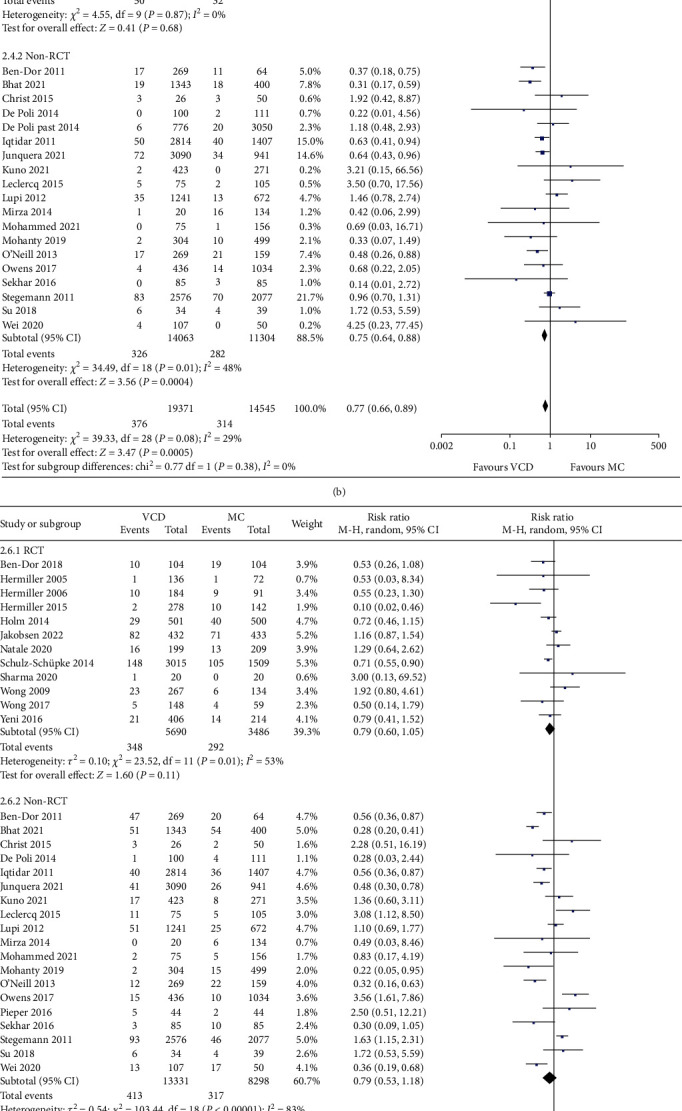
Forest plots comparing the (a) total vascular complications, (b) major vascular complications, (c) bleeding-related complications, and (d) bleeding-related complications omitting device failures between the VCD group and MC group.
3.5.2. Major Vascular Complications
A total of 29 studies reported major vascular complications. There was no serious complication occurred in the remaining 3 studies due to the small sample sizes. The major vascular complication rate was about 1.9% of VCD group and about 2.2% of MC group according to the quantitative synthesis. The difference reached statistical significance (RR: 0.77, fixed-effect model, 95% CI, 0.66 to 0.89, p = 0.0005; Figure 5(b)), with low degree of heterogeneity (I2 = 15%, p = 0.24 of Q test). However, results were significantly different between RCTs and non-RCTs. The result of the RCT subgroup showed no significant difference between VCD and MC in terms of major vascular complications (p = 0.68), whereas the non-RCT subgroup supported the evidence that VCD effectively reduced major vascular complications (p = 0.0004). The most common type of major complication in both two groups (VCD and MC) was major bleeding (41.8%), followed by large hematoma (20.4%) and pseudoaneurysm (17.0%).
3.5.3. Bleeding-Related Complications
Bleeding-related complications may effectively reflect the efficacy of postoperative hemostasis maintenance. A total of 28 studies provided relevant data. Similar to the result of total complications, bleeding-related complication rates were found to be lower with use of VCD compared with MC in cardiac interventional procedures, but did not reach statistical significance (RR: 0.77, random-effect model, 95% CI, 0.60 to 1.00, p = 0.05; Figure 5(c)). I2 was 77%, meaning a high degree of heterogeneity. However, when hemorrhagic complications caused by device failures in VCD group were removed, the result changed to favor of VCD group and reached statistical significance (RR: 0.53, random-effect model, 95% CI, 0.38 to 0.73, p = 0.0001; Figure 5(d)). Consistent results were observed in RCT subgroup (p < 0.00001) and non-RCT subgroup (p = 0.04), suggesting that VCDs could significantly improve hemostasis effects after successful device placements.
3.5.4. Sensitivity Analysis and Publication Bias
For above results, sensitivity analyses removing one study at a time did not find significant changes on overall effect test (p value) and heterogeneity (I2). No significant publication biases were detected by funnel plots and Egger's tests (Figures 4(d), 4(e), and 4(f)).
3.6. Patient-Reported Outcomes
A total of eight studies paid additional attention to the subjective feelings of patients. Participants received questionnaires after ambulation or before discharge that comprised several items: back pain and groin pain during bed rest, discomfort in diet, urination, and defecation during bed rest, walking discomfort after ambulation, satisfaction with closure process, as well as overall satisfaction. Five of the studies quantitatively compared differences between two groups using rating scales. Because of differences in scoring rules, the data were transformed and pooled; the final results showed that patients who received VCD had higher satisfaction and less pain after procedures than who received MC (SMD: − 0.93, random-effect model, 95% CI, − 1.53 to − 0.34, p = 0.002; Figure 6). No significant publication bias was observed (p = 0.314, Figure 4(g)). Respective analysis of RCTs and non-RCTs had the consistent result. Of the three studies not included in quantitative synthesis, one observed a significant reduction in the proportion of back pain caused by prolonged bed rest in VCD group (24.3% vs 47.9%), and the other two studies showed the slight advantage of VCDs.
Figure 6.
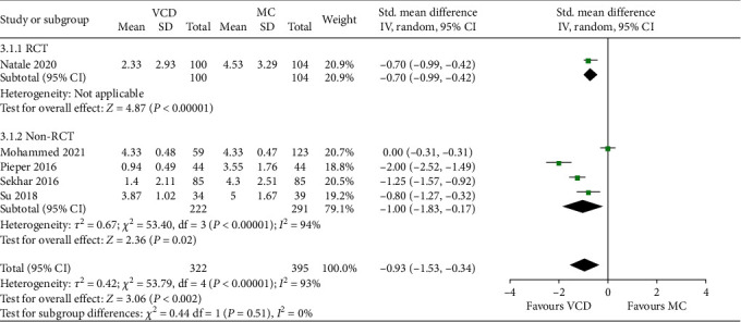
Forest plots comparing the patient-reported outcomes between the VCD group and MC group.
3.7. Device Failure Rates
For device failure rates of only VCD group, a total of 24 studies reported primary data, whereas the remaining studies were retrospective or propensity matching and did not report failures in original papers. Synthetic results showed that device failed at 278 of 8940 access sites for a total of 8677 participants, with a failure rate of approximately 3.1%. When device failed, either the inability to deploy the device or device deployment with inadequate hemostasis, it eventually required conversion to MC and increased the risk of bleeding-related complications.
3.8. Economic Benefits for Patients and Institutions
Two studies examined the costs of two closure strategies that involved passive approximator (Vascade) and active approximator (ProGlide). Both studies suggested that the use of VCDs resulted in significant cost savings for institutions and patients. Specifically, although patients had to pay for VCDs, the nursing expenses were saved due to fewer complications and shorter length of stay; meanwhile, the proportion of patients who required urinary catheter and pain medication after procedures was lower. Thus, population-level cost analysis revealed the advantages of VCDs. For example, one of the studies showed an average savings of $983.6 per patient undergoing cardiac catheterization using VCD.
3.9. Meta-Regression Analysis
There are some results of pooled analysis in this meta-analysis had high heterogeneity, but no significant change of heterogeneity could be observed by sensitivity analysis. Hence, meta-regression analyses were preformed to further search for the source of inconsistency between studies. Covariates included publication year, country where research was conducted, study design (RCT or observational study), operation type, VCD type (active or passive approximators), diagnosis or treatment, and vascular access site. The detailed results of the meta-regression analysis are presented in Table 3. Notably, only the analysis for total complications showed a decrease in τ square from 0.3168 to 0.2957, indicating that the above covariates could explain 6.7% of heterogeneity, whereas τ square of other indicators did not decrease. The final meta-regression results for all outcome indicators showed differences in included covariates were not the main factors affecting overall heterogeneity (p > 0.05).
Table 3.
Results of meta-regression analysis for outcome indicators.
| Variable | Slope coefficient | Standard error | Z value | p value | 95% CI | |
|---|---|---|---|---|---|---|
| Lower limit | Upper limit | |||||
| Total vascular complication | ||||||
| Publication year | − 0.0038882 | 0.0366959 | − 0.11 | 0.916 | − 0.0796248 | 0.0718485 |
| Research country | 0.2122738 | 0.1532003 | 1.39 | 0.179 | − 0.1039161 | 0.5284636 |
| Study design | 0.1899589 | 0.3187227 | 0.6 | 0.557 | − 0.4678523 | 0.8477701 |
| Operation type | − 0.1619395 | 0.170639 | − 0.95 | 0.352 | − 0.5141212 | 0.1902422 |
| VCD type | − 0.0748574 | 0.1508855 | − 0.5 | 0.624 | − 0.3862698 | 0.2365551 |
| Diagnosis or treatment | 0.0898802 | 0.2147782 | 0.42 | 0.679 | − 0.3534002 | 0.5331606 |
| Vascular access site | 0.0477251 | 0.2622489 | 0.18 | 0.857 | − 0.4935301 | 0.5889803 |
| Major vascular complication | ||||||
| Publication year | 0.0134347 | 0.0294913 | 0.46 | 0.653 | − 0.0478958 | 0.0747652 |
| Research country | 0.1285218 | 0.0682783 | 1.88 | 0.074 | − 0.0134707 | 0.2705144 |
| Study design | 0.0534072 | 0.1441656 | 0.37 | 0.715 | − 0.2464016 | 0.353216 |
| Operation type | − 0.2259866 | 0.1351589 | − 1.67 | 0.109 | − 0.5070649 | 0.0550916 |
| VCD type | 0.0388801 | 0.1573338 | 0.25 | 0.807 | − 0.2883134 | 0.3660736 |
| Diagnosis or treatment | 0.2951529 | 0.2025009 | 1.46 | 0.160 | − 0.1259707 | 0.7162765 |
| Vascular access site | − 0.1544536 | 0.2936646 | − 0.53 | 0.604 | − 0.7651627 | 0.4562555 |
| Bleeding-related complication | ||||||
| Publication year | 0.0071636 | 0.0433535 | 0.17 | 0.870 | − 0.0825199 | 0.0968471 |
| Research country | − 0.0502883 | 0.2334795 | − 0.22 | 0.831 | − 0.5332774 | 0.4327009 |
| Study design | 0.3204116 | 0.1969521 | 1.63 | 0.117 | − 0.087015 | 0.7278381 |
| Operation type | − 0.3396131 | 0.1908548 | − 1.78 | 0.088 | − 0.7344263 | 0.0552 |
| VCD type | − 0.2991253 | 0.2131044 | − 1.4 | 0.174 | − 0.7399653 | 0.1417147 |
| Diagnosis or treatment | 0.1150813 | 0.2458099 | 0.47 | 0.644 | − 0.3934151 | 0.6235778 |
| Vascular access site | − 0.2120466 | 0.290403 | − 0.73 | 0.473 | − 0.8127909 | 0.3886977 |
| TTH | ||||||
| Publication year | − 0.4921285 | 0.3435058 | − 1.43 | 0.195 | − 1.304391 | 0.3201336 |
| Research country | − 5.162946 | 3.383787 | − 1.53 | 0.171 | − 13.16433 | 2.83844 |
| Study design | 0.5445317 | 2.421016 | 0.22 | 0.828 | − 5.180262 | 6.269325 |
| Operation type | 1.148615 | 1.776338 | 0.65 | 0.538 | − 3.051757 | 5.348986 |
| VCD type | − 4.169431 | 3.268597 | − 1.28 | 0.243 | − 11.89843 | 3.559573 |
| Diagnosis or treatment | 1.495201 | 2.149798 | 0.7 | 0.509 | − 3.588264 | 6.578666 |
| Vascular access site | − 0.6847343 | 4.187115 | − 0.16 | 0.875 | − 10.58569 | 9.216219 |
| TTA ∗ | ||||||
| Publication year | − 0.3388484 | 0.2746872 | − 1.23 | 0.343 | − 1.520732 | 0.8430351 |
| Research country | − 1.871418 | 4.513303 | − 0.41 | 0.719 | − 21.2906 | 17.54776 |
| Study design | − 6.795713 | 1.759985 | − 3.86 | 0.061 | − 14.36832 | 0.7768918 |
| Operation type | − 2.313697 | 2.0215 | − 1.14 | 0.371 | − 11.01151 | 6.384116 |
| VCD type | − 6.795713 | 1.759985 | − 3.86 | 0.061 | − 14.36832 | 0.7768922 |
| Diagnosis or treatment | − 2.478133 | 1.655155 | − 1.5 | 0.273 | − 9.599688 | 4.643422 |
| Vascular access site | 2.048284 | 2.764677 | 0.74 | 0.536 | − 9.847159 | 13.94373 |
| TTD | ||||||
| Publication year | 0.0616289 | 0.6462428 | 0.1 | 0.939 | − 8.149665 | 8.272922 |
| Research country | − 0.3444556 | 3.792939 | − 0.09 | 0.942 | − 48.53832 | 47.84941 |
| Study design | − 0.1481234 | 1.674416 | − 0.09 | 0.944 | − 21.4236 | 21.12736 |
| Operation type | − 10.58069 | 11.3521 | − 0.93 | 0.522 | − 154.8228 | 133.6614 |
| VCD type | − 4.791256 | 5.455508 | − 0.88 | 0.541 | − 74.11005 | 64.52754 |
| Diagnosis or treatment | − 9.694279 | 11.71157 | − 0.83 | 0.560 | − 158.5038 | 139.1153 |
| Vascular access site | 1.529658 | 5.465324 | 0.28 | 0.826 | − 67.91387 | 70.97318 |
| Patient-reported outcome ∗∗ | ||||||
| Publication year | − 25.04569 | 23.21049 | − 1.08 | 0.476 | − 319.963 | 269.8716 |
| VCD type | − 38.13615 | 38.23929 | − 1 | 0.501 | − 524.0125 | 447.7402 |
| Vascular access site | − 172.319 | 97.29436 | − 1.77 | 0.327 | − 1408.561 | 1063.923 |
∗Stata suggested that colinearity between covariate study design and VCD type. ∗∗Some covariates were not included in this meta-regression analysis because sensitivity analysis had been performed.
4. Discussion
In this systematic review and meta-analysis, we comprehensively analyzed the performances of using VCDs versus conventional MC to close vascular access sites in cardiac interventional procedures. The main findings include the following: (1) VCDs significantly shorten the time of immediate hemostasis and postoperative bed rest, greatly increased the possibility of early discharge; (2) both showed similar results in terms of total vascular complications, but VCDs possibly reduced the risk of major complications and bleeding-related complications omitting device failures; (3) the use of VCDs increased patient satisfaction with the entire procedure; and (4) the use of VCDs contributes to cost saving for patients and hospitals.
The difference in hemostasis efficacy of the two methods is quite obvious. In most cases, complete hemostasis by VCDs takes only a few minutes, and fewer subsequent bleeding-related complications occur once the device success. Of course, VCDs have a certain failure rate, which is approximately 3.1% according to our analysis. Device failure rate has decreased with the development of technology and the operator experience, but it has not yet reached the desired perfection [41]. Another evidence of the hemostasis efficacy of VCDs is the reduction of TTA, which directly determines patient satisfaction. After successful hemostasis, conventional MC requires patients to remain on bed rest for 6-12 hours depending on the operation type [39, 50, 56]. According to previous studies, back pain, inconvenient diet, dysuria, and difficult defecation were the main causes of patient discomfort during this long period [51, 52]. Patients who received VCDs were allowed to early ambulate within 2 hours, thus avoiding these troubles. A problem with TTA is that Egger's test indicates a potential publication bias, although the bias demonstrated by the trim-and-full method does not affect the interpretation of the results. According to our analysis, the source of bias could be the study design of published papers, i.e., most of the included studies directly specify TTA in both groups without recording the actual situations.
Vascular complication is the focus of attention and the most controversial issue. Previous researches have suggested that VCDs may lead to an increase in femoral artery thrombosis, arteriovenous fistula, pseudoaneurysm, and other adverse events [29, 42, 43]. However, the results of this meta-analysis showed that VCDs did not cause additional injury and may even improve the severity of complications. Specifically, the distribution of vascular complication types was similar in both groups, whereas the major complications accounted for a relatively low proportion of the total complications in the VCD group, implying that VCDs are associated with reduced severity of adverse events, such as smaller hematoma and less groin bleeding. These minor complications are often self-healing without treatment. Our analysis confirms the safety and reliability of VCDs, and the robustness of these results is supported by the sensitivity analysis.
One notable point is that the analysis of major complications showed different results in RCTs and non-RCTs, with no significant difference between the two methods in the RCT subgroup, whereas the non-RCT subgroup favored VCD as reducing the risk of major complications. We carefully analyzed possible reasons that, first, non-RCTs might have unequal baseline patient characteristics due to study design limitations and, second, most included non-RCTs were not strictly double-blind, which might result in observer bias in assessing patients' complications. These reasons may contribute to the tendency of the results of non-RCTs to be positive. Therefore, from the perspective of evidence-based medicine, we cannot assume that VCD can reduce the risk of major complications.
Another interesting phenomenon is that, although VCDs were associated with lower bleeding-related complication rates according to this meta-analysis (4.0% vs. 5.2%), it did not reach statistical significance. One possible explanation is that VCDs indeed promote the efficiency of hemostasis, but the increased number of minor bleeding complications was driven by device failure [50]. We found evidence to support this explanation from the included studies, namely, that device failure increased the incidence of minor bleeding complications and partially offset the benefits of VCDs [35, 38, 41].
Regarding economic benefits, although there are no data that can be used for quantitative synthesis, all previous studies supported that VCDs can save costs. Notably, the cost analysis was based on the procedure success of VCDs, whereas patients would face more expensive costs once the device failed than MC. Therefore, it is important to improve device success rate and shorten the learning curve of operator in the future.
The high heterogeneity of multiple outcome indicators was observed in this meta-analysis, but neither sensitivity analysis omitting one study at a time nor meta-regression analysis found the source of inconsistency among studies. We considered that different proficiency of operators and characteristics of study population may be the reason for this result. Of course, it may also be related to the quality of included studies, that is, the accuracy and potential bias of the data. Larger real-world studies may be needed in the future to verify these conclusions.
5. Limitations
A limitation of this analysis is that high heterogeneity among included studies was found for most outcomes indicators. Although in most cases no factor was found to influence heterogeneity by meta-regression analysis, the effect of different baseline characteristics on outcomes cannot yet be fully assessed due to unclear reports such as race, operator experience and patient condition. Second, although included studies passed quality assessments, there were study characteristics that pose potential bias risk such as non-RCT, open-label design and related instrument manufacturer funding. Finally, duo to the lack of examination results such as ultrasound for access site, the assessment of vascular complications in some studies was based only on symptoms and patient perceptions, which may lead to potential bias.
6. Conclusions
The use of VCDs significantly shortens the hemostasis time and allows earlier ambulation and discharge, with the comparable safety as compared with MC. In addition, the use of VCDs achieves higher patient satisfaction and leads cost savings for patients and institutions.
Acknowledgments
This study was funded by the National Natural Science Foundation of China (Award number: 82000426) and the Natural Science Foundation of Shanxi Province, China (Award numbers: 201801D121222 and 201801D121337).
Data Availability
All the included studies data used to support the findings of this study are included within the article and have DOI numbers in the references.
Conflicts of Interest
The authors declare that there are no conflict of interests.
Authors' Contributions
Study concept and design were contributed by Rui Wang and Naidong Pang. Data search and extraction were contributed by Naidong Pang and Jia Gao. Formal analysis and investigation were contributed by Naidong Pang, Jia Gao, Binghang Zhang, and Nan Zhang. Writing-original draft preparation was contributed by Naidong Pang. Writing-review and editing were contributed by Jia Gao, Min Guo, and Meng Sun.
Supplementary Materials
Risk of bias graph of included RCTs in the meta-analysis was showed in the Supplementary Figure 1.
Firm-and-full analysis of potential publication bias of TTA was showed in the Supplementary Figure 2.
References
- 1.King S. B., 3rd., Meier B. Interventional treatment of coronary heart disease and peripheral vascular disease. Circulation . 2000;102(Supplement 4):IV-81–IV-86. doi: 10.1161/01.cir.102.suppl_4.iv-81. [DOI] [PubMed] [Google Scholar]
- 2.Davidson L. J., Davidson C. J. Transcatheter treatment of valvular heart disease. Journal of the American Medical Association . 2021;325(24):2480–2494. doi: 10.1001/jama.2021.2133. [DOI] [PubMed] [Google Scholar]
- 3.Patel M. R., Jneid H., Derdeyn C. P., et al. Arteriotomy closure devices for cardiovascular procedures. Circulation . 2010;122(18):1882–1893. doi: 10.1161/CIR.0b013e3181f9b345. [DOI] [PubMed] [Google Scholar]
- 4.Seldinger S. I. Catheter replacement of the needle in percutaneous arteriography. A new technique. Acta Radiologica . 2008;434:47–52. doi: 10.1080/02841850802133386. [DOI] [PubMed] [Google Scholar]
- 5.Steinbeck G., Sinner M. F., Lutz M., Müller-Nurasyid M., Kääb S., Reinecke H. Incidence of complications related to catheter ablation of atrial fibrillation and atrial flutter: a nationwide in-hospital analysis of administrative data for Germany in 2014. European Heart Journal . 2018;39(45):4020–4029. doi: 10.1093/eurheartj/ehy452. [DOI] [PMC free article] [PubMed] [Google Scholar]
- 6.Doyle B. J., Rihal C. S., Gastineau D. A., Holmes D. R., Jr. Bleeding, blood transfusion, and increased mortality after percutaneous coronary intervention: implications for contemporary practice. Journal of the American College of Cardiology . 2009;53(22):2019–2027. doi: 10.1016/j.jacc.2008.12.073. [DOI] [PubMed] [Google Scholar]
- 7.Mamas M. A., Ratib K., Routledge H., et al. Influence of arterial access site selection on outcomes in primary percutaneous coronary intervention: are the results of randomized trials achievable in clinical practice? JACC: Cardiovascular Interventions . 2013;6(7):698–706. doi: 10.1016/j.jcin.2013.03.011. [DOI] [PubMed] [Google Scholar]
- 8.Byrne R. A., Cassese S., Linhardt M., Kastrati A. Vascular access and closure in coronary angiography and percutaneous intervention. Nature Reviews Cardiology . 2013;10(1):27–40. doi: 10.1038/nrcardio.2012.160. [DOI] [PubMed] [Google Scholar]
- 9.Lawton J. S., Tamis-Holland J. E., Bangalore S., et al. 2021 ACC/AHA/SCAI Guideline for coronary artery revascularization: executive summary: a report of the American College of Cardiology/American Heart Association Joint Committee on Clinical Practice Guidelines. Circulation . 2022;145(3):e4–e17. doi: 10.1161/CIR.0000000000001039. [DOI] [PubMed] [Google Scholar]
- 10.Martin J. L., Pratsos A., Magargee E., et al. A randomized trial comparing compression, perclose proglide™ and Angio-Seal VIP™ for arterial closure following percutaneous coronary intervention: the cap trial. Catheterization and Cardiovascular Interventions . 2008;71(1):1–5. doi: 10.1002/ccd.21333. [DOI] [PubMed] [Google Scholar]
- 11.Duffin D. C., Muhlestein J. B., Allisson S. B., et al. Femoral arterial puncture management after percutaneous coronary procedures: a comparison of clinical outcomes and patient satisfaction between manual compression and two different vascular closure devices. The Journal of Invasive Cardiology . 2001;13(5):354–362. [PubMed] [Google Scholar]
- 12.Burke M. N., Hermiller J., Jaff M. R. StarClose® vascular closure system (VCS) is safe and effective in patients who ambulate early following successful femoral artery access closure-results from the RISE clinical trial. Catheterization and Cardiovascular Interventions . 2012;80:45–52. doi: 10.1002/ccd.23176. [DOI] [PubMed] [Google Scholar]
- 13.Slaughter P. M., Chetty R., Flintoft V. F., et al. A single center randomized trial assessing use of a vascular hemostasis device vs. conventional manual compression following PTCA: what are the potential resource savings? Catheterization and Cardiovascular Diagnosis . 1995;34(3):210–214. doi: 10.1002/ccd.1810340106. [DOI] [PubMed] [Google Scholar]
- 14.Koreny M., Riedmüller E., Nikfardjam M., Siostrzonek P., Müllner M. Arterial puncture closing devices compared with standard manual compression after cardiac catheterization: systematic review and meta-analysis. Journal of the American Medical Association . 2004;291(3):350–357. doi: 10.1001/jama.291.3.350. [DOI] [PubMed] [Google Scholar]
- 15.Carey D., Martin J. R., Moore C. A., Valentine M. C., Nygaard T. W. Complications of femoral artery closure devices. Catheterization and Cardiovascular Interventions . 2001;52(1):3–7. doi: 10.1002/1522-726x(200101)52:1<3::aid-ccd1002>3.0.co;2-g. [DOI] [PubMed] [Google Scholar]
- 16.Robertson L., Andras A., Colgan F., Jackson R., Cochrane Vascular Group Vascular closure devices for femoral arterial puncture site haemostasis. Cochrane Database of Systematic Reviews . 2016;3, article CD009541 doi: 10.1002/14651858.CD009541.pub2. [DOI] [PMC free article] [PubMed] [Google Scholar]
- 17.Nikolsky E., Mehran R., Halkin A., et al. Vascular complications associated with arteriotomy closure devices in patients undergoing percutaneous coronary procedures: a meta-analysis. Journal of the American College of Cardiology . 2004;44(6):1200–1209. doi: 10.1016/j.jacc.2004.06.048. [DOI] [PubMed] [Google Scholar]
- 18.Noori V. J., Eldrup-Jørgensen J. A systematic review of vascular closure devices for femoral artery puncture sites. Journal of Vascular Surgery . 2018;68(3):887–899. doi: 10.1016/j.jvs.2018.05.019. [DOI] [PubMed] [Google Scholar]
- 19.Dauerman H. L., Applegate R. J., Cohen D. J. Vascular closure devices: the second decade. Journal of the American College of Cardiology . 2007;50(17):1617–1626. doi: 10.1016/j.jacc.2007.07.028. [DOI] [PubMed] [Google Scholar]
- 20.Moher D., Liberati A., Tetzlaff J., Altman D. G., PRISMA Group Preferred reporting items for systematic reviews and meta-analyses: the PRISMA statement. PLoS Medicine . 2009;6(7, article e1000097) doi: 10.1371/journal.pmed.1000097. [DOI] [PMC free article] [PubMed] [Google Scholar]
- 21.Whiting P. F., Rutjes A. W. S., Westwood M. E., et al. QUADAS-2: a revised tool for the quality assessment of diagnostic accuracy studies. Annals of Internal Medicine . 2011;155(8):529–536. doi: 10.7326/0003-4819-155-8-201110180-00009. [DOI] [PubMed] [Google Scholar]
- 22.Luo D., Wan X., Liu J., Tong T. Optimally estimating the sample mean from the sample size, median, mid-range, and/or mid-quartile range. Statistical Methods in Medical Research . 2018;27(6):1785–1805. doi: 10.1177/0962280216669183. [DOI] [PubMed] [Google Scholar]
- 23.Wan X., Wang W., Liu J., Tong T. Estimating the sample mean and standard deviation from the sample size, median, range and/or interquartile range. BMC Medical Research Methodology . 2014;14(1):p. 135. doi: 10.1186/1471-2288-14-135. [DOI] [PMC free article] [PubMed] [Google Scholar]
- 24.Higgins J. P., Thompson S. G., Deeks J. J., Altman D. G. Measuring inconsistency in meta-analyses. BMJ . 2003;327(7414):557–560. doi: 10.1136/bmj.327.7414.557. [DOI] [PMC free article] [PubMed] [Google Scholar]
- 25.Higgins J. P., Thompson S. G. Quantifying heterogeneity in a meta-analysis. Statistics in Medicine . 2002;21(11):1539–1558. doi: 10.1002/sim.1186. [DOI] [PubMed] [Google Scholar]
- 26.Duval S., Tweedie R. Trim and fill: a simple funnel-plot–based method of testing and adjusting for publication bias in meta-analysis. Biometrics . 2000;56(2):455–463. doi: 10.1111/j.0006-341x.2000.00455.x. [DOI] [PubMed] [Google Scholar]
- 27.Sanborn T. A., Gibbs H. H., Brinker J. A., Knopf W. D., Kosinski E. J., Roubin G. S. A multicenter randomized trial comparing a percutaneous collagen hemotasis device with conventional manual compression after diagnostic angiography and angioplasty. Journal of the American College of Cardiology . 1993;22(5):1273–1279. doi: 10.1016/0735-1097(93)90529-a. [DOI] [PubMed] [Google Scholar]
- 28.Ben-Dor I., Craig P., Torguson R., et al. MynxGrip® vascular closure device versus manual compression for hemostasis of percutaneous transfemoral venous access closure: Results from a prospective multicenter randomized study. Cardiovascular Revascularization Medicine . 2018;19(4):418–422. doi: 10.1016/j.carrev.2018.03.007. [DOI] [PubMed] [Google Scholar]
- 29.Hermiller J., Simonton C., Hinohara T., et al. Clinical experience with a circumferential clip-based vascular closure device in diagnostic catheterization. The Journal of Invasive Cardiology . 2005;17(10):504–510. [PubMed] [Google Scholar]
- 30.Hermiller J. B., Simonton C., Hinohara T., et al. The StarClose® vascular closure system: interventional results from the CLIP study. Catheterization and Cardiovascular Interventions . 2006;68(5):677–683. doi: 10.1002/ccd.20922. [DOI] [PubMed] [Google Scholar]
- 31.Hermiller J. B., Leimbach W., Gammon R., et al. A prospective, randomized, pivotal trial of a novel extravascular collagen-based closure device compared to manual compression in diagnostic and interventional patients. The Journal of Invasive Cardiology . 2015;27(3):129–136. [PubMed] [Google Scholar]
- 32.Holm N. R., Sindberg B., Schou M., et al. Randomised comparison of manual compression and FemoSeal™ vascular closure device for closure after femoral artery access coronary angiography: the CLOSure dEvices used in everyday practice (CLOSE-UP) study. EuroIntervention . 2014;10(2):183–190. doi: 10.4244/EIJV10I2A31. [DOI] [PubMed] [Google Scholar]
- 33.Jakobsen L., Holm N. R., Maeng M., et al. Comparison of MynxGrip vascular closure device and manual compression for closure after femoral access angiography: a randomized controlled trial: the closure devices used in every day practice study, CLOSE-UP III trial. BMC Cardiovascular Disorders . 2022;22(1):p. 68. doi: 10.1186/s12872-022-02512-0. [DOI] [PMC free article] [PubMed] [Google Scholar]
- 34.AMBULATE Trial Investigators. Venous vascular closure system versus manual compression following multiple access electrophysiology procedures: the AMBULATE trial. JACC: Clinical Electrophysiology . 2020;6(1):111–124. doi: 10.1016/j.jacep.2019.08.013. [DOI] [PubMed] [Google Scholar]
- 35.Instrumental sealing of arterial puncture site—CLOSURE device vs manual compression (ISAR-CLOSURE) trial investigators. Comparison of vascular closure devices vs manual compression after femoral artery puncture: the ISAR-CLOSURE randomized clinical trial. Journal of the American Medical Association . 2014;312(19):1981–1987. doi: 10.1001/jama.2014.15305. [DOI] [PubMed] [Google Scholar]
- 36.Sharma S., Patel N., Jeevanantham V., Gupta K., Earnest M. B. Safety and efficacy study of the wound care 360° Site Seal® vascular closure device in percutaneous cardiac catheterization procedures. Vascular . 2021;29(2):228–236. doi: 10.1177/1708538120934573. [DOI] [PubMed] [Google Scholar]
- 37.Wong S. C., Laule M., Turi Z., et al. A multicenter randomized trial comparing the effectiveness and safety of a novel vascular closure device to manual compression in anticoagulated patients undergoing percutaneous transfemoral procedures: the CELT ACD trial. Catheterization and Cardiovascular Interventions . 2017;90(5):756–765. doi: 10.1002/ccd.26991. [DOI] [PubMed] [Google Scholar]
- 38.Wong S. C., Bachinsky W., Cambier P., et al. A randomized comparison of a novel bioabsorbable vascular closure device versus manual compression in the achievement of hemostasis after percutaneous femoral procedures: the ECLIPSE (Ensure's vascular closure device speeds hemostasis trial) JACC. Cardiovascular Interventions . 2009;2(8):785–793. doi: 10.1016/j.jcin.2009.06.006. [DOI] [PubMed] [Google Scholar]
- 39.Yeni H., Axel M., Örnek A., Butz T., Maagh P., Plehn G. Clinical and subclinical femoral vascular complications after deployment of two different vascular closure devices or manual compression in the setting of coronary intervention. International Journal of Medical Sciences . 2016;13(4):255–259. doi: 10.7150/ijms.14476. [DOI] [PMC free article] [PubMed] [Google Scholar]
- 40.Ben-dor I., Looser P., Bernardo N., et al. Comparison of closure strategies after balloon aortic valvuloplasty: suture mediated versus collagen based versus manual. Catheterization and Cardiovascular Interventions . 2011;78(1):119–124. doi: 10.1002/ccd.22940. [DOI] [PubMed] [Google Scholar]
- 41.Bhat K. G., Janardhanapillai R. K., Dabas A. K., Chadha D. S., Swamy A. J., Chadha A. S. Femoral artery access site closure with perclose suture mediated device in coronary interventions. Indian Heart Journal . 2021;73(2):180–184. doi: 10.1016/j.ihj.2020.12.014. [DOI] [PMC free article] [PubMed] [Google Scholar]
- 42.Christ M., von Auenmueller K. I., Liebeton J., et al. Using vascular closure devices following out-of-hospital cardiac arrest? International Journal of Medical Sciences . 2015;12(4):306–311. doi: 10.7150/ijms.11343. [DOI] [PMC free article] [PubMed] [Google Scholar]
- 43.De Poli F., Leddet P., Couppie P., Daessle J. M., Uhry S., Hanssen M. Femo Seal Evaluation Registry (FER). Prospective study of femoral arterial closure with a mechanical system on 100 patients who underwent angioplasty procedures. Annales de Cardiologie et d'Angéiologie . 2014;63(5):339–344. doi: 10.1016/j.ancard.2014.08.009. [DOI] [PubMed] [Google Scholar]
- 44.Leclercq F., Delseny D., Gervasoni R., et al. Les dispositifs de fermeture vasculaire percutanee a base de collagene ne diminuent pas les complications vasculaires et hemorragiques apres valvuloplastie percutanee. Archives of Cardiovascular Diseases . 2015;108(4):250–257. doi: 10.1016/j.acvd.2014.11.005. [DOI] [PubMed] [Google Scholar]
- 45.Lupi A., Rognoni A., Secco G. G., et al. Comparison of the Novel angio-seal Evolution With Angio-Seal STS Closure Device. The Journal of Invasive Cardiology . 2012;24(1):28–36. doi: 10.1177/1531003512442091. [DOI] [PubMed] [Google Scholar]
- 46.Mirza A. K., Steerman S. N., Ahanchi S. S., Higgins J. A., Mushti S., Panneton J. M. Analysis of vascular closure devices after transbrachial artery access. Vascular and Endovascular Surgery . 2014;48(7-8):466–469. doi: 10.1177/1538574414551576. [DOI] [PubMed] [Google Scholar]
- 47.Mohammed M., Ramirez R., Steinhaus D. A., et al. Comparative outcomes of vascular access closure methods following atrial fibrillation/flutter catheter ablation: insights from VAscular closure for cardiac ablation registry. Journal of Interventional Cardiac Electrophysiology . 2022;64(2):301–310. doi: 10.1007/s10840-021-00981-5. [DOI] [PubMed] [Google Scholar]
- 48.Mohanty S., Trivedi C., Beheiry S., et al. Venous access-site closure with vascular closure device vs. manual compression in patients undergoing catheter ablation or left atrial appendage occlusion under uninterrupted anticoagulation: a multicentre experience on efficacy and complications. Europace . 2019;21(7):1048–1054. doi: 10.1093/europace/euz004. [DOI] [PubMed] [Google Scholar]
- 49.O'Neill B., Singh V., Kini A., et al. The use of vascular closure devices and impact on major bleeding and net adverse clinical events (NACEs) in balloon aortic valvuloplasty: a sub- analysis of the BRAVO study. Catheterization and Cardiovascular Interventions . 2014;83(1):148–153. doi: 10.1002/ccd.24892. [DOI] [PMC free article] [PubMed] [Google Scholar]
- 50.Owens J. T., Bhatty S., Donovan R. J., et al. Usefulness of a nonsuture closure device in patients undergoing diagnostic coronary and peripheral angiography. International Journal of Angiology . 2017;26(4):228–233. doi: 10.1055/s-0037-1607037. [DOI] [PMC free article] [PubMed] [Google Scholar]
- 51.Pieper C. C., Thomas D., Nadal J., Willinek W. A., Schild H. H., Meyer C. Patient satisfaction after femoral arterial access site closure using the Exo Seal(®) vascular closure device compared to manual compression: a prospective intra-individual comparative study. Cardiovascular and Interventional Radiology . 2016;39(1):21–27. doi: 10.1007/s00270-015-1204-2. [DOI] [PubMed] [Google Scholar]
- 52.Sekhar A., Sutton B. S., Raheja P., et al. Femoral arterial closure using pro glide® is more efficacious and cost-effective when ambulating early following cardiac catheterization. IJC Heart & Vasculature . 2016;11(13):6–13. doi: 10.1016/j.ijcha.2016.09.002. [DOI] [PMC free article] [PubMed] [Google Scholar]
- 53.Stegemann E., Hoffmann R., Marso S., Stegemann B., Marx N., Lauer T. The frequency of vascular complications associated with the use of vascular closure devices varies by indication for cardiac catheterization. Clinical Research in Cardiology . 2011;100(9):789–795. doi: 10.1007/s00392-011-0313-4. [DOI] [PubMed] [Google Scholar]
- 54.Su S. F., Chang M. Y., Wu M. S., Liao Y. C. Safety and efficacy of using vascular closure devices for hemostasis on sheath removal after a transfemoral artery percutaneous coronary intervention. Japan Journal of Nursing Science . 2019;16(2):172–183. doi: 10.1111/jjns.12221. [DOI] [PubMed] [Google Scholar]
- 55.Wei X., Han T., Sun Y., et al. A retrospective study comparing the effectiveness and safety of EXOSEAL vascular closure device to manual compression in patients undergoing percutaneous transbrachial procedures. Annals of Vascular Surgery . 2020;62:310–317. doi: 10.1016/j.avsg.2019.06.031. [DOI] [PubMed] [Google Scholar]
- 56.Iqtidar A. F., Li D., Mather J., McKay R. G. Propensity matched analysis of bleeding and vascular complications associated with vascular closure devices vs standard manual compression following percutaneous coronary intervention. Connecticut Medicine . 2011;75(1):5–10. [PubMed] [Google Scholar]
- 57.Junquera L., Urena M., Muñoz-Garcia A., et al. Secondary femoral access hemostasis during transcatheter aortic valve replacement: impact of vascular closure devices. The Journal of Invasive Cardiology . 2021;33(8):E604–E613. doi: 10.25270/jic/20.00588. [DOI] [PubMed] [Google Scholar]
- 58.Kuno T., Claessen B. E., Guedeney P., et al. Outcomes of vascular closure device use after transfemoral coronary intervention: insights from the EXCEL trial. The Journal of Invasive Cardiology . 2021;33(8):E619–E627. doi: 10.25270/jic/20.00715. [DOI] [PubMed] [Google Scholar]
Associated Data
This section collects any data citations, data availability statements, or supplementary materials included in this article.
Supplementary Materials
Risk of bias graph of included RCTs in the meta-analysis was showed in the Supplementary Figure 1.
Firm-and-full analysis of potential publication bias of TTA was showed in the Supplementary Figure 2.
Data Availability Statement
All the included studies data used to support the findings of this study are included within the article and have DOI numbers in the references.


