Abstract
Background
Among Indigenous Australians, studies examining the clinical significance of airway bronchodilator responsiveness (BDR) are limited. In this retrospective study, we examined the nature of underlying lung disease in adult Indigenous patients with BDR referred for lung function testing (LFT) in the Top End Health Service region of the Northern Territory of Australia.
Methods
Presence or absence of BDR as per usual (FVC or FEV1 change pre to post ≥12% and ≥0.2L) and updated (2021 “>10% predicted) ATS/ERS criteria among Indigenous and non-Indigenous Australians was determined. The radiological findings in the Indigenous study participants with and without BDR were next assessed for the presence of underlying chronic airway/lung disease.
Results
We found that 123/742 (17%) Indigenous and 578/4579 (13%) non-Indigenous patients had a significant BDR. Indigenous patients with BDR were younger (mean difference 7 years), with a greater proportion of females (52 vs 32%), underweight (15 vs 4%) and current smokers (52 vs 25%). Indigenous patients with BDR displayed lower LFT values, and a higher proportion exhibited FVC BDR compared to non-Indigenous (34 vs 20%). Almost half (46%) of Indigenous patients with BDR had evidence of COPD and/or bronchiectasis on radiology. Adjusting for the presence of radiologic or spirometric evidence of COPD, the presence of BDR was similar between Indigenous and non-Indigenous patients (5–8 vs 7–11%), irrespective of which BDR criteria was used.
Conclusion
BDR was higher overall among Indigenous in comparison to non-Indigenous patients; however, a significant proportion of Indigenous patients demonstrating BDR had evidence of underlying COPD/bronchiectasis. This study highlights that although presence of BDR among Indigenous people may indicate asthma, it may also be observed among patients with COPD/bronchiectasis or could represent asthma/COPD/bronchiectasis overlap. Hence, a combination of clinical history, LFT and radiology should be considered for precise diagnosis of lung disease in this population.
Keywords: asthma, airway obstruction, first nations, radiology imaging, reversible airflow obstruction, spirometry
Plain Language Summary
Lung function testing (LFT) plays an important role in the clinical diagnosis of chronic respiratory conditions such as asthma, chronic obstructive pulmonary disease (COPD) and bronchiectasis. Bronchodilator responsiveness (BDR) may indicate the presence of asthma. Self-reported subjective population survey data suggest a higher prevalence of asthma among Indigenous Australians in comparison to non-Indigenous Australians. However, objective studies examining bronchial airway hyperreactivity among Indigenous people suggest this may not be the case. Indigenous people are known to have a high burden of chronic respiratory conditions such as COPD and bronchiectasis. Symptoms related to these conditions are similar to those of asthma. In this study, we assessed the presence of BDR in Indigenous and non-Indigenous patients undergoing LFT. Although overall BDR was observed to be higher among Indigenous patients in comparison to non-Indigenous patients, a significant proportion of Indigenous patients demonstrating BDR also had radiological evidence of underlying COPD and bronchiectasis. Moreover, when accounting for the presence of underlying chronic airway diseases such as COPD and bronchiectasis among the Indigenous patients, presence of BDR was no higher than non-Indigenous patients. The results of our study indicate BDR observed among both Indigenous and non-Indigenous patients could represent presence of asthma, asthma/COPD/bronchiectasis overlap, or could be observed among patients with COPD and/or bronchiectasis in isolation. Health professionals caring for Indigenous people with BDR should be aware of this.
Introduction
The global prevalence of asthma is estimated to be as high as 18%1 and to disproportionately affect some ethnic and socioeconomic groups.1,2 In day-to-day clinical practice, lung function tests (LFT) are often utilised in the accurate diagnosis and in the monitoring of chronic airway diseases, including asthma.3 The presence of bronchial airway hyperreactivity (AHR) either measured by bronchodilator responsiveness (BDR) on spirometry or by bronchial challenge testing with agents (eg, mannitol, histamine, metacholine) is the physiological “sine qua non” for asthma diagnosis.3,4 A diagnosis of asthma requires demonstration of AHR/BDR and a compatible clinical history that includes cough, wheeze, chest tightness and variable shortness of breath on exertion.3
In Australia, among the adult Indigenous population, previous reports have demonstrated that a self-reported prevalence of current or past asthma could be up to 16–27% (more so among older Indigenous adults), implying the prevalence of asthma in the Indigenous population is approximately 1.6 times higher than that found in the non-Indigenous Australian population.5,6 It should be emphasised, however, that these conclusions have been drawn from self-reported symptom-based surveys without the support of objective measures of AHR/BDR.5,6 In a population with a high prevalence of pulmonary diseases other than asthma, such as chronic obstructive pulmonary disease (COPD) and bronchiectasis, alongside a high prevalence of smoking,7–12 it is possible that self-reported asthma surveys could overestimate the prevalence of asthma.13,14 Moreover, the clinical symptoms of asthma (cough, wheezing, chest tightness and shortness of breath) are also very similar to other chronic respiratory conditions such as COPD and bronchiectasis.15,16 The few studies of AHR and wheezing performed among Indigenous Australians have not found a “high” prevalence of asthma.17,18 Indeed, a 2004 systematic review assessing asthma in the Indigenous Australian population concluded that there is a low level of hierarchy of evidence in the published literature (hierarchy level 1a to 3b=0 level of evidence).19 To complicate epidemiological studies of asthma is the fact that patients with a primary diagnosis of COPD or bronchiectasis may also have AHR,20–22 or presence of asthma/COPD overlap,23,24 and association of bronchiectasis with asthma.25
In the Northern Territory (NT) of Australia, approximately 30% of the population self-identify as Indigenous Australians, the highest proportion compared to all other Australian states and territories.26 Moreover, the Indigenous population residing in the Top End Health Service (TEHS) region of the NT of Australia are observed to have a high burden of chronic airway diseases, in particular, presence of COPD and bronchiectasis.7–11 Assessing LFT results, in particular for the presence of BDR, alongside radiology where available for patients would enable us to determine if the observed BDR is related to asthma, COPD or bronchiectasis, or due to an overlap of these concurrent respiratory conditions. Therefore, in this study, we aimed to characterise the nature of lung diseases with BDR amongst Indigenous people referred for LFT in our TEHS region over an eight-year period, in comparison to non-Indigenous people tested during the same period.
Methods
Study Setting
This study was conducted at the Respiratory and Sleep service based at the Royal Darwin hospital and Darwin private hospital in the TEHS region of the NT. The respiratory and sleep service visits around 20 remote Indigenous communities two to three times a year, providing respiratory outreach care for most Indigenous people in the TEHS region. Performing LFTs is a part of this service.7,27
Study Participants
The study participants were patients residing in the TEHS region who had undergone LFTs between 2012 and 2020. Also included were datasets from previously published studies of this region. These data sets included Indigenous and non-Indigenous people.28–33 Patients were referred for LFTs by primary health practitioners, respiratory specialists, and other specialist physicians, as a part of routine clinical care.
Inclusion Criteria
All patients aged 18 and above, both Indigenous and non-Indigenous who were identified to have had LFT which were graded as acceptable and reproducible for session quality, and were assessed for BDR during the study period were included.
Lung Function Tests and Radiology
All LFTs were performed according to the 2005-American thoracic society/European respiratory society (ATS/ERS) guidelines34 using a “EasyOne Pro®, (ndd Medical Technologies Inc. Zurich, Switzerland) spirometer and carbon monoxide diffusing capacity (DLCO) analyser.35 When feasible, all patients undergoing spirometry were asked to refrain from smoking for two to four hours prior to testing, and to avoid using airway directed inhaled pharmacotherapy for 12–24 hours. BDR was assessed 15–20 minutes after inhalation of 400 µg of salbutamol via a spacer.36,37 In the absence of validated spirometry reference norms, lung function for Indigenous study participants was assessed using third National health and nutrition examination survey regression equations (“other ethnicity”) during this study period.38 Further details in relation to LFTs are available from our previous reports.27,28 For patients who had undergone multiple LFTs, the first/earliest test that was acceptable for session quality was utilised. In order to define presence or absence of radiological evidence of underlying COPD and/or bronchiectasis, the reports from patients who had undergone radiology were reviewed. COPD/bronchiectasis was considered present if the reporting radiologist had noted the presence of either in their report.
Airflow Obstruction and BDR Definitions
The following spirometry criteria for BDR following administration of bronchodilator and for airflow obstruction (AO) were utilised.1,36,37,39,40
Indigenous Specific Sub-Group Analysis
In view of previous studies demonstrating a high prevalence of COPD and bronchiectasis amongst Indigenous Australians,7–11 a sub-set analysis of all Indigenous participants with BDR was undertaken to determine if there was radiological [Chest x-ray or chest computed tomography (CT) scan] evidence of COPD or bronchiectasis.
The following parameters were applied to classify airway disease among Indigenous study participants demonstrating BDR.
Presence of “potential asthma” was considered if the study patients demonstrated BDR3 in the absence of evidence of either COPD or bronchiectasis on radiology.
Presence of COPD was determined if the radiology demonstrated evidence of COPD, in particular presence of emphysema or bullous disease43 and spirometry demonstrating evidence of AO consistent with COPD [Global initiative for chronic obstructive lung disease] (GOLD) criteria - post-BD FEV1/FVC ratio <0.7).36
Presence of bronchiectasis was determined if the radiology showed evidence of bronchiectasis.44
Sub-Analysis for BDR >10% Predicted as per ATS/ERS 2021 Recommendation
In light of the recently updated guidelines for BDR (FEV1 and/or FVC predicted value change pre- to post-BD of >10% predicted),45 we conducted a further sub-set analysis within this study to assess the prevalence of BDR and prevalence of radiological abnormalities among Indigenous patients who fit this criterion.
Statistical Analysis
Clinical parameters were tested for normality via the Shapiro Wilks distribution test, with age, height, weight, body mass index (BMI) and smoking pack years displaying a non-parametric distribution, thus displayed as medians (interquartile ranges (IQR)) while lung function parameters (LFPs) were presented as means (95% confidence intervals (CIs)). Clinical characteristics were compared between Indigenous and non-Indigenous groups by Kruskal–Wallis rank test for non-parametrically distributed parameters and 2-tailed proportions z-test for categorical parameters for both the overall cohort, and the groups who displayed BDR. Clinical characteristics were also compared between BDR and non-BDR groups in the same fashion for Indigenous and non-Indigenous patients, respectively. LFPs were compared between Indigenous and non-Indigenous groups via 2-tailed Students t-test in both the overall cohort and the BDR group, and within Indigenous patients between those with normal or abnormal radiology results. Logistic regression models were developed to explore the univariate effects of clinical characteristics (ie, age, sex, BMI category, smoking status and radiology (Indigenous patients only)) on BDR outcome by Indigenous status (reporting odds ratios (ORs) (95% CIs)), with equivalence of ORs compared via post estimation commands. A multivariate logistic regression model was used to report the odds associated with Indigenous status after adjusting for age, sex, BMI category and smoking status. A subset analysis of the same testing was undertaken using BDR of >10% criteria, excluding logistic regression modelling. All data were analysed in STATA IC 15 (StataCorp, Texas) and alpha set to 0.05 throughout.
Ethical Considerations
This study was approved by the Human Research Ethics Committee of the NT, TEHS and Menzies School of Health Research (Reference: HREC 2019–3445), and was conducted according to the Declaration of Helsinki. This study was also conducted and reported according to strengthening of health research involving Indigenous people.46 Further details regarding setting and study patients are available from previous reports from our centre.27–33
Results
Of a total 1350 LFTs performed by Indigenous patients, 965 were assessed to fulfil session quality, while of 5547 LFTs performed by non-Indigenous patients, 5529 fulfilled session quality. Excluding patients who had multiple LFTs, this resulted in 742 LFTs for Indigenous and 4579 LFTs for non-Indigenous patients assessed for the presence of BDR.
Clinical and BDR Data Among Study Patients
Evidence of BDR was found in 123/742 (17%) Indigenous and 578/4579 (13%) non-Indigenous patients (≥12% and ≥0.2L change). Numerous demographic and clinical differences were noted between Indigenous and non-Indigenous patients with BDR (Table 1). Indigenous patients with BDR were typically female, reported current smoking, and were a mean seven years younger, with a BMI a mean 2.6 units lower compared to non-Indigenous patients. Both Indigenous and non-Indigenous patients with BDR had significantly lower BMI compared to their respective non-BDR patients. In addition, among non-Indigenous patients, those with BDR were significantly older (mean difference 5.4 years, p < 0.001), and included a higher proportion of males (p < 0.001).
Table 1.
Clinical Characteristics of Indigenous and Non-Indigenous Patients Who Met, or Did Not Meet BDR Criteria
| Clinical parameters | With BDR | ||
|---|---|---|---|
| Indigenous (n=123) | Non-Indigenous (n=578) | p-value | |
| Age (year) | 52.47 (45.03, 59.11) | 61.19 (51.48, 69.04) | <0.001* |
| Sex (male) | 59 (48%) | 394 (68%) | <0.001* |
| Height (m) | 1.64 (1.6, 1.7) | 1.7 (1.63, 1.76) | <0.001* |
| Weight (kg) | 70 (57, 82) | 81 (68, 98) | <0.001* |
| BMI (kg/m2) | 25.71 (20.45, 30.08) | 27.95 (24.01, 33.56) | <0.001* |
| Underweight (BMI < 18.5 kg/m2) | 18 (15%) | 20 (3%) | <0.001* |
| Normal weight (BMI 18.5 < 25 kg/m2) | 38 (31%) | 165 (29%) | 0.602 |
| Overweight (BMI 25 < 30 kg/m2) | 36 (29%) | 174 (30%) | 0.854 |
| Obese (BMI ≥ 30 kg/m2) | 31 (25%) | 219 (38%) | 0.008* |
| Smoking data reported | 123 (100%) | 134 (23%) | <0.001* |
| Current smoker | 64 (52%) | 33 (24%) | <0.001* |
| Former smoker | 45 (37%) | 56 (42%) | 0.393 |
| Never smoker | 14 (11%) | 45 (33%) | <0.001* |
| Pack years | 18.38 (4.9, 37.9) | 24.5 (9.5, 54.75) | 0.036* |
| Without BDR | |||
| Indigenous (n=619) | Non-Indigenous (n=4001) | p-value | |
| Age (year) | 51.45 (42.42, 59.39) | 55.52 (42.38, 65.77) | <0.001* |
| Sex (male) | 261 (42%) | 2311 (58%) | <0.001* |
| Height (m) | 1.65 (1.6, 1.73) | 1.7 (1.62, 1.77) | <0.001* |
| Weight (kg) | 76 (62, 94) | 85 (71, 101) | <0.001* |
| BMI (kg/m2) | 27.77 (22.66, 34.1) | 29.49 (25.34, 34.53) | <0.001* |
| Underweight (BMI < 18.5 kg/m2) | 60 (10%) | 92 (2%) | <0.001* |
| Normal weight (BMI 18.5 < 25 kg/m2) | 167 (27%) | 843 (21%) | 0.001* |
| Overweight (BMI 25 < 30 kg/m2) | 132 (21%) | 1168 (29%) | <0.001* |
| Obese (BMI ≥ 30 kg/m2) | 257 (42%) | 1894 (47%) | 0.009* |
| Smoking data reported | 615 (99%) | 987 (25%) | <0.001* |
| Current smoker | 301 (49%) | 181 (18%) | <0.001* |
| Former smoker | 205 (33%) | 398 (40%) | 0.005* |
| Never smoker | 109 (18%) | 408 (41%) | <0.001* |
| Pack years | 18 (5, 33.75) | 18.75 (5.95, 40) | 0.094 |
Notes: Data reported as median (IQR) for continuous parameters and number (%) for categorical parameters. p-value derived from Kruskal–Wallis test for continuous parameters and two-tailed z-test of proportions for categorical parameters. *Significance at p < 0.05.
Abbreviations: BDR, Bronchodilator responsiveness; BMI, Body mass index.
LFPs Among Indigenous and Non-Indigenous Patients Demonstrating BDR (≥12% and ≥0.2L Change)
Indigenous patients with BDR displayed significantly reduced LFPs in comparison to non-Indigenous patients with BDR (Table 2). Post-BD predicted values for FVC and FEV1, and absolute FEV1/FVC were a mean 19%, 22% and 3.4 points lower in the Indigenous group. Among Indigenous patients, 41 (33%) showed both FVC and FEV1 BDR, 42 (34%) showed an FVC response only, and 40 (32%) showed an FEV1 response only, while among the non-Indigenous patients, these were 232 (40%), 117 (20%) and 229 (40%), respectively, with a significant difference (p < 0.001) in the FVC only BDR.
Table 2.
Lung Function Parameters (LFPs) for Patients Displaying Bronchodilator Responsiveness (BDR) by Indigenous Status
| LFPs | Parameters | Indigenous (n=123) | Non-Indigenous (n=578) | p-value |
|---|---|---|---|---|
| FVC | Pre-BD absolute | 2.02 (1.89, 2.14) | 2.87 (2.79, 2.95) | <0.001* |
| Pre-BD predicted | 53.71 (51.03, 56.38) | 71.07 (69.61, 72.53) | <0.001* | |
| Post-BD absolute | 2.31 (2.2, 2.43) | 3.26 (3.17, 3.34) | <0.001* | |
| Post-BD predicted | 61.7 (59.28, 64.12) | 80.91 (79.49, 82.33) | <0.001* | |
| Change^ | 18.2 (15.05, 21.36) | 15.79 (14.53, 17.04) | 0.123 | |
| FEV1 | Pre-BD absolute | 1.27 (1.18, 1.36) | 1.92 (1.86, 1.99) | <0.001* |
| Pre-BD predicted | 42.31 (39.71, 44.91) | 62.28 (60.63, 63.94) | <0.001* | |
| Post-BD absolute | 1.53 (1.43, 1.63) | 2.26 (2.19, 2.34) | <0.001* | |
| Post-BD predicted | 51.33 (48.41, 54.24) | 73.35 (71.54, 75.16) | <0.001* | |
| Change^ | 21.51 (18.88, 24.14) | 19.35 (18.21, 20.49) | 0.122 | |
| FEV1/FVC | Pre-BD absolute | 0.64 (0.61, 0.66) | 0.66 (0.65, 0.67) | 0.044* |
| Pre-BD predicted | 79.71 (76.06, 83.35) | 86.55 (85.21, 87.9) | <0.001* | |
| Post-BD absolute | 0.65 (0.63, 0.68) | 0.68 (0.67, 0.69) | 0.008* | |
| Post-BD predicted | 81.77 (78.42, 85.12) | 89.18 (87.9, 90.46) | <0.001* | |
| Post-BD absolute <0.70 | 73 (59%) | 270 (47%) | 0.011* |
Notes: Data presented as mean (95% CI) for continuous parameters and number (%) for categorical parameters. p-value derived from two-tailed students t-test for continuous parameters and two-tailed z test of proportions for categorical parameters. ^Change - Mean percentage change in values pre- to post- BD. *Significance at p < 0.05.
Abbreviations: BD, Bronchodilator; FEV1, Forced expiratory volume in one second; FVC, Forced vital capacity.
LFPs and Radiology Data Among Indigenous Patients
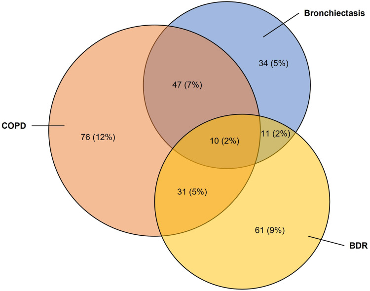
Among the 123 Indigenous patients demonstrating BDR, 113 (92%) had radiology reports available for review. Nearly half (46%) of the BDR group had evidence of chronic airway diseases such as COPD, bronchiectasis, or both, which was significantly more compared to the non-BDR group (30%, p = 0.001) (Table 3). Figure 1 illustrates the proportional overlap of BDR, COPD and Bronchiectasis among the 643 Indigenous patients with radiology available.
Table 3.
Radiology Results for Indigenous Patients (n = 643) with or Without Bronchodilator Responsiveness (BDR)
| Radiology | BDR (n=113) | No-BDR (n=530) | p-value |
|---|---|---|---|
| Chronic obstructive pulmonary disease (COPD) | 31 (27%) | 76 (14%) | 0.001* |
| Bronchiectasis | 11 (10%) | 34 (6%) | 0.209 |
| Combined COPD & Bronchiectasis | 10 (9%) | 47 (9%) | 0.995 |
| Any COPD or Bronchiectasis | 52 (46%) | 157 (30%) | 0.001* |
| Other abnormalityα | 26 (23%) | 129 (24%) | 0.764 |
| No abnormality | 35 (31%) | 244 (46%) | 0.003* |
Notes: p-value derived from two-tailed z-test of proportions. *Significance at p < 0.05. αPleural effusion, tracheobronchomegaly, lung mass, interstitial or lung opacity or fibrosis, malignancy, collapse, pneumonia, atelectasis, cavity, consolidation, goitre, other inflammation, ground glass, lung cysts.
Figure 1.
Venn diagram showing the overlap of COPD, BDR and Bronchiectasis among the 643 Indigenous patients with radiology available.
Abbreviations: BDR, bronchodilator responsiveness; COPD, chronic obstructive pulmonary disease.
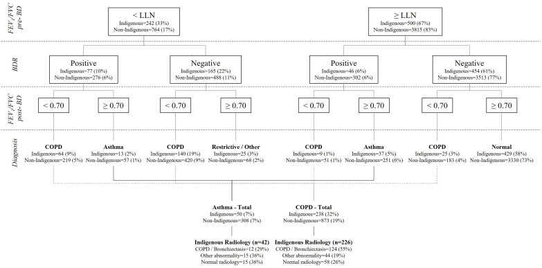
Spirometry results in 59% (n = 73) of Indigenous and 47% (n = 270) of non-Indigenous patients with BDR demonstrated evidence for a potential diagnosis of COPD (ie, post-BD spirometry FEV1/FVC ratio <0.7) (Figure 2). However, among the 50 Indigenous patients who did not display spirometric evidence of COPD, 29% (12/42 with radiology) showed evidence of COPD and/or bronchiectasis on radiology. Excluding patients with spirometric or radiographic evidence of COPD or bronchiectasis, 38 (5%) Indigenous patients exhibited BDR, which could be assigned solely to asthma and 308 (7%) non-Indigenous patients.
Figure 2.
Flow chart for plausible putative diagnosis of asthma among patients undergoing spirometry.
Abbreviations: BD, bronchodilator; BDR, bronchodilator responsiveness; COPD, chronic obstructive pulmonary disease; FEV1, Forced expiratory volume in one second; FVC, Forced vital capacity; LLN, lower limit of normal.
Univariate and Multivariate Analysis
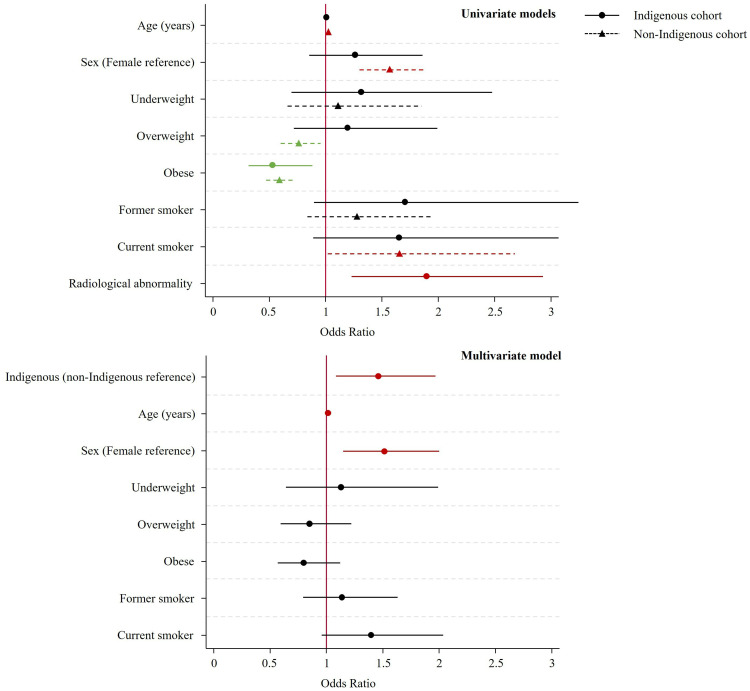
The use of logistic regression revealed significant differences in factors associated with BDR between the Indigenous and non-Indigenous groups. Increasing age, male sex and current smoking were associated with increased odds of BDR among non-Indigenous patients, while overweight and obesity were associated with decreased odds of BDR (Figure 3). Among Indigenous patients, only abnormal radiology was associated with increased odds of BDR (OR 1.9, 95% CI 1.23, 2.93), while only obesity was associated with decreased odds. Post-estimation tests of ORs, which were significant in the non-Indigenous group but non-significant in the Indigenous group, showed no significant difference in the effect of age (p = 0.114), sex (p = 0.330), overweight (p = 0.111) or current smoking (p = 0.997) on the odds of BDR between the groups.
Figure 3.
Odds ratios for univariate logistic regressions by Indigenous status, and multivariate logistic regression for factor effects on BDR. Red lines indicate significantly increased odds of BDR while green lines indicate significantly reduced odds of BDR. Normal BMI was used as the reference category for BMI. Radiology data was not available for the non-Indigenous cohort; therefore, these were excluded in the multivariate model.
Abbreviations: BDR, bronchodilator responsiveness; BMI, body mass index.
In the complete multivariate model, Indigenous status was significantly associated with BDR (OR 1.47, 95% CI 1.09, 1.97), as was age (OR 1.02, 95% CI 1.01, 1.03), and male sex (OR 1.52, 95% CI 1.15, 2.01).
Sub-Analysis for BDR >10% Predicted ATS/ERS Criteria
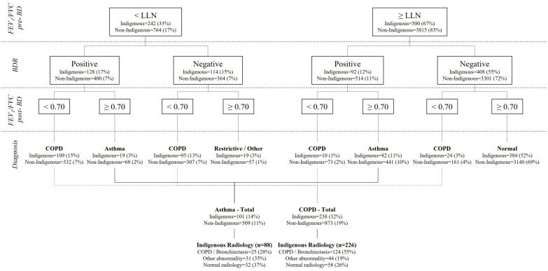
Utilising the 2021 updated BDR guidelines,41 220 (30%) Indigenous and 914 (20%) non-Indigenous patients fit the BDR criteria (p < 0.001 for difference of proportions). LFPs remained significantly lower among Indigenous patients compared to non-Indigenous patients in this sub-cohort (Table 4), and there was no significant difference on any LFPs when comparing between BDR “traditional” and BDR 2021 criteria (data not shown). Among Indigenous patients, COPD and/or bronchiectasis was identified in 46% and 26% of those with and without BDR, respectively (compared to 46% and 30% utilising the traditional BDR criteria) (Appendix 1). By spirometry criteria 54% (n = 119) of Indigenous and 44% (n = 405) of non-Indigenous patients with BDR demonstrated evidence to fulfil the criteria for a potential diagnosis of COPD (ie, post-BD spirometry FEV1/FVC ratio <0.7) (Figure 4). However, among the 101 Indigenous patients who did not display spirometric evidence of COPD, 28% (25/88 with radiology) showed evidence of COPD and/or bronchiectasis on radiology.
Table 4.
Lung function parameters (LFPs) for patients displaying bronchodilator responsiveness as per updated (>10% predicted) ATS/ERS criteria by Indigenous status
| LFPs | BDR | Indigenous (n=220) | Non-Indigenous (n=914) | p-value |
|---|---|---|---|---|
| FVC | Pre-BD absolute | 1.96 (1.86, 2.05) | 2.84 (2.77, 2.9) | <0.001* |
| Pre-BD predicted | 53.2 (51.12, 55.28) | 71.89 (70.73, 73.06) | <0.001* | |
| Post-BD absolute | 2.18 (2.09, 2.28) | 3.15 (3.08, 3.22) | <0.001* | |
| Post-BD predicted | 59.31 (57.3, 61.33) | 79.87 (78.7, 81.04) | <0.001* | |
| Change^ | 13.73 (11.8, 15.67) | 12.43 (11.57, 13.29) | 0.200 | |
| FEV1 | Pre-BD absolute | 1.26 (1.19, 1.34) | 1.92 (1.87, 1.98) | <0.001* |
| Pre-BD predicted | 43.51 (41.13, 45.89) | 63.42 (62.06, 64.78) | <0.001* | |
| Post-BD absolute | 1.47 (1.38, 1.55) | 2.21 (2.15, 2.27) | <0.001* | |
| Post-BD predicted | 50.23 (47.85, 52.61) | 73.01 (71.53, 74.49) | <0.001* | |
| Change^ | 17.43 (15.78, 19.08) | 16.36 (15.57, 17.14) | 0.239 | |
| FEV1/FVC | Pre-BD absolute | 0.64 (0.62, 0.66) | 0.67 (0.66, 0.68) | 0.021* |
| Pre-BD predicted | 80.55 (77.81, 83.29) | 86.9 (85.82, 87.98) | <0.001* | |
| Post-BD absolute | 0.66 (0.64, 0.68) | 0.69 (0.68, 0.7) | 0.004* | |
| Post-BD predicted | 82.93 (80.33, 85.53) | 89.9 (88.84, 90.95) | <0.001* | |
| Post-BD absolute <0.70 | 119 (54%) | 405 (44%) | 0.009* |
Notes: Data presented as mean (95% CI) for continuous parameters and number (%) for categorical parameters. p-value derived from two-tailed students t-test for continuous parameters and two-tailed z test of proportions for categorical parameters. ^Change - Mean percentage change in values pre- to post- BD. *Significance at p < 0.05.
Abbreviations: BD, Bronchodilator; FEV1, Forced expiratory volume in one second; FVC, Forced vital capacity.
Figure 4.
Flow chart for plausible putative diagnosis of asthma among patients undergoing spirometry utilising updated 2021 BDR guidelines.
Abbreviations: BD, bronchodilator; BDR, bronchodilator responsiveness; COPD, chronic obstructive pulmonary disease; FEV1, Forced expiratory volume in one second; FVC, Forced vital capacity; LLN, lower limit of normal.
Discussion
To the best of the authors’ knowledge, this is the first study to comprehensively assess chest radiological findings in a group of Indigenous Australian people with airway BDR. The key findings of our study were:
17% of Indigenous patients referred for lung function testing had evidence of BDR.
A high percentage (ie, 46%) of Indigenous patients with BDR had radiological evidence of chronic lung disease.
Presence of radiological abnormality (COPD/bronchiectasis) increased the odds of BDR among Indigenous patients.
When BDR “potential asthma” was adjusted for the presence of COPD and bronchiectasis it was no higher among Indigenous patients than non-Indigenous.
In this current study, LFPs of Indigenous and non-Indigenous Australian patients were compared in order to objectively assess the presence of BDR and its association with underlying chronic lung disease according to radiology in Indigenous patients. Within the study patients, a higher proportion of Indigenous (17%) compared to non-Indigenous patients (13%) demonstrated a significant BDR. Although, this may indeed truly reflect previous self-reported population survey results,5,47 demonstrating a higher prevalence of asthma (BDR) among Indigenous in comparison to non-Indigenous populations, a significant proportion of Indigenous patients with BDR had radiological evidence of underlying chronic airway diseases such as COPD or bronchiectasis (46%). Moreover, one-quarter of patients with COPD/bronchiectasis demonstrated BDR.20,21 A previous self-reported survey among Canadian Indigenous people observed that prevalence of asthma/COPD overlap could be higher among Indigenous people.48 The findings of our study indicate that among Indigenous Australians, conditions other than asthma or concurrent presence of asthma and COPD,23,24 or bronchiectasis alongside asthma25 could explain the apparently high rates of self-reported asthma in previous population surveys.
Spirometry has a critical role in clinical decision-making to support the accurate diagnosis of airway disease alongside clinical judgment, especially in the presence of concomitant airway diseases. In this vein, previous studies have shown a significant BDR with respect to FVC, could be associated with emphysema and small airways disease as opposed to asthma.49–51 Additionally, DLCO values have been observed to be higher among patients with asthma in comparison to patients with predominant COPD.52 In our study, we observed that Indigenous patients with BDR showed a greater trend towards improvement in FVC post-BD compared to non-Indigenous patients (34 vs 20%). Although, we did not examine the correlation of DLCO parameters in this study, we presume this could be the case among patients with radiological abnormalities in the BDR Indigenous group, especially among those with evidence of COPD.33 These findings tip the scale towards predominant COPD rather than asthma, or for the presence of potential asthma/COPD overlap among our Indigenous study patients with BDR.
Worldwide Indigenous populations suffer from a high burden of chronic respiratory conditions, in particular COPD and bronchiectasis, including higher prevalence of tobacco use.7–12,53,63 The hallmark symptoms of asthma are similar to those of other chronic lung diseases, as are the effects of smoking.1 Hence, it is not uncommon to either under or over diagnose the presence of asthma in clinical practice.13–16,64–68 Furthermore, it is reasonable to note that there could be biases in recall of underlying medical conditions among Indigenous people – recall that has predominantly been used in assessing the population prevalence of asthma in previous surveys.5,6 A recent study from our center found that only seven percent of Indigenous patients and 30% of non-Indigenous patients with COPD could accurately describe their respiratory condition, although 46 and 89%, respectively, were aware that “something” was wrong with their lungs.69 In the same study, 80% of Indigenous patients described shortness of breath, 60% described a cough and 10% described wheezing – thus it is easy to see how Indigenous patients may self-report a previous diagnosis of “asthma” in the presence of another underlying respiratory condition. When excluding the presence of COPD in the present study (via either radiology or spirometry), the prevalence of BDR dropped from 17% to 5% among Indigenous patients and from 13% to 7% among non-Indigenous patients (using the usual/traditional ≥12% and ≥0.2L criteria). This was remarkable given the Indigenous study patients came from a population purported to have a high prevalence of asthma.
This is the first study to assess BDR as per the updated 2021 ERS/ATS guidelines in an Indigenous population.45 Understandably, due to the lowered percentage change threshold from 12% to 10% and dropping the 200mL change requirement, this resulted in a higher prevalence of BDR. In the current study, 30% of Indigenous and 20% of non-Indigenous people met the new criteria,45 compared to 17% and 13% who met the existing usual/traditional criteria.42 Despite the significant increase in the number of individuals identified, no significant differences in any LFPs were noted when comparing the two BDR criteria. This could be due to the sample size in this study being not large enough to identify potentially small differences in these parameters, or that any such difference in parameters would be reduced anyway due to this being a referred population as compared to the general population. Nonetheless, despite utilising this new recommended criterion (ie, >10% BDR), when taking into consideration the evidence for presence of underlying chronic airway disease, presence of BDR reduces significantly resulting in a comparable rate between Indigenous and non-Indigenous patients (8% vs 11%).
Ascertaining the true prevalence of respiratory conditions is indeed difficult among Indigenous people due to geographical isolation, access to specialist health care and due to other social determinants. Moreover, presence of multiple concurrent respiratory disorders, including a high prevalence of COPD and bronchiectasis further adds to the complexity in the accurate diagnosis and in the management of Indigenous people presenting with respiratory disorders. In this study, we have demonstrated the potential causes for observing BDR among Indigenous patients in comparison to non-Indigenous patients undergoing LFTs. Despite overwhelming evidence in the literature to suggest Australian Indigenous people suffer from chronic respiratory disorders, there have been no previous reports demonstrating the clinical significance of observing BDR on spirometry in an Australian Indigenous population, especially from the Top End NT of Australia. Hence, we believe demonstrating these aspects in the current study is of significant value in addressing the gap in knowledge, and an invaluable addition to the existing literature. However, further prospective studies are warranted to determine accurate diagnostic and management pathways for Indigenous people presenting with chronic airway disorders.
Study Limitations
The authors acknowledge that a better characterisation of the patients’ respiratory diseases would have been possible if clinic history details, including prior clinical diagnosis of asthma and the findings on examination had been available for consideration. However, due to the retrospective nature of this study, this was not possible. Only Indigenous patients’ radiology was considered in this study, limiting the potential for comparison of the presence of concomitant airway disease in non-Indigenous patients. Furthermore, smoking data was missing for a large portion of the non-Indigenous patients, potentially biasing the regression results, which incorporated smoking data. This study’s participants were drawn from a referred population to a specialist respiratory service in the TEHS region of the NT of Australia; hence, the results are pertinent to the “Top End” but what, if any, relevance they have to the wider Indigenous Australian population is open to conjecture.
Conclusion
In this study, Indigenous patients were observed to have a higher frequency of BDR on spirometry in comparison to non-Indigenous patients. However, a significant proportion of Indigenous patients demonstrating BDR also had evidence of COPD and bronchiectasis. This may suggest that BDR on spirometry could be suggestive of potential asthma or asthma/COPD/bronchiectasis overlap or BDR could be present among patients with COPD and bronchiectasis in isolation. Hence, a more personalised approach should be adopted combining clinical/physical examinations, spirometry and radiology in the accurate diagnosis of airway disease among Indigenous Australians, which may have long-term therapeutic implications and overall better outcome.
Acknowledgment
We sincerely thank all the respiratory technologists, especially Ms Ara Joy Perez from Darwin Respiratory and Sleep Health, Darwin Private Hospital, Darwin, Australia for her help with data collection for this study. We also thank Ms Amelia Skaczkowskit, Flinders University, Northern Territory Medical Program medical student for helping with data collection. We also extend our sincere appreciation to our remote community Indigenous health workers, especially Mr Izaak Thomas (Australian Indigenous Luritja descendent) from the respiratory chronic respiratory disease co-ordination division in approving this research addressing much needed data in the diagnosis and management of adult Indigenous patients with respiratory disorders and for the appropriateness and respect in relation to the Indigenous context represented in this study.
Abbreviations
AHR, Airway hyperreactivity; AO, Airflow obstruction; ATS, American thoracic society; BMI, Body mass index; BD, Bronchodilator; BDR, Bronchodilator responsiveness; CI, Confidence interval; COPD, Chronic obstructive pulmonary disease; DLCO, carbon monoxide diffusing capacity; ERS, European respiratory society; FVC, Forced vital capacity; FEV1, Forced expiratory volume in one second; LFP, Lung function parameter; LFT, Lung function test; NT, Northern Territory; ORs, Odds ratios; TEHS, Top End Health Service.
Ethics Approval and Informed Consent
This study was approved by the Human Research Ethics Committee of the Northern Territory, Top End Health Service and Menzies School of Health Research (Reference: HREC 2019-3445), and was conducted according to the Declaration of Helsinki.
Author Contributions
All authors made significant contribution to the work reported, whether that is in the conception, study design, execution, acquisition of data, analysis and interpretation, or in all these areas. Have drafted or written, or substantially revised or critically reviewed the article. Have agreed on the journal to which the article will be submitted. Reviewed and agreed on all versions of the article before submission, during revision, the final version accepted for publication, and any significant changes introduced at the proofing stage. Agree to take responsibility and be accountable for the contents of the article.
Disclosure
All authors declare no conflicts of interest for this study.
References
- 1.Global Initiative for Asthma. Global strategy for asthma management and prevention; 2021. Available from: www.ginasthma.org. Accessed May 22, 2022.
- 2.To T, Stanojevic S, Moores G, et al. Global asthma prevalence in adults: findings from the cross-sectional world health survey. BMC Public Health. 2012;12:204. doi: 10.1186/1471-2458-12-204 [DOI] [PMC free article] [PubMed] [Google Scholar]
- 3.Saglani S, Menzie-Gow AN. Approaches to asthma diagnosis in children and adults. Front Pediatr. 2019;7:148. doi: 10.3389/fped.2019.00148 [DOI] [PMC free article] [PubMed] [Google Scholar]
- 4.Chapman DG, Irvin CG. Mechanisms of airway hyper-responsiveness in asthma: the past, present and yet to come. Clin Exp Allergy. 2015;45(4):706–719. doi: 10.1111/cea.12506 [DOI] [PMC free article] [PubMed] [Google Scholar]
- 5.Cunningham J. Socioeconomic status and self-reported asthma in Indigenous and non-Indigenous Australian adults aged 18–64 years: analysis of national survey data. Int J Equity Health. 2010;9:18. doi: 10.1186/1475-9276-9-18 [DOI] [PMC free article] [PubMed] [Google Scholar]
- 6.Australian Institute of Health and Welfare 2020. Asthma. Cat. no. ACM 33. Canberra: AIHW. Available from: https://www.aihw.gov.au/reports/chronic-respiratory-conditions/asthma. Accessed May 22, 2022. [Google Scholar]
- 7.Kruavit A, Fox M, Pearson R, Heraganahally S. Chronic respiratory disease in the regional and remote population of the Northern Territory Top End: a perspective from the specialist respiratory outreach service. Aust J Rural Health. 2017;25:275–284. [DOI] [PubMed] [Google Scholar]
- 8.Heraganahally S, Wasgewatta SL, McNamara K, et al. Chronic obstructive pulmonary disease in Aboriginal patients of The Northern Territory of Australia: a landscape perspective. Int J Chron Obstruct Pulmon Dis. 2019;14:2205–2217. [DOI] [PMC free article] [PubMed] [Google Scholar]
- 9.Heraganahally SS, Timothy TP, Sorger L. Chest computed tomography findings among adult Indigenous Australians in the Northern Territory of Australia. J Med Imaging Radiat Oncol. 2021. doi: 10.1111/1754-9485.13295 [DOI] [PubMed] [Google Scholar]
- 10.Heraganahally SS, Wasgewatta SL, McNamara K, et al. Chronic obstructive pulmonary disease with and without bronchiectasis in Aboriginal Australians – a comparative study. Int Med J. 2020;50(12):1505–1513. doi: 10.1111/imj.14718 [DOI] [PubMed] [Google Scholar]
- 11.Mehra S, Chang AB, Lam CK, et al. Bronchiectasis among Australian Aboriginal and non-Aboriginal patients in the regional and remote population of the Northern Territory of Australia. Rural Remote Health. 2021;21(2):6390. doi: 10.22605/RRH6390 [DOI] [PubMed] [Google Scholar]
- 12.Colonna E, Maddox R, Cohen R, et al. Review of tobacco use among Aboriginal and Torres Strait Islander peoples. Austr Indigenous Health Bulletin. 2020;20(2): Available from: https://aodknowledgecentre.ecu.edu.au/learn/specific-drugs/tobacco/. [Google Scholar]
- 13.Aaron SD, Boulet LP, Reddel HK, Gershon AS. Underdiagnosis and overdiagnosis of asthma. Am J Respir Crit Care Med. 2018;198(8):1012–1020. [DOI] [PubMed] [Google Scholar]
- 14.LindenSmith J, Morrison D, Deveau C, Hernandez P. Overdiagnosis of asthma in the community. Can Respir J. 2004;11(2):111–116. [DOI] [PubMed] [Google Scholar]
- 15.Kavanagh J, Jackson DJ, Kent BD. Over- and under-diagnosis in asthma. Breathe. 2019;15(1):e20–e27. doi: 10.1183/20734735.0362-2018 [DOI] [PMC free article] [PubMed] [Google Scholar]
- 16.Tay TR, Lee JWY, Hew M. Diagnosis of severe asthma. Med J Aust. 2018;209(2):S3–S10. doi: 10.5694/mja18.00125 [DOI] [PubMed] [Google Scholar]
- 17.Veale AJ, Peat JK, Tovey ER, Salome CM, Thompson JE, Woolcock AJ. Asthma and atopy in four rural Australian aboriginal communities. Med J Aust. 1996;165(4):192–196. doi: 10.5694/j.1326-5377.1996.tb124923.x [DOI] [PubMed] [Google Scholar]
- 18.Cooksley NA, Atkinson D, Marks GB, et al. Prevalence of airflow obstruction and reduced forced vital capacity in an Aboriginal Australian population: the cross-sectional BOLD study. Respirology. 2015;20(5):766–774. doi: 10.1111/resp.12482 [DOI] [PubMed] [Google Scholar]
- 19.Dawson AP. Asthma in the Australian Indigenous population: a review of the evidence. Rural Remote Health. 2004;4:238. doi: 10.22605/RRH238 [DOI] [PubMed] [Google Scholar]
- 20.Janson C, Malinovschi A, Amaral AFS, et al. Bronchodilator reversibility in asthma and COPD: findings from three large population studies. Eur Respir J. 2019;54:1900561. doi: 10.1183/13993003.00561-2019 [DOI] [PubMed] [Google Scholar]
- 21.Nogrady SG, Evans WV, Davies BH. Reversibility of airways obstruction in bronchiectasis. Thorax. 1978;33(5):635–637. doi: 10.1136/thx.33.5.635 [DOI] [PMC free article] [PubMed] [Google Scholar]
- 22.Radovanovic D, Santusa P, Blasi F, et al. A comprehensive approach to lung function in bronchiectasis. Respir Med. 2018;145:120–129. [DOI] [PubMed] [Google Scholar]
- 23.Mekov E, Nuñez A, Sin DD, et al. Update on asthma-COPD overlap (ACO): a narrative review. Int J Chron Obstruct Pulmon Dis. 2021;16:1783–1799. doi: 10.2147/COPD.S312560 [DOI] [PMC free article] [PubMed] [Google Scholar]
- 24.Hosseini M, Almasi-Hashiani A, Sepidarkish M, Maroufizadeh S. Global prevalence of asthma-COPD overlap (ACO) in the general population: a systematic review and meta-analysis. Respir Res. 2019;20(1):229. doi: 10.1186/s12931-019-1198-4 [DOI] [PMC free article] [PubMed] [Google Scholar]
- 25.Matsumoto H. Bronchiectasis in severe asthma and asthmatic components in bronchiectasis. Respir Investig. 2022;60(2):187–196. doi: 10.1016/j.resinv.2021.11.004 [DOI] [PubMed] [Google Scholar]
- 26.Australian bureau of statistics. Estimates of aboriginal and torres strait Islander Australians. ABS, Canberra, Australia; 2016. [Google Scholar]
- 27.Schubert J, Kruavit A, Mehra S, Wasgewatta S, Chang AB, Heraganahally SS. Prevalence and nature of lung function abnormalities among Indigenous Australians referred to specialist respiratory outreach clinics in the Northern Territory. Intern Med J. 2019;49(2):217–224. [DOI] [PubMed] [Google Scholar]
- 28.Heraganahally SS, Howarth T, White E, Sorger L, Biancardi E, Ben Saad H. Lung function parameters among Australian Aboriginal “apparently healthy” adults: an Australian Caucasian and global lung function initiative (GLI-2012) various ethnic norms comparative study. Expert Rev Respir Med. 2020;23:1–11. doi: 10.1080/17476348.2021.1847649 [DOI] [PubMed] [Google Scholar]
- 29.Heraganahally SS, Howarth T, Mo L, Sorger L, Saad HB. Critical analysis of spirometric patterns in correlation to chest computed tomography among adult Indigenous Australians with chronic airway diseases. Expert Rev Respir Med. 2021;15(9):1229–1238. doi: 10.1080/17476348.2021.1928496 [DOI] [PubMed] [Google Scholar]
- 30.Heraganahally SS, Ponneri TR, Howarth TP, Saad HB. The effects of inhaled airway directed pharmacotherapy on decline in lung function parameters among Indigenous Australian adults with and without underlying airway disease. Int J Chron Obstruct Pulmon Dis. 2021;16:2707–2720. [DOI] [PMC free article] [PubMed] [Google Scholar]
- 31.Heraganahally S, Howarth TP, White E, Ben Saad H. Implications of using the GLI-2012, GOLD and Australian COPD-X recommendations in assessing the severity of airflow limitation on spirometry among an Indigenous population with COPD: an Indigenous Australians perspective study. BMJ Open Respir Res. 2021;8:e001135. doi: 10.1136/bmjresp-2021-001135 [DOI] [PMC free article] [PubMed] [Google Scholar]
- 32.Heraganahally SS, Howarth T, Sorger L, Ben Saad H. Sex differences in pulmonary function parameters among Indigenous Australians with and without chronic airway disease. PLoS One. 2022;17(2):e0263744. doi: 10.1371/journal.pone.0263744 [DOI] [PMC free article] [PubMed] [Google Scholar]
- 33.Howarth TP, Saad HB, Perez AJ, Atos CB, White E, Heraganahally SS. Comparison of diffusing capacity of carbon monoxide (DLCO) and total lung capacity (TLC) between Indigenous Australians and Australian Caucasian adults. PLoS One. 2021;16(4):e0248900. doi: 10.1371/journal.pone.0248900 [DOI] [PMC free article] [PubMed] [Google Scholar]
- 34.Miller MR, Hankinson J, Brusasco V, et al. Standardisation of spirometry. Eur Respir J. 2005;26:319–338. [DOI] [PubMed] [Google Scholar]
- 35.NDD Medical Technologies. EasyOne Pro. Andover MA 01810, USA; 2017. [Google Scholar]
- 36.Global initiative for chronic obstructive lung disease. Global strategy for the diagnosis, management, and prevention of chronic obstructive pulmonary disease; 2021. Available from: http://www.goldcopd.org. Accessed May 22, 2022.
- 37.Sim YS, Lee JH, Lee WY, et al. Spirometry and bronchodilator test. Tuberc Respir Dis. 2017;80(2):105–112. doi: 10.4046/trd.2017.80.2.105 [DOI] [PMC free article] [PubMed] [Google Scholar]
- 38.Hankinson JL, Odencrantz JR, Fedan KB. Spirometric reference values from a sample of the general US population. Am J Respir Crit Care Med. 1999;159(1):179–187. [DOI] [PubMed] [Google Scholar]
- 39.Guezguez F, Knaz H, Anane I, Bougrida M, Ben Saad H. The ‘clinically significant’ bronchodilator responsiveness (BDR) in children: a comparative study between six definitions of scholarly societies and a mini-review. Expert Rev Respir Med. 2021;15(6):823–832. doi: 10.1080/17476348.2021.1906653 [DOI] [PubMed] [Google Scholar]
- 40.Guezguez F, Ben Saad H. What constitutes a ”clinically significant” bronchodilator response in children? Eur Respir J. 2020;55(5):2000207. doi: 10.1183/13993003.00207-2020 [DOI] [PubMed] [Google Scholar]
- 41.Chhabra SK. Clinical application of spirometry in asthma: why, when and how often? Lung India. 2015;32(6):635–637. doi: 10.4103/0970-2113.168139 [DOI] [PMC free article] [PubMed] [Google Scholar]
- 42.Johnson JD, Theurer WM. A stepwise approach to the interpretation of pulmonary function tests. Am Fam Physician. 2014;89(5):359–366. [PubMed] [Google Scholar]
- 43.Pipavath SNJ, Schmidt RA, Takasugi JE, Godwin JD. Chronic obstructive pulmonary disease: radiology-pathology correlation. J Thorac Imaging. 2009;24(3):171–180. [DOI] [PubMed] [Google Scholar]
- 44.Gaillard F, Weerakkody Y. Bronchiectasis. Reference article. Available from: Radiopaedia.org. Accessed August 1, 2022.
- 45.Stanojevic S, Kaminsky DA, Miller M, et al. ERS/ATS technical standard on interpretive strategies for routine lung function tests. Eur Respir J. 2021;23:2101499. doi: 10.1183/13993003.01499-2021 [DOI] [PubMed] [Google Scholar]
- 46.National Health and Medical Research Council. Ethical conduct in research with Aboriginal and Torres Strait Islander Peoples and communities: guidelines for researchers and stakeholders. Canberra: Commonwealth of Australia; 2018. Available from: https://www.nhmrc.gov.au. Accessed May 22, 2022. [Google Scholar]
- 47.Brock C, McGuane J. Determinants of asthma in Indigenous Australians: insights from epidemiology. Austr Indigenous Health Bulletin. 2018;18(2):12–20. [Google Scholar]
- 48.Koleade A, Farrell J, Mugford G, Gao Z. Prevalence and risk factors of ACO (Asthma-COPD Overlap) in Aboriginal people. J Environ Public Health. 2018;9. doi: 10.1155/2018/4657420 [DOI] [PMC free article] [PubMed] [Google Scholar]
- 49.Ben Saad H, Préfaut C, Tabka Z, Zbidi A, Hayot M. The forgotten message from gold: FVC is a primary clinical outcome measure of bronchodilator reversibility in COPD. Pulm Pharmacol Ther. 2008;21(5):767–773. [DOI] [PubMed] [Google Scholar]
- 50.Schermer T, Heijdra Y, Zadel S, et al. Flow and volume responses after routine salbutamol reversibility testing in mild to very severe COPD. Respir Med. 2007;101(6):1355–1362. doi: 10.1016/j.rmed.2006.09.024 [DOI] [PubMed] [Google Scholar]
- 51.Vigna M, Aiello M, Bertorelli G, Crisafulli E, Chetta A. Flow and volume response to bronchodilator in patients with COPD. Acta Biomed. 2018;89(3):332–336. doi: 10.23750/abm.v89i3.5631 [DOI] [PMC free article] [PubMed] [Google Scholar]
- 52.Dias RM, Chacur FH, Carvalho SR, Neves DD. Which functional parameters can help differentiate severe asthma from COPD? Rev Port Pneumol. 2010;16(2):253–272. [PubMed] [Google Scholar]
- 53.Ospina MB, Voaklander DC, Stickland MK. Prevalence of asthma and chronic obstructive pulmonary disease in Aboriginal and non-Aboriginal populations: a systematic review and meta-analysis of epidemiological studies. Can Respir J. 2012;19(6):355–360. doi: 10.1155/2012/825107 [DOI] [PMC free article] [PubMed] [Google Scholar]
- 54.Bird Y, Moraros J, Mahmood R, Esmaeelzadeh S, Kyaw Soe NM. Prevalence and associated factors of COPD among Aboriginal peoples in Canada: a cross-sectional study. Int J Chron Obstruct Pulmon Dis. 2017;12:1915–1922. doi: 10.2147/COPD.S138304 [DOI] [PMC free article] [PubMed] [Google Scholar]
- 55.Sze DFL, Howarth TP, Lake CD, Ben Saad H, Heraganahally SS. Differences in the spirometry parameters between indigenous and non-indigenous patients with COPD: a matched control study. Int J Chron Obstruct Pulmon Dis. 2022;17:869–881. [DOI] [PMC free article] [PubMed] [Google Scholar]
- 56.Heraganahally SS, Silva SAMS, Howarth TP, Kangaharan N, Majoni SW. Comparison of clinical manifestation among Australian Indigenous and non‐ Indigenous patients presenting with pleural effusion. Int Med J. 2021. doi: 10.1111/imj.15310 [DOI] [PubMed] [Google Scholar]
- 57.Heraganahally SS, Mortimer N, Howarth T, et al. Utility and outcomes among Indigenous and non-Indigenous patients requiring domiciliary oxygen therapy in the regional and rural Australian population. Aust J Rural Health. 2021;29(6):918–926. doi: 10.1111/ajr.12782 [DOI] [PubMed] [Google Scholar]
- 58.Heraganahally SS, Ghimire RM, Howarth T, Kankanamalage OM, Palmer D, Falhammar H. Comparison and outcomes of emergency department presentations with respiratory disorders among Australian indigenous and non-indigenous patients. BMC Emerg Med. 2022;22:11. doi: 10.1186/s12873-022-00570-3 [DOI] [PMC free article] [PubMed] [Google Scholar]
- 59.Seyedshahabedin MM, Howarth TP, Mo L, Biancardi E, Heraganahally SS. Flexible bronchoscopy indications and outcomes between Indigenous and non-Indigenous patients in the Northern Territory of Australia. Int Med J. 2022. doi: 10.1111/imj.15865 [DOI] [PubMed] [Google Scholar]
- 60.Heraganahally SS, Rajaratnam B, Silva SAAS, et al. Obstructive sleep apnoea and cardiac disease among aboriginal patients in the Northern Territory of Australia. Heart Lung Circ. 2021. doi: 10.1016/j.hlc.2021.01.007 [DOI] [PubMed] [Google Scholar]
- 61.Heraganahally SS, Kruavit A, Oguoma VM, et al. Sleep apnoea among Australian Aboriginal and Non- Aboriginal patients in the Northern Territory of Australia– a comparative study. Sleep. 2020;43(3):zsz248. doi: 10.1093/sleep/zsz248 [DOI] [PubMed] [Google Scholar]
- 62.La-Grappe D, Massey L, Kruavit A, et al. Sleep disorders among Aboriginal Australians with Machado-Joseph Disease: quantitative results from a multiple methods study to assess the experience of people living with the disease and their caregivers. Neurobiol Sleep Circadian Rhythms. 2022. doi: 10.1016/j.nbscr.2022.100075 [DOI] [PMC free article] [PubMed] [Google Scholar]
- 63.Howarth TP, Gentin N, Reyes-Chicuellar N, et al. Sleep quality and obstructive sleep apnoea in Indigenous and non-Indigenous Australian children. Sleep Med. 2022;98:68–78. doi: 10.1016/j.sleep.2022.06.014 [DOI] [PubMed] [Google Scholar]
- 64.Kann K, Long B, Koyfman A. Clinical mimics: an emergency medicine-focused review of asthma mimics. J Emerg Med. 2017;53(2):195–201. doi: 10.1016/j.jemermed.2017.01.005 [DOI] [PubMed] [Google Scholar]
- 65.Reddel HK. Common conditions that mimic asthma. Med J Aust. 2022;216(7):337–340. doi: 10.5694/mja2.51467 [DOI] [PMC free article] [PubMed] [Google Scholar]
- 66.King CS, Moores LK. Clinical asthma syndromes and important asthma mimics. Respir Care. 2008;53(5):568–80;discussion 580–2. [PubMed] [Google Scholar]
- 67.Heraganahally SS, Ghataura AS, Er XY, Heraganahally S, Biancardi E. Excessive dynamic airway collapse: a COPD/Asthma mimic or a treatment emergent consequence of inhaled corticosteroid therapy - case series and brief literature review. Clin Pulm Med. 2020;27:175–182. [Google Scholar]
- 68.Rogliani P, Ora J, Puxeddu E, Cazzola M. Airflow obstruction: is it asthma or is it COPD? Int J Chron Obstruct Pulmon Dis. 2016;11:3007–3013. doi: 10.2147/COPD.S54927 [DOI] [PMC free article] [PubMed] [Google Scholar]
- 69.Pal A, Howarth TH, Rissel C, et al. COPD disease knowledge, self-awareness and reasons for hospital presentations among a predominately Indigenous Australian cohort– a study to explore preventable hospitalisation. BMJ Open Resp Res. 2022;9:e001295. doi: 10.1136/bmjresp-2022-001295 [DOI] [PMC free article] [PubMed] [Google Scholar]


