

Abstract
Forensic identification of human remains is crucial for legal, humanitarian, and civil reasons. Wide heterogeneity in sphenoid sinus morphology can be used for personal identification. This study aimed to propose a new protocol for personal identification based on three-dimensional (3D) reconstruction of sphenoid sinus CT images using Iterative Closest Point (ICP) algorithm. Seven hundred thirty-two patients which consisted of 348 females and 384 males were retrospectively included. The study sample includes 732 previous images as a source point set and 743 later ones as a scene target set. The sphenoid sinus computed tomography (CT) images were processed on a workstation (Dolphin imaging) to obtain 3D images and stored as a file format of Stereo lithography (.STL). Then, a Python library vtkplotter was used to transform the STL format to PLY format, which was adapted to Point Cloud Library (PCL). The ICP algorithm was used for point clouds matching. The metric Rank-N recognition rate was used for evaluation. The scene target set of 743 individuals was compared with the source point set of 732 individual models and achieved Rank-1 accuracy of 96.24%, Rank-2 accuracy of 99.73%, and Rank-3 accuracy of 100%. Our results indicated that the 3D point cloud registration of sphenoid sinuses was useful for assessing personal identification in forensic contexts.
Keywords: Personal identification, Iterative Closest Point (ICP) algorithm, Point Cloud Library (PCL), Sphenoid sinus, Computed Tomography (CT) images
Introduction
Personal identification of unidentified bodies is crucial for civil, ethical, and juridical reasons [1]. Recently, the anatomical characterization of paranasal sinuses has shown to be valuable for biometrics and individual identification [2, 3]. Sphenoid sinuses, one of the paranasal sinuses, are two pneumatized areas positioned in the body of sphenoid and are separated by an intersphenoidal septum. Their pneumatization can be seen after birth and starts to progress inferiorly and posteriorly around 15–18 months, becoming evident at around 4 years old. Their expansion will reach the full extent at 12 years old and the final formation at puberty [4]. Their variability in pneumatization ranges from absent to extensive [5] and is highly variable among different individuals [4]. Sizes, shapes, and pneumatization types vary from one person to another, even between true twins [6]. Furthermore, the sphenoid sinus is the most inaccessible part of the face, locating inside the sphenoid bone and involving several different structures. The deep anatomical location makes it difficult to approach and can be beneficial in the case of forensic identification due to it is well protected from traumatic degradation resulting from external causes [7]. Therefore, the wide heterogeneity in sphenoid sinus morphology can be used for personal identification.
The uniqueness of anatomical and biological features represents the premise for personal identification in forensics classified by the DVI and INTERPOL guidelines as “secondary methods.” From this point of view, imaging can be indispensable support for personal identification [8]. Recently, CT images are used in forensics more and more frequently and that of paranasal sinuses demonstrated particularly to be of great help in personal identification procedures [3, 8–10], which includes the usage of sphenoid sinuses [3, 8].
Recently, several studies have proposed anatomical classification systems for forensic identification based on the morphology of the 2D CT images of sphenoid sinus [3, 11]. The visual comparison of sphenoid sinus achieved 100% accuracy on a small dataset with less than 60 patients or cases. Those methods demonstrated that sphenoid sinus is a reliable evidence for forensic identification. However, the subjective visual comparison relies on a certain level of expertise. And the classification is too time-consuming and error‐prone. Furthermore, the 2D images have low recognition rate compared with 3D images due to insufficient information [12]. Variations of sphenoid sinus can be noticed in different positions and orientations arising from the clinical examination, thus adding difficulty to the process of comparison. Due to the limitations of 2D images, 3D images of the sphenoid sinus have been used for forensic identification [7, 8].
Recently, deep learning algorithms have quickly become essential in the field of medical image analysis. Compared to the traditional methods, deep learning techniques are more efficient in extracting compact information and leading towards significantly improved performance of the medical image analysis system [7]. In the study of Souadih et al. [7], the deep learning methods were used for forensic identification based on the 3D sphenoid sinus images. They achieved 100% accuracy in a dataset that consists of 72 patients with only 13 patients who did twice CT. However, the deep learning methods should be pre-trained on big data. One hurdle presented in applying deep learning techniques to medical imaging is the lack of samples [13]. The Iterative Closest Points (ICP) algorithm is the mainstream algorithm used in the process of accurate registration of 3D point cloud data which requires no pre-training. It is used to find the rigid transformation between the scene point set and the target point set so that the two matching data satisfy the optimal match under some kind of metric criterion [14]. The purpose of the present study was to propose a new protocol by using the ICP algorithm for personal identification from 3D sphenoid sinus images. A schematic representation of the proposed method is shown in Fig. 1.
Fig. 1.

Overview of the proposed method
Materials and methods
Study Population
The data used in the present study were obtained from participants undergoing routine examination at the West China Hospital of Sichuan University. Subjects were above 18 years old with a history of chronic illness; trauma, physical deformity, or any surgical procedure that might affect sphenoid sinus were excluded from the study. The data were collected from 732 patients which consisted of 348 females and 384 males, who underwent at least twice high-resolution computed tomography (CT) scan of head. The study sample included 732 previous images that were treated as the source point set and 743 later ones as the scene target set. The present study was a retrospective study with approval by the Ethics Committee of Sichuan University, and a waiver of the requirement for informed consent was obtained.
Image Acquisition
Multidetector CT was carried out on SOMATOM Definition AS 128 slice CT scanner (Siemens, Erlangen, Germany). After obtaining the scout projection, the area of scanning was defined to include the region of paranasal sinuses. The scanning protocol was as follows: collimation of 1 mm, reconstruction interval of 1 mm, tube voltage of 120 kV, tube current of 110 mA, and scanning time of 0.3 s. It was assumed that the scans provided a comparable and sufficient image quality concerning the evaluation of sphenoid sinus because all the scans were of high-resolution thin-slice MDCT imaging. Image data were processed on a workstation (Dolphin imaging, (version 11.5; Chatsworth, CA)) to obtain three-dimensional (3D) images (Supplementary Video 1) and stored as a file format of Stereo lithography (.STL). According to the difference of CT values between gas and surrounding tissues, probes or “seeds” were inserted into each sinus in horizontal, sagittal, and coronal sections, increasing in size to permeate the entire volume. Then, the 3D sphenoid sinus digital model was segmented. The process of reestablishing the images is shown in Fig. 2. Then, a Python library vtkplotter was used to transform the STL format to PLY format, which was adapted to Point Cloud Library (PCL) for point cloud processing tasks. Figure 3 displays the point cloud of two separate individuals.
Fig. 2.

The process on the workstation of Dolphin imaging to establish 3D images of the sphenoid sinus. a Horizontal position, b sagittal position, c coronal position
Fig. 3.
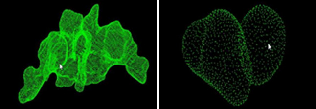
The point cloud of sphenoid sinus from two different individuals
Processing
The point cloud matching was achieved by the ICP algorithm in the PCL library. The ICP algorithm takes two point clouds as an input and returns the rigid transformation (rotation matrix R and translation vector T) that best aligns the point clouds.
Two point sets were denoted as X and P; let X be the source point set, and let P be the target point set:
The objective function:
The ICP algorithm which corrects the bias field and segments the image into different clusters with an iterative approach can be summarized into the following steps:
Step 1: Select initial transformation (t) and rotation (R).
Step 2: Initialize the fuzzy partition memberships functions.
Step 3: Calculate the centroid removal coordinates of two sets of point clouds according to the following function:
Step 4: The rotation matrix is calculated by using the singular value decomposition according to the following function:
Step 5: Calculate the translation vector:
Step 6: Repeat the above steps until convergence when or reaching the maximum number of iterations of 50 for the first time and 100 for the second time.
Sphenoidal Sinus Recognition
In the recognition stage of our method, each point in one point cloud from the target point database was compared with all points in each individual from the source point database by rotation and translation in the same spatial system. A distance matrix between the two groups (as source point and target point) was calculated and denoted as FitnessScore. The threshold of FitnessScore was set as 1. The matched individuals were ranked from low to high based on FitnessScore. The metric RANK-N recognition rate was used for evaluation. The RANK-N indicates that the target is in the top-N most probable individuals in the identification FitnessScore ascending order. The recognition rate is the number of RANK-N that accounts for the proportion of the total number of query samples. The process of recognition is shown in Fig. 4.
Fig. 4.

The process for point cloud matching
Results
The age of all participants was ranged from 18 to 92 years old, and the average was 45.97 years old, as shown in Fig. 5. The interval between the source point set and the scene target set was ranged from 1 day to 9.5 years; the average was 1.0 year, as shown in Fig. 6.
Fig. 5.

Age distribution of included participants
Fig. 6.
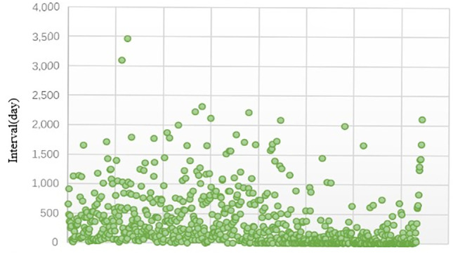
Interval between
source point set and scene target set
The scene target set of 743 individuals was compared with the source point set of 732 individual models. Using the present method, we have achieved Rank-1 accuracy of 96.24%, Rank-2 accuracy of 99.73%, and Rank-3 accuracy of 100%.
Discussion and Conclusions
Personal identification is performed through comparison between biological data obtained from the cadaver and antemortem material, and identifying the body may have belonged in life from one or more missing persons [1]. The sphenoid sinus is a double sinus located within the body of the sphenoid bone and separates the two sinuses from each other by the bony septum. However, they are asymmetric sinuses. Many important neurovascular structures such as the pituitary gland, internal carotid artery, optic nerve, maxillary nerve, and pterygoid nerve surround the sinus [15]. Its deep anatomical location makes it difficult to approach and well protected from traumatic degradation, but can be beneficial in the case of forensic identification. The extreme variable in degrees and directions contribute to the radiologic identification, especially if dental records, fingerprints, or DNA samples are not available [7]. The CT allows precise evaluation craniofacial of bones and the extent of their pneumatization and is an excellent imaging method used for the assessment of sinuses’ anatomy [16].
We could make useful measurements of its volume anatomy without the influence of different head-position by using the segmentation of 3D CT images of the sphenoid sinus [17]. Compared to the previous studies, this study used 3D point cloud to present the 3D shapes of sphenoidal sinus, which can provide more accurate 3D information without the influence of different head-position. In the present study, image data were processed on a workstation (Dolphin imaging) to obtain 3D images and stored as a file format of Stereo lithography (.STL). As already pointed out, the goal of the segmentation process performed is to exploit the deep anatomical characterization of the sphenoid sinus for body identification purposes. Then, a Python library vtkplotter was used to transform the STL format to PLY format, which was adapted to the PCL-based ICP algorithm.
The purpose of point cloud registration is to find a 3D rigid body transformation, so that the 3D coordinates of the point cloud at different angles can be correctly matched. This is a technique widely used in areas such as computer vision, medical image analysis, and pattern recognition. The purpose of registration between two point sets is to find their correspondence and then to transform one point set to its counterpart. A good algorithm for point set registration should match the two point sets correctly, and be robust to noisy points, outliers, and missing points, etc. [18]. The Iterative Closest Points (ICP) algorithm is the mainstream algorithm used in the process of accurate registration of 3D point cloud data [14]. The most prominent contribution is the ICP algorithm proposed by Besl and McKay [19]. In this method, the transformation parameters of two point sets are calculated through the relationship between the corresponding matching points of two point sets to satisfy the given convergence precision, and finally, the translation and rotation parameters between the two points are obtained to complete the registration process. In the present study, the ICP algorithm was used for point cloud matching which is essentially automated and quantitative. The scene target set of 743 individuals was compared with the source point set of 732 individual models, and achieved Rank-1 accuracy of 96.24% and Rank-2 accuracy of 99.73%. And the identification accuracy of 100% was reached at Rank-3. Compared to the previous studies, this method can also be validly applied to forensic identification with an excellent accuracy. Meanwhile, this method does not require pre-training and much expert experience. And the present dataset is much larger than that of previous studies. Since the previous studies addressed a fully automated computer-assisted identification technique by deep learning [7], based on our large dataset, deep learning-based point cloud matching method should be considered for forensic identification in future study.
In conclusion, the present study proposed a new method for personal identification using an ICP algorithm based on 3D sphenoid sinus images. Our results indicated that the 3D point cloud registration of sphenoid sinuses was useful for personal identification in forensic contexts.
Availability of Data and Material
The data and material that support the findings of this study are available from the corresponding author upon reasonable request.
Code Availability
Not applicable.
Declarations
Ethics Approval
The present study was a retrospective study with approval by the Ethics Committee of Sichuan University and a waiver of the requirement for informed consent was obtained.
Consent to Participate
Not applicable.
Consent for Publication
Not applicable.
Conflict of Interest
The authors declare no competing interests.
Footnotes
Xiaoai Dong and Fei Fan contributed equally to this work.
Publisher's Note
Springer Nature remains neutral with regard to jurisdictional claims in published maps and institutional affiliations.
Contributor Information
Kui Zhang, Email: cheungque@163.com.
Ji Zhang, Email: zhangj@scu.edu.cn.
Zhenhua Deng, Email: dengzhenhua@scu.edu.cn.
References
- 1.Ciaffi R, Gibelli D, Cattaneo C. Forensic radiology and personal identification of unidentified bodies: a review. Radiol Med. 2011;116(6):960–968. doi: 10.1007/s11547-011-0677-6. [DOI] [PubMed] [Google Scholar]
- 2.Souza LA, Jr, Marana AN, Weber SAT. Automatic frontal sinus recognition in computed tomography images for person identification. Forensic Sci Int. 2018;286:252–264. doi: 10.1016/j.forsciint.2018.03.029. [DOI] [PubMed] [Google Scholar]
- 3.Deloire L, Diallo I, Cadieu R, Auffret M, Alavi Z, Ognard J, Ben Salem D. Post-mortem X-ray computed tomography (PMCT) identification using ante-mortem CT-scan of the sphenoid sinus. J Neuroradiol. 2019;46(4):248–255. doi: 10.1016/j.neurad.2018.08.003. [DOI] [PubMed] [Google Scholar]
- 4.Guldner C, Pistorius SM, Diogo I, Bien S, Sesterhenn A, Werner JA. Analysis of pneumatization and neurovascular structures of the sphenoid sinus using cone-beam tomography (CBT) Acta Radiol. 2012;53(2):214–219. doi: 10.1258/ar.2011.110381. [DOI] [PubMed] [Google Scholar]
- 5.Laury AM, Oyesiku NM, Hadjipanayis CG, Delgaudio JM, Wise SK. Incidental sinonasal findings identified during preoperative evaluation for endoscopic transsphenoidal approaches. Am J Rhinol Allergy. 2013;27(3):202–205. doi: 10.2500/ajra.2013.27.3871. [DOI] [PMC free article] [PubMed] [Google Scholar]
- 6.Chaiyasate S, Baron I, Clement P. Analysis of paranasal sinus development and anatomical variations: a CT genetic study in twins. Clin Otolaryngol. 2007;32(2):93–97. doi: 10.1111/j.1365-2273.2007.01404.x. [DOI] [PubMed] [Google Scholar]
- 7.Souadih K, Belaid A, Ben Salem D, Conze PH. Automatic forensic identification using 3D sphenoid sinus segmentation and deep characterization. Med Biol Eng Comput. 2020;58(2):291–306. doi: 10.1007/s11517-019-02050-6. [DOI] [PubMed] [Google Scholar]
- 8.Cappella A, Gibelli D, Cellina M, Mazzarelli D, Oliva AG, De Angelis D, Sforza C, Cattaneo C. Three-dimensional analysis of sphenoid sinus uniqueness for assessing personal identification: a novel method based on 3D–3D superimposition. Int J Legal Med. 2019;133(6):1895–1901. doi: 10.1007/s00414-019-02139-5. [DOI] [PubMed] [Google Scholar]
- 9.Ruder TD, Kraehenbuehl M, Gotsmy WF, Mathier S, Ebert LC, Thali MJ, Hatch GM. Radiologic identification of disaster victims: a simple and reliable method using CT of the paranasal sinuses. Eur J Radiol. 2012;81(2):e132–e138. doi: 10.1016/j.ejrad.2011.01.060. [DOI] [PubMed] [Google Scholar]
- 10.Gibelli D, Cellina M, Cappella A, Gibelli S, Panzeri MM, Oliva AG, Termine G, De Angelis D, Cattaneo C, Sforza C. An innovative 3D–3D superimposition for assessing anatomical uniqueness of frontal sinuses through segmentation on CT scans. Int J Legal Med. 2019;133(4):1159–1165. doi: 10.1007/s00414-018-1895-4. [DOI] [PubMed] [Google Scholar]
- 11.Auffret M, Garetier M, Diallo I, Aho S, Ben Salem D. Contribution of the computed tomography of the anatomical aspects of the sphenoid sinuses to forensic identification. J Neuroradiol. 2016;43(6):404–414. doi: 10.1016/j.neurad.2016.03.007. [DOI] [PubMed] [Google Scholar]
- 12.Mou Q-n, Ji L-l, Liu Y, Zhou P-r, Han M-q, Zhao J-m, Cui W-t, Chen T, Du S-y, Hou Y-x, and Guo Y-c, Three-dimensional superimposition of digital models for individual identification, Forensic Sci Int 318, 110597, 2021. [DOI] [PubMed]
- 13.Yang L and Chakraborty R, "A GMM Based Algorithm To Generate Point-Cloud And Its Application To Neuroimaging," in 2020 IEEE 17th International Symposium on Biomedical Imaging Workshops (ISBI Workshops), 2020, pp. 1–4.
- 14.He Y, Liang B, Yang J, Li S, He J. An Iterative Closest Points Algorithm for Registration of 3D Laser Scanner Point Clouds with Geometric Features. Sensors (Basel) 2017;17(8):1862. doi: 10.3390/s17081862. [DOI] [PMC free article] [PubMed] [Google Scholar]
- 15.Reino AJ, Factors in the pathogenesis of tumors of the sphenoid and maxillary sinuses: a comparative study, Laryngoscope 110(10 Pt 2 Suppl 96), 1–38, 2000. [DOI] [PubMed]
- 16.Uthman AT, Al-Rawi NH, Al-Naaimi AS, Tawfeeq AS, and Suhail EH, Evaluation of frontal sinus and skull measurements using spiral CT scanning: an aid in unknown person identification, Forensic Sci Int 197(1–3), 124 e1–7, 2010. [DOI] [PubMed]
- 17.Kawarai Y, Fukushima K, Ogawa T, Nishizaki K, Gunduz M, Fujimoto M, Masuda Y. Volume quantification of healthy paranasal cavity by three-dimensional CT imaging. Acta Otolaryngol Suppl. 1999;540:45–49. [PubMed] [Google Scholar]
- 18.Zhu H, Guo B, Zou K, Li Y, Yuen KV, Mihaylova L, and Leung H, A Review of Point Set Registration: From Pairwise Registration to Groupwise Registration, Sensors (Basel) 19(5)2019. [DOI] [PMC free article] [PubMed]
- 19.Besl PJ, McKay ND. A method for registration of 3-D shapes. IEEE Transactions on Pattern Analysis and Machine Intelligence. 1992;14(2):239–256. doi: 10.1109/34.121791. [DOI] [Google Scholar]
Associated Data
This section collects any data citations, data availability statements, or supplementary materials included in this article.
Data Availability Statement
The data and material that support the findings of this study are available from the corresponding author upon reasonable request.
Not applicable.