André Barros-Carvalho and Eurico Morais-de-Sá highlight work from Calvi and colleagues that demonstrates a role for PP1 in establishing embryo polarity via regulation of PAR-2 localization.
Abstract
How cells spatially organize their plasma membrane, cytoskeleton, and cytoplasm remains a central question for cell biologists. In this issue of JCB, Calvi et al. (2022. J. Cell Biol. https://doi.org/10.1083/jcb.202201048) identify PP1 phosphatases as key regulators of C. elegans anterior–posterior polarity, by counterbalancing aPKC-mediated phosphorylation of PAR-2.
Ancient sailors relied on a compass to navigate the sea. Millennials now use a phone app to move across the world. Proteins and organelles lack these tools, and so navigating the cellular space was made possible by evolution of a machinery that sets intracellular asymmetries. The central PARts of animal cell polarity were identified in the Caenorhabditis elegans embryo through screens for partitioning defective mutants. Their identification pioneered the understanding of a network that establishes the complementary localization of anterior (aPARs: atypical PKC [aPKC; PKC-3 in C. elegans], PAR-3, PAR-6, and the small GTPase CDC42) and posterior (PAR-1, PAR-2, LGL-1, and CHIN-1) proteins (1). Polarization of the C. elegans one-cell embryo defines a distinct fate and size of the two cells (AB and P1) produced after the first division, and is a robust model to uncover fundamental principles of cell polarization. In fact, the majority of the PAR network is highly conserved, adapted to regulate polarity in a range of processes, including stem cell division, cell migration, and epithelial apical–basal polarity (1). In a new study published in this issue of JCB, Calvi et al. (2) present protein phosphatase 1 (PP1) as a new PARt in C. elegans polarization, raising the importance of dephosphorylation in the dynamic behavior of PAR polarity.
Anterior–posterior polarization of the C. elegans embryo stems from mutual antagonistic interactions that restrict the activity of two opposing kinases—PKC-3 and PAR-1—to each side of the cortex. PKC-3 phosphorylates PAR-2, which represses binding of multivalent PAR-1 and PAR-2 complexes to the plasma membrane (3, 4). While polarity kinases are conserved, the RING finger protein PAR-2 is worm specific. Nevertheless, its ability to recruit PAR-1 (4), and subsequently exclude aPARs from the posterior, makes PAR-2 a central player in C. elegans polarization. Fertilization marks symmetry breaking through two redundant pathways linked to the paternal centrosome and associated microtubules: first, the centrosome induces actomyosin flows that displace PAR-3/PAR-6/PKC-3 oligomers toward the future anterior domain, clearing space for PAR-2/PAR-1 cortical loading (1); and second, microtubules emanating from the centrosome bind PAR-2, protecting it from PKC-3 phosphorylation (4). Disruption of cortical flows or mutations in PAR-2 microtubule-binding sites are insufficient on their own to disrupt the asymmetric establishment of PAR domains (4). This redundancy could be explained by a mechanism that enables phosphorylation-inhibited PAR-2 to recover its membrane binding ability even if only one of these pathways is operational. To solve this enigma, Calvi et al. (2) shifts our attention from polarity kinases toward protein phosphatases.
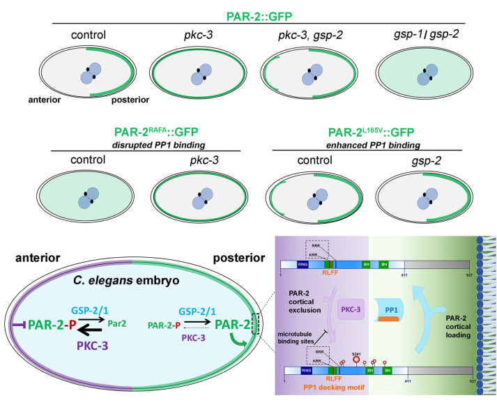
An import piece of the puzzle came from an earlier screen that identified GSP-2, one of two catalytic subunits of C. elegans PP1, as a suppressor of embryonic lethality in pkc-3 temperature sensitive mutants (pkc-3ts; 5). pkc-3ts mutants exhibit several polarity defects—PAR-2 distribution along the entire cell cortex; defective furrow positioning; and at the two-cell stage, AB and P1 cells divide synchronously, rather than asynchronously. Calvi et al. (2) show that RNAi-mediated depletion of GSP-2 in pkc-3ts mutants restores all these phenotypes. Thus, GSP-2 could antagonize PKC-3 function by reverting the phosphorylation of its substrates. However, GSP-2 depletion alone has no major impact on cell polarization. This hinted at another C. elegans PP1 catalytic subunit, GSP-1. GSP-1 depletion did not suppress embryonic lethality in pkc-3ts mutants, but co-depletion with GSP-2 led to PAR-2 mislocalization almost entirely to the cytoplasm. Assuming that the anterior-directed actomyosin flows and microtubule protection from PKC-3 phosphorylation remain unaffected, this suggests PP1 phosphatases could directly control PAR-2’s ability to bind the plasma membrane.
The hypothesis that PP1 could dephosphorylate PAR-2 to remove the phosphorylation-inhibited binding to the plasma membrane was gaining strength. Calvi et al. (2) spotted important clues in a two-hybrid screen that reported an interaction between GSP-2 and the PAR-2 N-terminal region (6). So how does PAR-2 bind PP1? Binding of PP1 to its substrates and regulatory subunits is normally mediated by short linear motifs, such as the RVxF motif (7). Through a yeast two-hybrid, the authors identify a degenerate PP1-docking motif in the N-terminal region of PAR-2, RLFF, which physically interacts with PP1. The significance of this PP1–PAR2 interaction is then elegantly shown in vivo, since mutations in the PP1-docking motif (par-2RAFA) fail to bind the cortex and reproduce the polarity defects of gsp-1/gsp2 RNAi and of par-2 mutants that mimic PKC-3 phosphorylation (3). Importantly, reducing PKC-3 activity restores gfp::par-2RAFA posterior localization and polarity establishment, which provides compelling evidence that the PP1–PAR2 interaction is necessary to antagonize PAR-2 phosphorylation by PKC-3.
Calvi et al. (2) then devised a strategy to enhance PAR-2 binding to PP1. A single mutation converted the degenerate RLFF motif into an optimal RVxF motif (gfp::par-2[L165V]), which led to ectopic membrane enrichment of PAR-2 before and during polarity establishment. These defects are rescued by gsp-2 depletion, and so, likely result from shifting the balance toward excessive PP1-mediated PAR-2 dephosphorylation. Though this indicates that PP1 activity toward PAR-2 must be perfectly tuned, it was intriguing that gfp::par-2L165V animals are homozygous viable. One explanation comes from the observation that the anterior PAR-2 domains are partially corrected during mitosis. This implicates other mechanisms that restricts PAR-2 localization to the posterior during mitosis, and which may relate to a cell-cycle regulated brake in PP1 activity (7). However, how PP1 activity is modulated during polarity establishment and maintenance in the C. elegans embryo, as it goes through meiosis, interphase, and mitosis, remains an exciting open question.
Calvi et al. (2) do not provide direct answers for when and where PP1 activity is turned on/off, and both GSP-1 and GSP-2 localize uniformly in the embryo’s cytoplasm. Thus, the most parsimonious model for the PAR-2 phosphorylation gradient would couple asymmetric PKC-3 kinase activity to uniform PP1 activity (Fig. 1). This is reminiscent to the polarization of phosphorylated MEX-5 by the posterior PAR-1 kinase and uniform PP2 phosphatase activity (8). However, Calvi et al. (2) also propose the possibility that anteriorly enriched PLK-1 could be a negative PP1 regulator to promote PAR-2 phosphorylation at the anterior. This is based in two observations. First, gfp::par-2L165V bipolarity phenocopies PAR-2 localization in plk-1 temperature-sensitive mutants. Second, Calvi et al. (2) show a genetic interaction whereby reducing GSP-2 activity in plk-1 mutants rescues PAR-2 mislocalization. Nevertheless, a direct link between PLK-1 and PP1 was not detected. Further work is necessary to untangle the function of PLK-1 on PP1 from its other roles, including the regulation of aPARs before and after symmetry breaking (9, 10).
Figure 1.
PP1 regulates PAR-2 asymmetry in the one-cell C. elegans embryo. An overview of PAR-2 localization in genetic modifications presented in Calvi et al. (2) is shown on top. A model to generate an anterior–posterior asymmetry of phosphorylated PAR-2 based on uniform PP1 activity counterbalancing asymmetric PKC-3 is shown at the bottom. Binding of the degenerate RVxF motif of PAR-2 to PP1 may collaborate with microtubule-dependent protection from PKC-3 phosphorylation to promote cortical loading of PAR-2 at the posterior (close-up). BH, Basic-and-Hydrophobic motif.
This study cements PP1-aPKC as a key antagonistic phosphatase-kinase pair that regulates animal cell polarity. PP1 reverses PKC-3–mediated phosphorylation of PAR-2 and cooperates with the protection provided by microtubules emanating from the centrosome to ensure PAR-2 cortical loading at the posterior of the C. elegans embryo (Fig. 1). PP1 also dephosphorylates other aPKC targets, including orthologues of the C. elegans polarity network, such as Drosophila Lgl, and Par-3 in mammalian cells (11, 12). It will be critical to dissect how the opposing roles of aPKC/PKC-3 and PP1 are integrated over multiple targets, some of which act as aPKC/PKC-3 antagonists themselves, to establish and maintain polarity. Solving this intricate web of protein interactions and understanding how the spatiotemporal regulation of PP1 activity contributes to the reactions that polarize the C. elegans embryo will enlighten the cellular adaptation that polarizes several different systems.
Acknowledgments
Work in the E. Morais-de-Sá lab is supported by FCT—Fundação para a Ciência e a Tecnologia, I.P., under the project PTDC/BIA-CEL/1511/2021. A. Barros-Carvalho is supported by a PhD fellowship from FCT (2021.07215.BD).
The authors declare no competing financial interests.
References
- 1.Lang, C.F., and Munro E.. 2017. Development. 10.1242/dev.139063 [DOI] [PMC free article] [PubMed] [Google Scholar]
- 2.Calvi, I., et al. 2022. J. Cell. Biol. 10.1083/jcb.202201048 [DOI] [PMC free article] [PubMed] [Google Scholar]
- 3.Hao, Y., et al. 2006. Dev. Cell. 10.1016/j.devcel.2005.12.015 [DOI] [PMC free article] [PubMed] [Google Scholar]
- 4.Motegi, F., et al. 2011. Nat. Cell Biol. 10.1038/ncb2354 [DOI] [Google Scholar]
- 5.Fievet, B.T., et al. 2013. Nat. Cell Biol. 10.1038/ncb2639 [DOI] [PMC free article] [PubMed] [Google Scholar]
- 6.Koorman, T., et al. 2016. Nat. Cell Biol. 10.1038/ncb3300 [DOI] [Google Scholar]
- 7.Nilsson, J. 2019. J. Cell Biol. 10.1083/jcb.201809138 [DOI] [Google Scholar]
- 8.Griffin, E.E., et al. 2011. Cell. 10.1016/j.cell.2011.08.012 [DOI] [Google Scholar]
- 9.Dickinson, D.J., et al. 2017. Dev. Cell. 10.1016/j.devcel.2017.07.024 [DOI] [PMC free article] [PubMed] [Google Scholar]
- 10.Reich, J.D., et al. 2019. Curr. Biol. 10.1016/j.cub.2019.04.058 [DOI] [Google Scholar]
- 11.Traweger, A., et al. 2008. Proc. Natl. Acad. Sci. USA. 10.1073/pnas.0804102105 [DOI] [Google Scholar]
- 12.Moreira, S., et al. 2019. Cell Rep. 10.1016/j.celrep.2018.12.060 [DOI] [PubMed] [Google Scholar]


