Abstract
The spleen is an infrequent metastatic organ of solid tumours, the prevalence of which ranges between 2.3% and 7.1% in populations with cancer as determined through autopsy. The most common sources of metastasis are breast, lung, colorectal and ovarian carcinoma and melanoma. Isolated metastasis of the spleen is rarely reported with only 93 cases from all sources having been reported up to 2007. Therefore, isolated splenic metastasis from primary lung cancer is exceedingly rare with only 11 cases reported to date.
Herein, we report a rare case of isolated splenic metastasis in a 49-yr-old female 3 months after lobectomy for an undifferentiated large cell carcinoma in the right lung (pT2aN0M0). The only symptom the patient presented with was continuous high fever, which had never been previously reported. This patient presented diagnostic challenges due to the presentation of high fever, leukoapenia after chemotherapy and the cystic splenic mass, all of which led to the initial consideration of splenic abscess. The patient's high fever resolved rapidly after splenectomy and splenic metastasis was confirmed by pathological findings.
We also reviewed all 11 reported previously cases and summarised the characteristics and appropriate management of isolated splenic metastasis from lung cancer.
Keywords: Fever, lung cancer, splenectomy, splenic metastasis
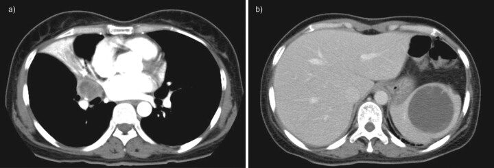
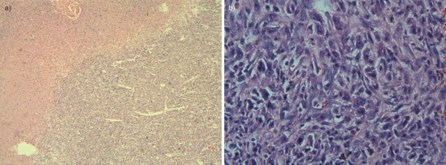
A 49-yr-old nonsmoking female was admitted to our hospital (Changzheng Hospital, Shanghai, China) in April 2009 complaining of cough, blood-stained sputum and recurrent low-grade fever for 4 months. A computed tomography (CT) scan demonstrated stenosis in the bronchus of the middle lobe of the right lung, and obstructive pneumonitis in the same lobe (fig. 1a). Bronchoscopy demonstrated neoplasia in the bronchial lumen of the right middle lobe. Positron emission tomography (PET) detection showed no metastases. Following treatment with antibiotics the patient’s fever resolved and she underwent lobectomy of the right middle and lower lobe and lymphadenectomy on April 22, 2009. Pathological examination demonstrated an undifferentiated large cell carcinoma 4.0×3.0×3.0 cm in size, with negative surgical margins and negative lymph nodes. The post-surgical stage was defined as T2aN0M0, according to the new staging system released by the International Association for the Study of Lung Cancer in 2009 [1]. Following surgery the patient was referred for chemotherapy (vinorelbine with cisplatin) for three cycles during June 2009 and July 2009. However, on August 3, 2009 the patient suddenly presented with a high fever and was admitted to the emergency room of our hospital. A routine blood test showed that the white blood count (WBC) was 2.7×10⁹ L−1 (neutrophils 1.2×10⁹ L−1). Although the chemotherapy was thought to be responsible for myelosuppression and a secondary infection, no other symptoms existed to indicate the site of infection. The patient was admitted and treated with granulocyte colony-stimulating factors and antibiotics. However, even though strong, broad spectrum anti-infection strategies were administered and the WBC recovered the patient’s high fever was not resolved. Surprisingly, the abdominal ultrasonic inspection revealed a solid, cystic mass in the spleen 8.2×8.1 cm in size. A further abdominal CT scan also showed a 9.5×8.0 cm splenic mass with complex solid and cystic components (fig. 1b). Splenic abscess was initially considered due to the manifestation of high fever only. However, percutaneous splenic puncture guided by ultrasound showed little fluid, which was cultured with no bacteria. Due to her history of lung cancer, splenic metastasis was considered; however, a chest CT scan showed no relapse in the primary site. A metastatic workup (scans of the brain, liver, adrenals and bones) was performed revealing no other metastatic disease. Splenectomy was performed on September 21, 2009 before the high fever could cause deterioration in her condition. Splenectomy revealed a 9.0×6.5×12.5 cm cavitated, necrotic mass in the middle pole of the spleen. Pathology of the mass demonstrated undifferentiated metastatic carcinoma compatible with the lung primary carcinoma (fig. 2). Following splenectomy the patient's high fever resolved rapidly and she was discharged in a good condition. She received another cycle of chemotherapy (paclitaxel with cisplatin) 1 month later. Because the patient could not tolerate the adverse effects, she refused to receive any further chemotherapy. Follow-up to date has not demonstrated any evidences of recurrent metastatic disease.
Figure 1.
a) Chest computed tomography (CT) scan showing a mass and obstructive pneumonitis in the right upper lobe. b) An abdominal CT scan showing a splenic mass 3 months after lobectomy.
Figure 2.
Histology slides showing undifferentiated large cell carcinoma in the spleen compatible with the lung. a) The juncture of metastatic carcinoma and normal splenic parenchyma and b) metastatic carcinoma only.
DISCUSSION
The spleen is an infrequent metastatic organ of solid tumours, the prevalence of which ranges between 2.3% and 7.1% in populations with cancer, as determined through autopsy [2]. This is probably due to its high density of immune-system cells, its role in “immune surveillance” and its high concentration of angiogenesis inhibition factor [3, 4]. However, isolated splenic metastasis is rarely reported. A recent review found only 93 reported cases of isolated splenic metastases up to 2007 [5]. Therefore, isolated splenic metastasis of primary lung cancer is exceedingly rare. To date, only 11 cases have been reported in published literature (table 1) [3, 4, 6–14].
Table 1. Incidence of isolated splenic metastasis from lung cancer.
| First author [Ref.] | Primary tumour type | Interval from primary tumour to metastasis | Follow-up | Splenectomy | Symptoms of metastasis |
| Klein [6] | Bronchioalveolar carcinoma | 20 months | Died 49 months after splenectomy | Yes | Abdominal pain |
| Edelman [7] | Lung poorly differentiated adenocarcinoma | 0 | No data | Asymptomatic | |
| Macheers [8] | Lung large cell, undifferentiated | 0 | Died 1 month after splenectomy | Yes | Asymptomatic |
| Gupta [9] | Right hilar neoplasm | 0 | Died 8 weeks after splenectomy | Yes | Splenic rupture |
| Kinoshita [10] | Lung squamous cell | 14 months | Died 27 months after splenectomy | Yes | Asymptomatic |
| Takada [11] | Bronchopulmonary carcinoid | 8 yr | Yes | Abdominal pain | |
| Massarweh [3] | Lung adenocarcinoma | 0 | Yes | Splenic rupture | |
| Schmidt [12] | Lung moderately differentiated adenocarcinoma | 4 yr | Yes | Asymptomatic | |
| Pramesh [13] | Lung squamous cell | 2 months | No | Asymptomatic | |
| Sanchez-Romero [4] | Lung adenocarcinoma | 0 | Yes | Abdominal pain | |
| Van Hul [14] | Lung adenocarcinoma | 2 yr | Yes | Asymptomatic |
Splenic metastasis from lung cancer was found to be most often associated with carcinoma of the left lung, perhaps due to preferentially higher blood flow compared with the right lung [10]. This finding is supported by several recently reported cases [3, 4, 12, 13]. However in our patient, the primary lung cancer was in the right lung. Among the previous cases, only one was found to have the primary lesion in the right-hand side [9]. As shown in table 1, the most frequent histological type of lung cancer with isolated splenic metastasis is adenocarcinoma (n = 5), followed by squamous carcinoma (n = 2) and large cell undifferentiated carcinoma (n = 2, if our case is included). The length of time between detection of primary lung cancer to isolated splenic metastasis ranges from 0 yrs to 8 yrs. The interval from primary tumour to splenic metastasis is probably associated with the histological type of the cancer. Generally speaking, adenocarcinoma and large cell undifferentiated carcinoma occur earlier in splenic metastasis.
Isolated splenic metastasis from lung cancer is most often incidentally detected by ultrasonography or CT scanning during the regular follow-up of patients with cancer. Among all cases reviewed, ∼55% (six out of 11) are asymptomatic when detected (table 1), which is similar the cases reported (60%) by Comperat et al. [5] regarding isolated splenic metastasis from all sources. Other symptoms include abdominal pain (three out of 11) and spontaneous splenic rupture (two out of 11). Surprisingly, continuous high fever was the only symptom of splenic metastasis in our patient. Because the metastasis occurred in the spleen which is an immune organ, fever could be regarded as a paraneoplastic manifestation. However, paraneoplastic syndrome mainly presents with endocrine, neurological, mucocutaneous or haematological symptoms; thus, due to the presentation of only fever paraneoplastic manifestation seems unlikely. As the splenic mass grew so fast that colliquative necrosis occurred inside the mass, necrotic material could be the endogenous pyrogen. We prefer to regard fever as the consequence of endogenous pyrogen. To our knowledge, fever as the presentation of splenic metastasis from lung cancer has not been previously reported.
Splenectomy was performed in most of the reviewed cases (table 1), either due to the symptoms of metastasis or as a part of therapeutic strategies. Although, at present, no guidelines are available for the application of splenectomy in treatment of isolated splenic metastasis, it is remarkable that long-term remission can be achieved by splenectomy alone in patients with isolated splenic metastasis from lung cancer [6, 10]. Lee et al. [15] thought splenectomy should be recommended because most splenic metastases appeared within the parenchyma, representing probable haematogenous spread. In our case, splenectomy showed good effects not only in resection of the metastasis, but also in resolving the high fever, which had causing the patient to deteriorate. According to 2009 National Comprehensive Cancer Network guidelines of nonsmall cell lung cancer [16], when solitary metastasis of adrenal and brain occurs without recurrence of metastasis in the primary site following surgical resection of the initial disease, resection of the brain or adrenal lesion is recommended. Since the spleen is not a frequent organ in which metastases of lung cancer occurs, splenectomy as ?>the therapeutic strategy when splenic metastasis occurs is not discussed in the guidelines. However, if we follow the therapeutic principle of solitary brain or adrenal metastasis, splenectomy is also a good option for isolated splenic metastasis. Systemic chemotherapy could have been considered after splenectomy since it is thought that it extends disease-free survival or overall survival. Additionally, if clinical assessment before initial treatments showed resectable lung lesion and isolated splenic metastasis, double surgical resection (splenectomy followed by resection of lung lesion) could be recommended to: avoid further metastatic disease; provide the potential of a cure or extend survival; and avoid the complications, such as painful splenomegaly and splenic rupture. Systemic adjuvant platin-based chemotherapy could be considered in order to reduce metastastic risk and increase survival.
Footnotes
Provenance
Submitted article, peer reviewed.
Statement of Interest
None declared.
REFERENCES
- 1.Giroux DJ, Rami-Porta R, Chansky K, et al. The ISALC Lung Cancer Staging Project: data elements for the prospective project. J Thorac Oncol 2009; 4: 679–683. [DOI] [PubMed] [Google Scholar]
- 2.Berge T. Splenic metastases. Frequencies and patterns. Acta Pathol Microbiol Scand A 1974; 82: 499–506. [PubMed] [Google Scholar]
- 3.Massarweh S, Dhingra H. Unusual sites of malignancy: case 3. Solitary splenic metastasis in lung cancer with spontaneous rupture. J Clin Oncol 2001; 19: 1574–1575. [DOI] [PubMed] [Google Scholar]
- 4.Sanchez-Romero A, Oliver I, Costa D, et al. Giant splenic metastasis due to lung adenocarcinoma. Clin Transl Oncol 2006; 8: 294–295. [DOI] [PubMed] [Google Scholar]
- 5.Comperat E, Bardier-Dupas A, Camparo P, et al. Splenic metastases: clinicopathologic presentation, differential diagnosis, and pathogenesis. Arch Pathol Lab Med 2007; 131: 965–969. [DOI] [PubMed] [Google Scholar]
- 6.Klein B, Stein M, Kuten A, et al. Splenomegaly and solitary spleen metastasis in solid tumors. Cancer 1987; 60: 100–102. [DOI] [PubMed] [Google Scholar]
- 7.Edelman AS, Rotterdam H. Solitary splenic metastasis of an adenocarcinoma of the lung. Am J Clin Pathol 1990; 94: 326–328. [DOI] [PubMed] [Google Scholar]
- 8.Macheers SK, Mansour KA. Management of isolated splenic metastases from carcinoma of the lung: a case report and review of the literature. Am Surg 1992; 58: 683–685. [PubMed] [Google Scholar]
- 9.Gupta PB, Harvey L. Spontaneous rupture of the spleen secondary to metastatic carcinoma. Br J Surg 1993; 80: 613– [DOI] [PubMed] [Google Scholar]
- 10.Kinoshita A, Nakano M, Fukuda M, et al. Splenic metastasis from lung cancer. Neth J Med 1995; 47: 219–223. [DOI] [PubMed] [Google Scholar]
- 11.Takada T, Takami H. Solitary splenic metastasis of a carcinoid tumor of the lung eight years postoperatively. J Surg Oncol 1998; 67: 47–48. [DOI] [PubMed] [Google Scholar]
- 12.Schmidt BJ, Smith SL. Isolated splenic metastasis from primary lung adenocarcinoma. South Med J 2004; 97: 298–300. [DOI] [PubMed] [Google Scholar]
- 13.Pramesh CS, Prabhudesai SG, Parasnis AS, et al. Isolated splenic metastasis from non small cell lung cancer. Ann Thorac Cardiovasc Surg 2004; 10: 247–248. [PubMed] [Google Scholar]
- 14.Van Hul I, Cools P, Rutsaert R. Solitary splenic metastasis of an adenocarcinoma of the lung 2 years postoperatively. Acta Chir Belg 2008; 108: 462–463. [DOI] [PubMed] [Google Scholar]
- 15.Lee SS, Morgenstern L, Phillips EH, et al. Splenectomy for splenic metastases: a changing clinical spectrum. Am Surg 2000; 66: 837–840. [PubMed] [Google Scholar]
- 16.National Comprehensive Cancer Network (NCCN). Non-small Cell Lung Cancer: Clinical Practice Guidelines in Oncology. 2010. www.nccn.org/professionals/physician_gls/f_guidelines.asp. [Google Scholar]


