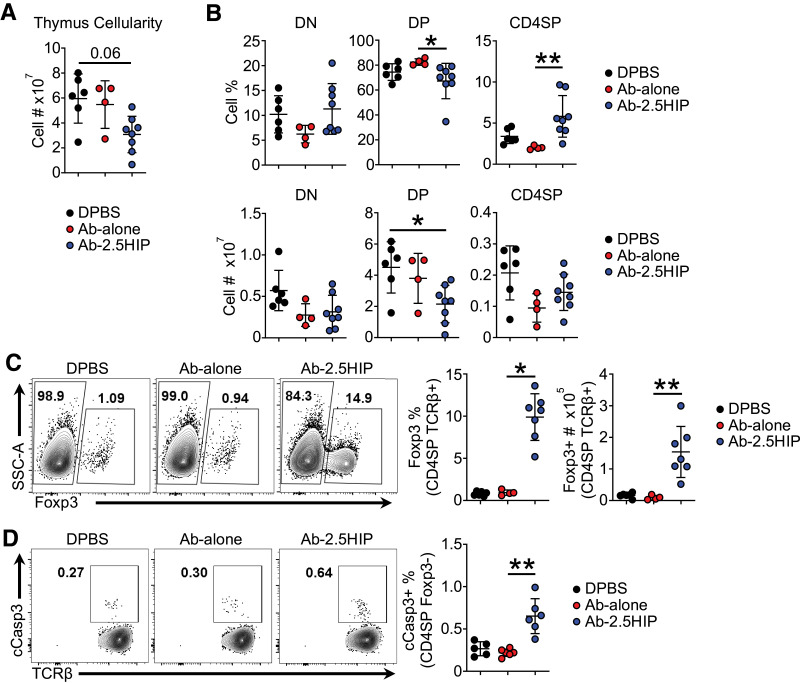
Figure 2.
Anti–Langerin-2.5HIP alters thymocyte development, increasing thymocyte apoptosis and Foxp3+ thymocyte population in BDC2.5 neonatal thymus. A: Total thymus cell count (n = 4–8 mice per group). B: Frequency and total number of thymocytes in the CD4−CD8− (DN), CD4+CD8+ (DP), and CD4+CD8− (CD4SP) stages of thymocyte development 72 h posttreatment. C: Representative flow plot and quantification of Foxp3+ cell percentage and number within CD4SP TCRβ+ thymocytes (n = 4–7 mice per group). SSC-A, side scatter area. D: Representative flow plot of apoptotic cell percentage within CD4SP TCRβ+ Foxp3− thymocytes (n = 5–6 mice per group). Data were pooled from at least two independent experiments and are reported as mean ± SD. Kruskal-Wallis and Dunn tests. *P < 0.05, **P < 0.01.

