PLOS Neglected Tropical Diseases logoLink to PLOS Neglected Tropical Diseases
. 2022 Sep 9;16(9):e0010693. doi: 10.1371/journal.pntd.0010693

Epidemiology of infection by pulmonary non-tuberculous mycobacteria in French Guiana 2008–2018

Milène Chaptal 1,2,*, Claire Andrejak 3, Timothée Bonifay 4, Emmanuel Beillard 5, Geneviève Guillot 6, Stéphanie Guyomard-Rabenirina 7, Magalie Demar 8, Sabine Trombert-Paolantoni 9, Veronique Jacomo 10, Emilie Mosnier 1, Nicolas Veziris 11, Felix Djossou 1, Loïc Epelboin 1,12; French Guiana PNTM working group
Editor: Joseph M Vinetz13
PMCID: PMC9491559  PMID: 36084148

Abstract

Introduction

Unlike diseases caused by Mycobacterium tuberculosis, M. leprae and M. ulcerans, the epidemiology of pulmonary non-tuberculous mycobacteria (PNTM) has not received due attention in French Guiana. The main objective of the current study was to define the incidence of these PNTM infections: NTM pulmonary diseases (NTM-PD) and casual PNTM isolation (responsible of latent infection or simple colonization). The secondary objectives were to determine species diversity and geographic distribution of these atypical mycobacteria.

Methods

A retrospective observational study (2008–2018) of French Guiana patients with at least one PNTM positive respiratory sample in culture was conducted. Patients were then classified into two groups: casual PNTM isolation or pulmonary disease (NTM-PD), according to clinical, radiological and microbiological criteria defined by the American Thoracic Society / Infectious Disease Society of America (ATS / IDSA) in 2007.

Results

178 patients were included, out of which 147 had casual PNTM isolation and 31 had NTM-PD. Estimated annual incidence rate of respiratory isolates was 6.17 / 100,000 inhabitants per year while that of NTM-PD was 1.07 / 100,000 inhabitants per year. Among the 178 patients, M. avium complex (MAC) was the most frequently isolated pathogen (38%), followed by M. fortuitum then M. abscessus (19% and 6% of cases respectively), the latter two mycobacteria being mainly found in the coastal center region. Concerning NTM-PD, two species were mainly involved: MAC (81%) and M. abscessus (16%).

Discussion/Conclusion

This is the first study on the epidemiology of PNTM infections in French Guiana. PNTM’s incidence looks similar to other contries and metropolitan France and NTM-PD is mostly due to MAC and M.abscessus. Although French Guiana is the French territory with the highest tuberculosis incidence, NTM should not be overlooked.

Introduction

Mycobacteria are widespread acid-fast bacilli (AFB) belonging to the genus Mycobacterium which include over 190 species. Non-tuberculous mycobacteria (NTM) have been poorly studied, while contrastingly, Mycobacterium tuberculosis complex and Mycobacterium leprae, human causative agents of Tuberculosis and Leprosy, have been studied in great detail. NTM are ubiquitous bacteria found in soil and water sources. Human contamination usually occurs by inhalation or skin-penetration [1]. NTMs mostly affect lungs and also others organs such as skin and soft tissues, bones, the lymphatic system etc. Buruli ulcer, caused by Mycobacterium ulcerans, is well known in French Guiana, with the tissue destruction resulting in skin ulcers and in severe cases causing bone infection [2]. Respiratory strains, called pulmonary-NTM (PNTM), can colonize respiratory airways without invading pulmonary tissues and can cause either an indolent infection or progress to an invasive disease. The unfortunate main complication is the progression to chronic respiratory failure. Among PNTM infections, the diagnostic criteria of the American Thoracic Society / Infectious Disease Society of America (ATS / IDSA) in 2007, enabled to distinguish PNTM isolation, including colonization and indolent infection, from true NTM pulmonary disease (NTM-PD) [3]. Latest guidelines in 2020 kept the same criteria, supplementing it with the precision of an minimal interval between two samples [4].

French Guiana is a tropical French overseas territory. It is located on the northeast Atlantic coast of the South American continent, between Brazil and Suriname. There are marked environmental and socio-demographical differences with mainland France. This territory is scarcely populated (about 250,000 inhabitants for 83,500 km2) and mostly covered by the Amazon forest [5]. French Guiana is the French region with the highest Tuberculosis incidence (32.5 per 100,000 inhabitants in 2017) [6]. Although the epidemiology of Tuberculosis, Leprosy and Buruli ulcer in French Guiana has been studied extensively, the literature on the epidemiology of NTM-PD is scarce [2,711].

Considering the scarcity of knowledge on PNTM infections in French Guiana, we conducted a retrospective and descriptive epidemiological study from 2008 to 2018 on patients living in the region. The main objective was to define the incidence of PNTM infections. Secondary objectives were to determine species diversity and geographic distribution of these atypical mycobacteria in French Guiana.

Methods

Ethics statement

This is a retrospective, non-interventional study and the data were anonymized for all patients. We only collected data necessary for the purpose of our study. The data collected will remain in an archive for 15 years. This research was in compliance with the law "Informatique et Libertés" of January 6, 1978 as amended and the law No. 2018–493 of June 20, 2018 on the protection of personal data. Is also in compliance with European Parliament guidelines in April 27, 2016. Finally, data were transferred and collected in accordance with the reference methodology MR003 of the Commission Nationale de l’Informatique et des Libertés (CNIL) for which the Centre Hospitalier de Cayenne has signed a compliance commitment. The database was declared to the CNIL and approved on April 19, 2019 (declaration number: 2212828v0).

Study design

We performed an observational and descriptive study on patients from the three French Guiana general hospitals: Cayenne, Kourou and Saint Laurent du Maroni.

Inclusion and exclusion criteria and case definition

All patients living in French Guiana and having at least one NTM positive culture of a respiratory sample were included between January 1, 2008 and December 31, 2018. Patient identification was based on respiratory samples obtained from all laboratories analyzing mycobacteria in French Guiana, namely: Pasteur Institute of French Guiana, Pasteur Institute of Guadeloupe, and private laboratories in mainland France Cerba and Biomnis. All medical records were reviewed and patients were classified into 2 categories: patients with diagnostic criteria of the ATS / IDSA 2007 fulfilled and patients who did not fulfill all the criteria but had casual PNTM isolation. The criteria are as follows: 1) clinical and radiological criteria: presence of symptoms, imaging compatible with cavities, nodules, micronodules associated with bronchiectasis, and the exclusion of other diagnoses. 2) microbiological criteria: presence of at least two separate sputum or gastric tubing or one positive bronchoalveolar lavage in culture. Excluded cases were sampling errors (M. tuberculosis or isolated extra-pulmonary NTM) and patients who could not be categorized after collegial discussion between lung and infectious disease specialists of the Cayenne general hospital.

Data collection and analysis

The data was collected and anonymized on Excel. For the analysis, we used the software Stata (version 12). Incidence assessment was based on the census of the National Institute of Statistics and Economic Studies (INSEE). For binary qualitative variables, Chi2 or Fisher test were used. Finally, we used Mapinfo software (version 12) to make a distribution map of respiratory isolates, according to the geographical definition of the 4 regions in French Guiana: Coastal center, Savannas, West and East French Guiana.

Results

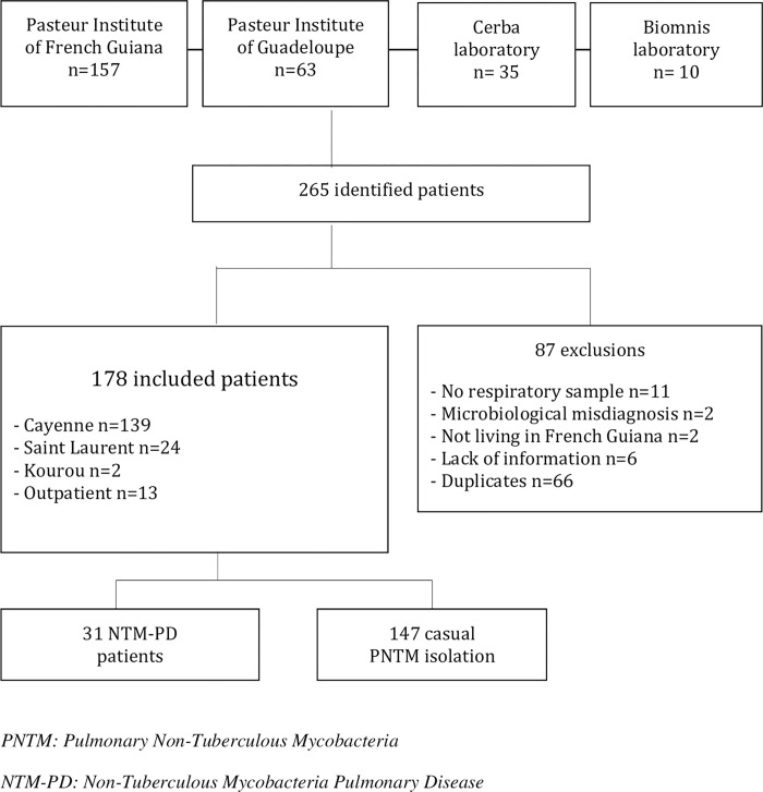
265 patients were identified between January 1st, 2008 to December 31st, 2018. 178 patients had a positive respiratory sample in culture (Fig 1). Thirty-one NTM-PD patients (17%) met the ATS / IDSA diagnostic criteria. Demographic characteristics are summarized in Table 1.

Fig 1. Study flow chart.

Fig 1

Table 1. Demographic characteristics and comorbidities.

Caracteristics Patients
N = 178 %
Demographic data
Men 108/178 61
Children (< 18y) 3/178 2
Age (mean, y), (min–max) 49 9–90
Native country:
    French Guiana 34/155 22
    Neighboring countries (Brazil, Surinam, Guyana) 66/155 43
    Haiti 31/155 20
Precarous living conditions * 96/149 64
No heath insurance coverage 86/144 60
Comorbidities
Chronic pulmonary disease 57/174 33
Chronic Obstructive Pulmonary Disease 31/ 174 17
Bronchiectasies 8/ 174 5
Tuberculosis history 27/174 16
Heart disease history 35/174 20
Arterial hypertension 28/174 16
Diabetes 12/174 7
Cerebrovascular disease** 9/174 5
HIV Infection 77/167 46
    CD4 < 200 49/72 68
    CD4<50 28/72 39
Immunosuppressive therapy 7/178 4
Solid tumors 4/178 2
Undernutrition (BMI<18,5) 37/163 23
Gastroesophageal reflux or gastritis 49/175 28
Smoking 85/148 57
CCI score ***
    Low 46/176 26
    Medium 92/176 52
    High 38/176 22

* Homeless, Prisoners, Jobless, No health insurance coverage, without social subsidies

** Cerebrovascular disease = 7 stroke, 1 cerebral amyloidosis et 1 mental retardation

*** CCI = Charlson Comorbidity Index simplify Low (score = 0)- medium (1–2)- High (3 or more) among: diabetes, chronic pulmonary disease, connective tissue disease, AIDS, moderate or severe kidney or liver failure, cerebrovascular disease, hemiplegia, dementia, solid tumors, hemopatologic maligniancies.

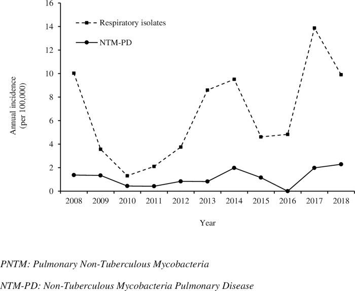
Mean annual incidence over the study period was 6.17 / 100,000 inhabitants / year for overall PNTM positive respiratory sample and 1.07 cases / 100,000 inhabitants / year for NTM-PD. Incidence of NTM-PD remained stable over the years with a slight increase since 2017. The number of casual PNTM isolation tended to increase from 2013 onwards (Fig 2).

Fig 2. Incidence of PNTM infections in French Guiana (2008–2018).

Fig 2

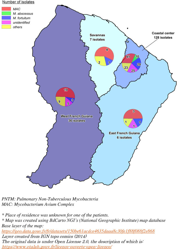
Culture positive samples were mainly obtained from sputum (64%). Bronchoalveolar lavage (BAL) was performed in 35% of NTM-PD (11 out of 31 patients). No pleural fluid, protected brushing, trans-bronchial or surgical lung biopsy were analyzed. The main strain identified was Mycobacterium avium complex (MAC), which was seen in 68 patients (38%), followed by M. fortuitum in 34 patients (19%) and M. abscessus group in 10 patients (6%) (Tables 2 and 3). MAC and M. abscessus were significantly associated with pulmonary disease (p<0.001 and p = 0.02 respectively). M.fortuitum and unidentified and non-pathogenic mycobacteria were significantly associated with a lack of disease (p<0.001, p = 0.01 and p = 0.03 respectively). 31 out of 147 casual PNTM isolation were unidentified species (Table 2). Geographical distribution of PNTM was heterogeneous, with a predominance of Rapid Growing Mycobacteria (RGM) in the coastal central region while MAC showed an even distribution across the region (Fig 3).

Table 2. Identified PNTM strains.

PNTM species NTM-PD n = 31 (17%) Casual PNTM isolation n = 147 (83%) Overall N = 178 (100%)
Slow Growing Mycobacteria (SGM):
M. avium 16 (52%) 18 (12%) 34 (19%)
M. intracellulare 9 (29%) 25 (17%) 34 (19%)
M. asiaticum 0 2 (1%) 2 (1%)
M. interjectum 0 3 (2%) 3/ (2%)
M. kansasii 0 3/ (2%) 3 (2%)
M. lentiflavum 0 1 (1%) 1 (1%)
M. genavense 1 (3%) 0 1 (1%)
M. scrofulaceum 0 7 (5%) 7 (4%)
M. simiae 0 1 (1%) 1 (1%)
M. szulgai 0 2 (1%) 2 (1%)
M. xenopi 0 1 (1%) 1 (1%)
M. gordonae 0 7 (5%) 7 (4%)
M. celatum 0 2 (1%) 2 (1%)
Rapid Growing Mycobacteria (RGM):
M. abscessus 5 (16%) 5 (3%) 10 (6%)
M. fortuitum 0 34 (23%) 34 (19%)
M. smegmatis 0 4 (3%) 4 (2%)
M. mucogenicum 0 1 (1%) 1 (1%)
Unidentified 0 31 (21%) 31 (17%)

Table 3. Significance of main PNTM group in French Guiana.

PNTM species NTM-PD n = 31 (17%) Casual PNTM isolation n = 147 (83%) Overall n = 178 OR 95% CI p
M. avium complex 25 (81) 43 (29) 68 (38) 10, 08 3,65–31,75 < 0,001
Other SGM* 1 (4) 29 (28) 30 (17) 0,10 0,01–0,69 0,008
M. abscessus complex 5 (16) 5 (3) 10 (6) 5,46 1,15–25,23 0,02
M. fortuitum 0 34 (23) 34 (19) 0 0–0,42 <0,001
Other RGM** 0 5 (3) 5 (3) 0 0–12,51 0,55
Unidentified 0 31 (21) 31 (17) 0 0–0,47 0,01
Non pathogenic $ 0 17 (12) 17 (10) 0 0–0,80 0,03
M. tuberculosis coinfection 2 (7) 5 (4) 7 (4) 1,97 0,17–12,74 0,35

* Slow Growing Mycobacteria

** Rapid Growing Mycobacteria

$ M.lentiflavum, M.scrofulaceum, M.simiae, M.gordonae, M.mucogenicum

Fig 3. Distribution of respiratory isolates of PNTM in French Guiana.

Fig 3

Map was created using BdCarto NGI’s (National Geographic Institute) map database. Base layer of the map: https://geo.data.gouv.fr/fr/datasets/150be61acdce4635daaa8c30fe1f88f088f2e868. Layer created from IGN topo comics (2014). The original data is under Open License 2.0, the description of which is: https://www.etalab.gouv.fr/licence-ouverte-open-licence/.

Discussion

To the best of our knowledge, this is the first study describing the incidence and microbiological epidemiology of infections linked to NTM with respiratory expression in French Guiana.

The average annual incidence over the study period was 6.17 / 100,000 inhabitants / year for overall PNTM positive respiratory sample and 1.07 / 100,000 inhabitants / year for NTM-PD. The incidence in French Guiana is lower compared to mainland France incidence, which showed a range between 0.72 to 0.74 / 100,000 inhabitants / year for NTM-PD between 2001 and 2003, followed by 1.3 to 13.6 / 100,000 inhabitants in 2016. It is also important to mention that the range of the incidence in mainland France was dependent on the region that was studied [12,13]. The differences in the incidences could be explained by different study periods, inclusion criteria and NTM-PD definitions. The current study used the 2007 ATS/IDSA criteria. All patients were discussed and classified into either NTM-PD or casual NTM isolation group, which could perhaps underestimate our NTM-PD incidence. However, similar incidence for respiratory specimen and NTM-PD was found in Brazil in 2005 (5.3 and 1.0 / 100,000 inhabitants / year respectively) and Canada (6.5 / 100,000 inhabitants / year on average between 1990 and 2006) also with an increasing in incidence over time [14].

Our findings also showed an increasing trend for NTM isolates from 2013 onwards. This finding could be reflective of the improved diagnostic methods, since the culture on liquid medium of Mycobacteria was only available from 2012 onwards at Pasteur Institute of French Guiana. Nevertheless, 17% of NTM were unidentified. From 2008 to 2015 the identification of NTM was performed at the Pasteur Institute of Guadeloupe and private laboratories using the GenoType Mycobacterium CM and AS kits. This technique unfortunately, does not allow the identification of all species of NTM. More acurate identification of isolates by 16S rDNA and hsp65 sequencing, in future studies, would be helpful in discovering new species, similar to the finding in French Polynesia [15]. Some strains were sent to the Referential National Center (RNC) in Paris for antibiogram susceptibility.

This study showed an increasing trend in the number of NTM-PD from 2017 onwards. Other studies had shown an increasing incidence of NTM-PD between 2008 and 2015 [16]. NTM-PD incidence increases for elderly patients [17]. However, French Guiana’s population is much younger compared to mainland France (median age 28.5 versus 40 in mainland France in 2015). Precariousness is also notably more important (unemployment rate 21.5% in 2015) along with a constantly increasing birth rate [18]. Among the studied population, 46% of patients had HIV infection. French Guiana remains the French territory with the highest tuberculosis declaration rate in 2017 reaching 32.5 / 100,000 inhabitants (2 times higher than in Ile de France and Mayotte Island in the Indian Ocean, 6 times more than in other French regions) [6,19]. These high number might be associated with a significant migratory flow and high HIV infection rate (number of HIV positive findings in 2015: 743 versus 89 per million people in mainland France).

Even in areas where tuberculosis is of major importance, infection with non-tuberculous mycobacteria needs to be considered [20]. Real incidence of NTM-PD may be underestimated, since NTM-PD is not a reportable disease, contrary to tuberculosis. Hence, the creation of a surveillance network of mycobacteria, would probably allow for more precise results.

Additionally, there is a possibility of underestimation of the results due to significant migratory flow in French Guiana, and the likely difficult access to health care [18].

In our study, the main species found in respiratory isolates were MAC, followed by M. fortuitum and M. abscessus. NTM-PD were due to MAC, M. abscessus and M. genavense. MAC is the first NTM found worldwide [21,22] and showed an even distribution across the different regions of French Guiana (Fig 3). Similar results were found in the study by Streit et al [10]. His and Cadelis et al’s teams have shown similar mycobacterial epidemiology in the French West Indies (Guadeloupe and Martinique Islands)[23]. These are tropical French overseas territories, separating the Atlantic Ocean and the Caribbean Sea. The results could be explained by environmental similitaries: climate, atmospheric water vapor, soil, or water exposure. Nevertheless, it differs from the findings of a study in Reunion island, another French island in the Indian Ocean, where M. simiae was seen in 15% of cases of PNTM, while only one isolate was found in this study [24]. M. simiae seems to be more prevalent in contaminated water supplies. Underlying diseases such as cystic or non-cystic fibrosis bronchiectasis and diabetes mellitus were commonly described in others studies, however, they were not frequent in this study [10,24,25].

Regional differences support the hypothesis that habitus and environment are involved in the epidemiology of NTM. In French Guiana, M. abscessus was isolated in patients only in the coastal center region whereas in mainland France the proportion of M. xenopi was more important in Paris [12].

Respiratory mycobacteriosis in French Guiana in this study were due to MAC, M. abscessus and M. genavense. Contrary to mainland France where M. xenopi and M. kansasii were the second and third species to have a clinical impact after MAC [12,26].

M. xenopi and M. kansasii are more prevalent in Chronic Obstructive Pulmonary Disease (COPD) which concerned only 33% of patients of this study [27,28].

M. xenopi is rarely found in South America compared to M. kansasii due to the continental and regional differences [22,29]. M. kansasii seems to be prevelant in industrial regions and is also associated with lifestyle of the patients. For e.g. same species and similar distributions these species can be seen in Brazil and French Guiana. However, a great variability in the distribution of M. kansasii was seen. This variability in distribution was seen due to regional differences (presence of heavy and mainly mining industries) and lifestyle (swimming pools, whirlpool baths, hammam etc) [3032]. In Argentina, the most important pathogen was MAC particularly because of HIV co-infected patients [33]. In Colombia, MAC and M. abscessus were predominant as well [34]. In the current study, 16% of patients of this study had a history of tuberculosis and 46% had HIV infection.

Atypical mycobacteria differences in isolates seem to be correlated with water vapor exposure, which is a more important factor in tropical territories compared to Europe. [35,36]. Water vapour exposure was reported in studies in the USA, where NTM-PD were more predominant on the South coast and Hawaii [37,38]. As suggested by Prevots et al, the use of municipal water could explain why PNTM were more often isolated on the coastal central area, as the main city of Cayenne is located here in French Guiana, where there is significant use of municipal water [36]. French Guiana’s soil is rich in manganese, clay, and the coastal swampy soils have an acidic pH, as described by Joseph Falkinham [39,40]. The temperature of the sea water in French Guiana is quite stable, generally around 27–28°C, while the water in the rivers and creeks is a little cooler. Because of the high temperature throughout the year, water heaters are used less compared to mainland France. In coastal towns and villages, residential areas are well equipped with running water. However, in the slums of the main towns, rainwater is often the primary water source. In the remote communities far from the coast, rainwater collection tanks and small boreholes are used for the daily water supply [41].

M. abscessus is the second or third mycobacteria responsible for diseases on the American continent [29]. It is often associated to underlying pulmonary diseases such as bronchiectasis or COPD [42,43]. In French Guiana, while M. abscessus was the second specie to have a clinical impact, however bronchiectasis and COPD was seen in only 5 and 17% (respectively) of our population. Rapid Growing Mycobacteria (RGM) were predominant in the coastal center region. M. abscessus was found only in the coastal central region. Adjemian and al showed a highest prevalence of M. abscessus around the tropical climate [44]. Because of antibiotic resistance, the pulmonary disease caused by M. abscessus remains difficult to treat. As previously described, M. fortuitum was found in 19% of patients, and an associated with pulmonary disease was not seen [21].

There are several limitations to this study. Since this is a retrospective study, missing data is an unfortunate but an inherent limitation of the study design. As prevalences could not be assessed due to the missing data, we thereby focused on incidences. The incidences were calculated on an average annual incidence. Another limitation could be the lack of compulsory notification of NTM samples to public instances which would adversely affect the reporting of the number of cases. Nevertheless, the collection of respiratory samples was done exhaustively, wherein we included every laboratory analyzing these bacteria. Secondly, every chest scan was re-read by a pulmonologist but not systematically by a radiologist during the study, which could have lead to a classification bias. Thirdly, we used the ATS/IDSA 2007 because the ATS/IDSA 2020 criteria were published after the study had been completed. Nevertheless, 50% of NTM-PD patients had a positive protected sample in addition to their expectorated sputum samples and could agree with the 2020 ATS/ERS/IDSA/ESCMID criteria as well.

Conclusion

Although the incidence of tuberculosis remains the highest in French Guiana, NTM should be given due consideration since its incidence is comparable to other countries and metropolitan France. The microbial epidemiology of NTM in French Guiana is different in comparison to the mainland France due to geographical, environmental and clinical differences, however it is similar to South America and the Caribbean territories. MAC, M. fortuitum and M. abscessus are the most frequently found species. MAC and M. abscessus are mainly responsible for diseases linked to NTM-PD and could be responsible for the adverse social and economic consequences due to the various difficulties associated with treating these diseases in a comparatively more precarious and immunosuppressed population than in mainland France.

Acknowledgments

The autor thanks Mathieu Nacher (Inserm 1424, French Guiana), Hugo Testaert (Pneumology Department, University Hospital of Guadeloupe) and Sadia Khan (Bordeaux University, INSERM, Bordeaux Population Health Research Center, team: EPICENE, UMR1219, Bordeaux, France) for correcting the English. The autor thanks Gilles Chaptal for the realization of the map of French Guiana. The autor also thanks the French Guiana PNTM working group: Philippe Abboud, Houari Aissaoui, Alain Berlioz-Arthaud, Bastien Bidaud, Denis Blanchet, Anne-Marie Bourbigot, Mathilde Boutrou, Jean-Michel Cauvin, Fabrice Flament, Claire Grenier, Hatem Kallel, Anthony Le Labourier, Dominique Louvel, Aude Lucarelli, Aba Mahamat, Alessia Melzani, Balthazar N’tab, Richard Naldjinan-Kodbaye, Milko Sobeski, Antoine Talarmin, Stéphanie Thomas, Valentine Travers, Vincent Vantilke, Tania Vaz, Guillaume Vesin, Gaëlle Walter.

Data Availability

All relevant data are within the manuscript.

Funding Statement

The author(s) received no specific funding for this work.

References

  • 1.Mougari F, Cambau E. Mycobactéries et eau. Rev Francoph Lab. 2014. Mar;2014(460):69–74. [Google Scholar]
  • 2.Douine M, Gozlan R, Nacher M, Dufour J, Reynaud Y, Elguero E et al. Mycobacterium ulcerans infection (Buruli ulcer) in French Guiana, South America, 1969–2013: an epidemiological study. Lancet Planet Health. 2017. May;1(2):e65–73. doi: 10.1016/S2542-5196(17)30009-8 [DOI] [PubMed] [Google Scholar]
  • 3.Griffith DE, Aksamit T, Brown-Elliott BA, Catanzaro A, Daley C, Gordin F et al. An Official ATS/IDSA Statement: Diagnosis, Treatment, and Prevention of Nontuberculous Mycobacterial Diseases. Am J Respir Crit Care Med. 2007. Feb 15;175(4):367–416. doi: 10.1164/rccm.200604-571ST [DOI] [PubMed] [Google Scholar]
  • 4.Daley CL, Iaccarino JM, Lange C, Cambau E, Wallace RJ, Andrejak C et al. Treatment of nontuberculous mycobacterial pulmonary disease: an official ATS/ERS/ESCMID/IDSA clinical practice guideline. Eur Respir J. 2020. Jul;56(1):2000535. doi: 10.1183/13993003.00535-2020 [DOI] [PMC free article] [PubMed] [Google Scholar]
  • 5.INSEE 2015-dossier complet. https://www.insee.fr/fr/statistiques/2011101?geo=DEP-973.
  • 6.INVS- epidémiologie de la tuberculose-2017. http://invs.santepubliquefrance.fr/Dossiers-thematiques/Maladies-infectieuses/Infections-respiratoires/Tuberculose/Donnees-epidemiologiques.
  • 7.Domergue V, Clyti E, Sainte-Marie D, Huber F, Marty C, Couppié P. La lèpre en Guyane française: étude rétrospective de 1997 à 2006. Med Trop 2007. 68(33–37). [PubMed] [Google Scholar]
  • 8.Couppié P, Dufour J, Marty C, Edouard S, Nacher M, Sainte-Marie D. Augmentation du nombre de cas de lèpre en Guyane depuis 2006. Ann Dermatol Vénéréologie. 2012. Dec;139(12):B197–8. [Google Scholar]
  • 9.Legrand E, Sola C, Verdol B, Rastogi N. Genetic diversity of mycobacterium avium recovered from AIDS patients in the caribbean as studied by a consensus IS1245-RFLP method and pulsed-field gel electrophoresis. Res Microbiol. 2000. May;151(4):271–83. doi: 10.1016/s0923-2508(00)00147-9 [DOI] [PubMed] [Google Scholar]
  • 10.Streit E, Millet J, Rastogi N. Nontuberculous Mycobacteria in Guadeloupe, Martinique, and French Guiana from 1994 to 2012. Tuberc Res Treat. 2013;2013:1–8. [DOI] [PMC free article] [PubMed] [Google Scholar]
  • 11.Adenis A, Aznar C, Dufour J, Nacher M, Hanf M, Couppié P et al. What Is AIDS in the Amazon and the Guianas? Establishing the Burden of Disseminated Histoplasmosis. Am J Trop Med Hyg. 2011. Feb 4;84(2):239–40. doi: 10.4269/ajtmh.2011.10-0251 [DOI] [PMC free article] [PubMed] [Google Scholar]
  • 12.Dailloux M, Abalain ML, Laurain C, Lebrun L, Loos-Ayav C, Lozniewski A et al. Respiratory infections associated with nontuberculous mycobacteria in non-HIV patients. Eur Respir J. 2006. Dec 1;28(6):1211–5. doi: 10.1183/09031936.00063806 [DOI] [PubMed] [Google Scholar]
  • 13.Schildkraut JA, Gallagher J, Morimoto K, Lange C, Haworth C, Floto RA et al. Epidemiology of nontuberculous mycobacterial pulmonary disease in Europe and Japan by Delphi estimation. Respir Med. 2020. Nov;173:106164. doi: 10.1016/j.rmed.2020.106164 [DOI] [PubMed] [Google Scholar]
  • 14.Hernández-Garduño E, Rodrigues M, Elwood RK. The incidence of pulmonary non-tuberculous mycobacteria in British Columbia, Canada. Int J Tuberc Lung Dis Off J Int Union Tuberc Lung Dis. 2009. Sep;13(9):1086–93. [PubMed] [Google Scholar]
  • 15.Phelippeau M, Aboubaker Osman D, Musso D, Drancourt M. Epidemiology of Nontuberculous Mycobacteria in French Polynesia. Land GA, editor. J Clin Microbiol. 2015. Dec;53(12):3798–804. doi: 10.1128/JCM.01560-15 [DOI] [PMC free article] [PubMed] [Google Scholar]
  • 16.Winthrop KL, Marras TK, Adjemian J, Zhang H, Wang P, Zhang Q. Incidence and Prevalence of Nontuberculous Mycobacterial Lung Disease in a Large U.S. Managed Care Health Plan, 2008–2015. Ann Am Thorac Soc. 2020. Feb;17(2):178–85. doi: 10.1513/AnnalsATS.201804-236OC [DOI] [PMC free article] [PubMed] [Google Scholar]
  • 17.Henkle E, Hedberg K, Schafer S, Novosad S, Winthrop KL. Population-based Incidence of Pulmonary Nontuberculous Mycobacterial Disease in Oregon 2007 to 2012. Ann Am Thorac Soc. 2015. May;12(5):642–7. doi: 10.1513/AnnalsATS.201412-559OC [DOI] [PMC free article] [PubMed] [Google Scholar]
  • 18.Floraline Cratère. La Guyane, une région jeune et cosmopolite- https://www.insee.fr/fr/statistiques/3695893. INSEE. 2019;(35).
  • 19.CIRE Antilles-Guyane. BVS- Bulletin de veille sanitaire- infections sexuellement transmissibles VIH. 2016;(3). [Google Scholar]
  • 20.Bensi E, Panunto P, Ramos M. Incidence of tuberculous and non-tuberculous mycobacteria, differentiated by multiplex PCR, in clinical specimens of a large general hospital. Clinics. 2013. Feb 28;68(2):179–83. doi: 10.6061/clinics/2013(02)oa10 [DOI] [PMC free article] [PubMed] [Google Scholar]
  • 21.Zweijpfenning S, Ingen J, Hoefsloot W. Geographic Distribution of Nontuberculous Mycobacteria Isolated from Clinical Specimens: A Systematic Review. Semin Respir Crit Care Med. 2018. Jun;39(03):336–42. doi: 10.1055/s-0038-1660864 [DOI] [PubMed] [Google Scholar]
  • 22.Hoefsloot W, van Ingen J, Andrejak C, Ängeby K, Bauriaud R, Bemer P et al. The geographic diversity of nontuberculous mycobacteria isolated from pulmonary samples: an NTM-NET collaborative study. Eur Respir J. 2013. Dec;42(6):1604–13. doi: 10.1183/09031936.00149212 [DOI] [PubMed] [Google Scholar]
  • 23.Cadelis G, Ducrot R, Bourdin A, Rastogi N. Predictive factors for a one-year improvement in nontuberculous mycobacterial pulmonary disease: An 11-year retrospective and multicenter study. Small PLC, editor. PLoS Negl Trop Dis. 2017. Aug 7;11(8):e0005841. doi: 10.1371/journal.pntd.0005841 [DOI] [PMC free article] [PubMed] [Google Scholar]
  • 24.Coolen-Allou N, Touron T, Belmonte O, Gazaille V, Andre M, Allyn J et al. Clinical, Radiological, and Microbiological Characteristics of Mycobacterium simiae Infection in 97 Patients. Antimicrob Agents Chemother. 2018. May 14;62(7):e00395–18. [DOI] [PMC free article] [PubMed] [Google Scholar]
  • 25.El Sahly HM, Septimus E, Soini H, Septimus J, Wallace RJ, Pan X et al. Mycobacterium simiae Pseudo-outbreak Resulting from a Contaminated Hospital Water Supply in Houston, Texas. Clin Infect Dis. 2002. Oct;35(7):802–7. doi: 10.1086/342331 [DOI] [PubMed] [Google Scholar]
  • 26.Provoost J, Valour F, Gamondes D, Roux S, Freymond N, Perrot E et al. A retrospective study of factors associated with treatment decision for nontuberculous mycobacterial lung disease in adults without altered systemic immunity. BMC Infect Dis. 2018. Dec;18(1):659. doi: 10.1186/s12879-018-3559-x [DOI] [PMC free article] [PubMed] [Google Scholar]
  • 27.Andrejak C, Lescure FX, Pukenyte E, Douadi Y, Yazdanpanah Y, Laurans G et al. Mycobacterium xenopi pulmonary infections: a multicentric retrospective study of 136 cases in north-east France. Thorax. 2009. Jan 8;64(4):291–6. doi: 10.1136/thx.2008.096842 [DOI] [PubMed] [Google Scholar]
  • 28.Shitrit D, Baum GL, Priess R, Lavy A, Shitrit ABG, Raz M et al. Pulmonary Mycobacterium kansasii infection in Israel, 1999–2004: clinical features, drug susceptibility, and outcome. Chest. 2006. Mar;129(3):771–6. doi: 10.1378/chest.129.3.771 [DOI] [PubMed] [Google Scholar]
  • 29.Prevots DR, Marras TK. Epidemiology of Human Pulmonary Infection with Nontuberculous Mycobacteria. Clin Chest Med. 2015. Mar;36(1):13–34. [DOI] [PMC free article] [PubMed] [Google Scholar]
  • 30.Kim JH, Seo KW, Shin Y, Oh JS, Jun JB, Jeong J et al. Risk factors for developing Mycobacterium kansasii lung disease: A case-control study in Korea. Medicine (Baltimore). 2019. Feb;98(5):e14281. doi: 10.1097/MD.0000000000014281 [DOI] [PMC free article] [PubMed] [Google Scholar]
  • 31.Chobot S, Malis J, Sebáková H, Pelikán M, Zatloukal O, Palicka P et al. Endemic incidence of infections caused by Mycobacterium kansasii in the Karviná district in 1968–1995 (analysis of epidemiological data—review). Cent Eur J Public Health. 1997. Dec;5(4):164–73. [PubMed] [Google Scholar]
  • 32.Bailey WC, Brown M, Buechner HA, Weill H, Ichinose H, Ziskind M. Silico-mycobacterial disease in sandblasters. Am Rev Respir Dis. 1974. Aug;110(2):115–25. doi: 10.1164/arrd.1974.110.2.115 [DOI] [PubMed] [Google Scholar]
  • 33.Imperiale B, Zumárraga M, Gioffré A, Di Giulio B, Cataldi A, Morcillo N. Disease caused by non-tuberculous mycobacteria: diagnostic procedures and treatment evaluation in the North of Buenos Aires Province. Rev Argent Microbiol. 2012. Mar;44(1):3–9. doi: 10.1590/S0325-75412012000100002 [DOI] [PubMed] [Google Scholar]
  • 34.Llerena C, Valbuena A, Zabaleta AP. Micobacteriosis identificadas en el Laboratorio Nacional de Referencia de Colombia entre 2012 y 2016. Biomédica. 2018. Aug 1;38:87–94. [DOI] [PubMed] [Google Scholar]
  • 35.Adjemian J, Olivier KN, Seitz AE, Falkinham JO, Holland SM, Prevots DR. Spatial Clusters of Nontuberculous Mycobacterial Lung Disease in the United States. Am J Respir Crit Care Med. 2012. Sep 15;186(6):553–8. doi: 10.1164/rccm.201205-0913OC [DOI] [PMC free article] [PubMed] [Google Scholar]
  • 36.Prevots DR, Adjemian J, Fernandez AG, Knowles MR, Olivier KN. Environmental Risks for Nontuberculous Mycobacteria. Individual Exposures and Climatic Factors in the Cystic Fibrosis Population. Ann Am Thorac Soc. 2014. Sep;11(7):1032–8. doi: 10.1513/AnnalsATS.201404-184OC [DOI] [PMC free article] [PubMed] [Google Scholar]
  • 37.Adjemian J, Olivier KN, Seitz AE, Holland SM, Prevots DR. Prevalence of Nontuberculous Mycobacterial Lung Disease in U.S. Medicare Beneficiaries. Am J Respir Crit Care Med. 2012. Apr 15;185(8):881–6. doi: 10.1164/rccm.201111-2016OC [DOI] [PMC free article] [PubMed] [Google Scholar]
  • 38.Adjemian J, Frankland TB, Daida YG, Honda JR, Olivier KN, Zelazny A et al. Epidemiology of Nontuberculous Mycobacterial Lung Disease and Tuberculosis, Hawaii, USA. Emerg Infect Dis. 2017. Mar;23(3):439–47. doi: 10.3201/eid2303.161827 [DOI] [PMC free article] [PubMed] [Google Scholar]
  • 39.F.Colmet. Etude préliminaire des sols de Guyane. https://horizon.documentation.ird.fr/exl-doc/pleins_textes/divers15-11/010010641.pdf. 1953;
  • 40.Falkinham JO. Environmental Sources of Nontuberculous Mycobacteria. Clin Chest Med. 2015. Mar;36(1):35–41. doi: 10.1016/j.ccm.2014.10.003 [DOI] [PubMed] [Google Scholar]
  • 41.Mansotte F, Margueron T, Maison D. L’alimentation en eau potable en Guyane: problématique et solutions appropriées: Santé Publique. 2010. May 17;Vol. 22(2):181–92. [PubMed] [Google Scholar]
  • 42.Roux AL, Catherinot E, Ripoll F, Soismier N, Macheras E, Ravilly S et al. Multicenter Study of Prevalence of Nontuberculous Mycobacteria in Patients with Cystic Fibrosis in France. J Clin Microbiol. 2009. Dec 1;47(12):4124–8. doi: 10.1128/JCM.01257-09 [DOI] [PMC free article] [PubMed] [Google Scholar]
  • 43.Ford ES, Horne DJ, Shah JA, Wallis CK, Fang FC, Hawn TR. Species-Specific Risk Factors, Treatment Decisions and Clinical Outcomes for Laboratory Isolates of Less Common Nontuberculous Mycobacteria in Washington State. Ann Am Thorac Soc. 2017. Apr 7;AnnalsATS.201609-731OC. doi: 10.1513/AnnalsATS.201609-731OC [DOI] [PMC free article] [PubMed] [Google Scholar]
  • 44.Adjemian J, Olivier KN, Prevots DR. Epidemiology of Pulmonary Nontuberculous Mycobacterial Sputum Positivity in Patients with Cystic Fibrosis in the United States, 2010–2014. Ann Am Thorac Soc. 2018. Jul;15(7):817–26. doi: 10.1513/AnnalsATS.201709-727OC [DOI] [PMC free article] [PubMed] [Google Scholar]
PLoS Negl Trop Dis. doi: 10.1371/journal.pntd.0010693.r001

Decision Letter 0

Joseph M Vinetz

30 May 2021

Dear Dr. Chaptal,

Thank you very much for submitting your manuscript "Epidemiology of infection by pulmonary non-tuberculous mycobacteria in French Guiana 2008-2018." for consideration at PLOS Neglected Tropical Diseases. As with all papers reviewed by the journal, your manuscript was reviewed by members of the editorial board and by several independent reviewers. In light of the reviews (below this email), we would like to invite the resubmission of a significantly-revised version that takes into account the reviewers' comments.

We cannot make any decision about publication until we have seen the revised manuscript and your response to the reviewers' comments. Your revised manuscript is also likely to be sent to reviewers for further evaluation.

When you are ready to resubmit, please upload the following:

[1] A letter containing a detailed list of your responses to the review comments and a description of the changes you have made in the manuscript. Please note while forming your response, if your article is accepted, you may have the opportunity to make the peer review history publicly available. The record will include editor decision letters (with reviews) and your responses to reviewer comments. If eligible, we will contact you to opt in or out.

[2] Two versions of the revised manuscript: one with either highlights or tracked changes denoting where the text has been changed; the other a clean version (uploaded as the manuscript file).

Important additional instructions are given below your reviewer comments.

Please prepare and submit your revised manuscript within 60 days. If you anticipate any delay, please let us know the expected resubmission date by replying to this email. Please note that revised manuscripts received after the 60-day due date may require evaluation and peer review similar to newly submitted manuscripts.

Thank you again for your submission. We hope that our editorial process has been constructive so far, and we welcome your feedback at any time. Please don't hesitate to contact us if you have any questions or comments.

Sincerely,

Joseph M. Vinetz

Deputy Editor

PLOS Neglected Tropical Diseases

Joseph Vinetz

Deputy Editor

PLOS Neglected Tropical Diseases

***********************

Reviewer's Responses to Questions

Key Review Criteria Required for Acceptance?

As you describe the new analyses required for acceptance, please consider the following:

Methods

-Are the objectives of the study clearly articulated with a clear testable hypothesis stated?

-Is the study design appropriate to address the stated objectives?

-Is the population clearly described and appropriate for the hypothesis being tested?

-Is the sample size sufficient to ensure adequate power to address the hypothesis being tested?

-Were correct statistical analysis used to support conclusions?

-Are there concerns about ethical or regulatory requirements being met?

Reviewer #1: the analysis is simple and does not require additional work

Reviewer #2: The objectives of the study are clearly defined and presented. The authors used an appropriate study design to reach the objectives of the study, the population that was used was adequately described and the sample size, while quite small, is appropriate for such a rare phenotype. The authors made use of appropriate statistical models and all ethical considerations for the study has been met.

Reviewer #3: The methods were a retrospective review of non-tuberculous mycobacterial disease (specifically pulmonary in origin) in French Guiana in toto. Three major hospitals participated in the inquiry. This was done by first doing an inquiry of mycobacterial cultures from 2008-2018; once patients were identified they went through a second inquiry to look at radiographic evidence of disease and the plurality of the cultures (and of course their source).

The study population was appropriately studied based on the specific aims of the authors and based on the organisms in question the population studied would be limited in number.

Statistic analysis was done via STATA, and demographic collected via Microsoft Excel; this kind of analysis is on par with other studies of the same caliber.

The authors state that this their studies were notified to the "National Commission for Data Protection", but it is unknown if this study was approved by a governing board (such as an institutional review board or IRB) for human protections or if there was any study protocols which took safeguards into account for this project. There is no indication that there are ethical issues, but this should be explained in the Methods section of the manuscript.

--------------------

Results

-Does the analysis presented match the analysis plan?

-Are the results clearly and completely presented?

-Are the figures (Tables, Images) of sufficient quality for clarity?

Reviewer #1: the results are sufficiently clear given the simplicity of the analysis

Reviewer #2: The results as presented does match the analysis plan. The results are clearly presented and are complete, while all figures and tables are of sufficient quality. There are some editorial issues with the figures and tables that I will point out under the Editorial and Data Presentation Modification section of this review.

Reviewer #3: The results were interesting and they are clearly presented. This is especially true since they mirror what is seen in other tropical/subtropical areas of the planet that have NTM infections.

Tables were adequate, however there should be legends for both the tables and figures. For the figures: Figure 1 appeared to be of poor resolution and thus was blurry to read; Figure 2 had line markers that were difficult to see, and thus using a different scheme for the appearance of the two lines would be better; Figure 3 was satisfactory, yet the font color for the numbers within the pie charts were difficult to read against the background and thus should be more uniform with a darker color.

--------------------

Conclusions

-Are the conclusions supported by the data presented?

-Are the limitations of analysis clearly described?

-Do the authors discuss how these data can be helpful to advance our understanding of the topic under study?

-Is public health relevance addressed?

Reviewer #1: (No Response)

Reviewer #2: The conclusions that the authors draw for their study are supported by the data. They do acknowledge that there are some limitations to the study and these study limitations are clearly and frankly discussed. The impact on public health is not discussed in detail and the authors do not really discuss how the results of their study advances what is known about NTMs. However, the study is the first to investigate and report on the prevalence of NTMs in French Guiana, it does have significant public health issues for the region.

Reviewer #3: The final conclusions are supported by the data presented and the limitations were appropriate for this kind of study. The real issue with this section was the Discussion. It was not presented in a way that the reader could easily follow. Also, the vernacular seemed to be less put together contrasted with what was seen in the rest of the manuscript. This made the issue of understanding the conclusions within the discussion more problematic and thus less impactful. A good example of this is noted on Pages 8-10. The authors go from documenting Canadian rates of NTM prevalence and then diving into France and what is seen there to coastal Brazil and then back to France when it comes to their MTB prevalence. Also there is no data discussed about what is transpiring in other nations such as the United States, Mexico or other countries of South America. Furthermore, there are minimal mentioning of association with other diseases which are more frequently associated with NTM mentioned in the manuscript (as was illustrated in Table 1). One would ask the authors if there was any data to suggest that M. xenopi is seen in more temperate/cooler climates compared to M. abscessus which is seen in more tropical/subtropical climates? Data from Rebecca Prevots group at National Institutes of Health (USA) and Theodore Marras' group at University of Toronto have both looked at this in the past. Both are in the references, but their data is important to review and thus make the argument for the rigor of what is documented in these data.

Finally, would include any supporting conclusions about whether soil composition, potable water methods used in French Guiana or other factors could be supporting the results seen in this investigation. This is touched upon, but there is no real hypothesis and literature review regarding this topic. Manuscripts by Joseph Falkinham's group has documented this well and has reasoning for potential hypotheses that would make the case stronger for these data.

--------------------

Editorial and Data Presentation Modifications?

Use this section for editorial suggestions as well as relatively minor modifications of existing data that would enhance clarity. If the only modifications needed are minor and/or editorial, you may wish to recommend “Minor Revision” or “Accept”.

Reviewer #1: (No Response)

Reviewer #2: There are a few issues with the way that the results are presented.

1) In table 1, the 1.1.1.1.1.1. in the CCI score (in column 2) should be removed.

2) In table 1, the ** denoting cerebrovascular disease mentions 6 stroke, 1 cerebral amyloidosis and 1 mental retardation. This adds up to 8, but in the table, it indicates that there are 9 cerebrovascular disease. Please reconcile what is in the legend with what is in the table.

3) There needs to be consistency between the labeling and heading and titles used in the table. In table 2, "Pulmonary NTM" is used while in table 3, "PNTM" is used. Please choose one and stick with it.

4) In the tables, the number of patients in each category (n=31 for NTM-PD and 147 for Causal PNTM) is already stated in the heading, so there is no need to have the number in each cell as well. For example, instead of writing 16/31 (52%), it is sufficient to just write 16 (52%).

5) Table 3 shows a comparison between the NTM-PD and Causal PNTM, however this is not very clear from the manuscript. Please make it clear in the text that this what is shown in table 3. Also, this is should be discussed more in the results.

6) In table 3, some of the p-values are in bold. I assume that these indicate significant differences between the two groups. However, there are some p-values which are less than 0.05 are not shown in bold. This needs to be fixed or changed in the table.

7) Figure 3: The pie charts in the chart key are confusing and not needed. These seem to denote the number of isolates, which are indicated as 20, 10 and 2. This does not correspond to the pie charts on the figure. Why are some of the pie charts on the figure bigger than the others?

8) Figure 3: The total number of isolated indicated on the table is 177, while in the tables the total number of isolates in the study is indicated as 178. Please check and fix. I noticed that in figure 3, the total number of MAC is 67, while in the tables it is 68.

Reviewer #3: As mentioned above, there needs to be a better approach to the figures/tables as they are the fastest way to convey data to the readers of PLOS NTD. In addition, the discussion needs to be more streamlined in it's message and use appropriate English.

--------------------

Summary and General Comments

Use this section to provide overall comments, discuss strengths/weaknesses of the study, novelty, significance, general execution and scholarship. You may also include additional comments for the author, including concerns about dual publication, research ethics, or publication ethics. If requesting major revision, please articulate the new experiments that are needed.

Reviewer #1: The article is an interesting addition to the literature. however, there is room for improvement

the introduction should explicitly mention that the particular amazonian ecosystem makes the question interesting at least in my view this is what the added value of the paper is

in line with this i find the discussion of the differences between locations canada france etc hard to follow. it may be helped by a comparative table of the ranking of different studies in different sites (tropical non tropical) proportion of hiv perhaps

the authors mention that "This could be one of the explanations of the

low proportion of M. kansasii found in French Guiana since this species is the adage of HIVpositive

patients with CD4> 200 / mm3 (18). " i think this is wrong because most hiv patients in french guiana have cd4 counts above 200

although it is a minor point the english language should be improved.

Reviewer #2: In general the article is well-written with and easy to understand. There are concerns about the figures and tables that I highlighted. Some additional comments:

1) Please make sure that all abbreviations are adequately defined. For example, on page 7, the abbreviation INSEE is used. Please define this abbreviation the first time it is used.

2)On page 10, the authors talk about a decrease in the number of HIV positive cases and says that it remains high with 743 million inhabitants (versus 89 million in mainland France). Do the authors mean that there were 743 million people wit HIV in French Guiana vs 89 Million in mainland France? This cannot be possible. Please rephrase the sentence so that it explains these numbers.

Reviewer #3: I have been asked to review Chaptal et al. "Epidemiology of infection by pulmonary non-tuberculous mycobacteria in French

Guiana 2008-2018." This is a retrospective review of cases of non-tuberculous mycoabcteria (NTM) in the French territory of Guiana in South America. This was done in collaboration with academic institutions in both the territory and in France as well as with a consortium of NTM researchers across the Atlantic. Since this is an emerging area of not only infectious diseases, but also in epidemiology, this manuscript has an important impact message to the readers of the journal. Thus, the significance of the study is not only appropriate, but also is necessary as mycobacteria are ubiquitous in the environment as well as becoming studied more frequently as a cause of human disease. The study methods and execution were sound. The real weaknesses were not with the study, but how the data was put together and illustrated. This was evident in the Figures/Tables and the Discussion.

Thus, the manuscript requires major revision of the following:

1) addition of an ethical statement regarding the use of patient sensitive information and how that information was reviewed and safeguarded. This is generally important when dealing with data mined from patient charts and thus patient information should not be compromised.

2) Figures and tables need to be more clear and the data illustrated in a more appropriate form. All figures/tables should come with legends explaining the data.

3) The Discussion section appeared to not be written in the same format as the rest of the manuscript and thus needs major revision and rewording to accomplish the illustration of what these data demonstrate. Someone proficient in the scientific use of English would be helpful to have on hand to proofread the manuscript. Lastly, the data here are important to document, however the hypotheses as to why these data are the way the way they are is critical to what the authors are trying to accomplish. A more intense literature review and understanding of what is happening in environment and making the correlation with the clinical human disease is paramount.

What is documented here is also documented in the above sections.

--------------------

PLOS authors have the option to publish the peer review history of their article (what does this mean?). If published, this will include your full peer review and any attached files.

If you choose “no”, your identity will remain anonymous but your review may still be made public.

Do you want your identity to be public for this peer review? For information about this choice, including consent withdrawal, please see our Privacy Policy.

Reviewer #1: No

Reviewer #2: No

Reviewer #3: No

Figure Files:

While revising your submission, please upload your figure files to the Preflight Analysis and Conversion Engine (PACE) digital diagnostic tool, https://pacev2.apexcovantage.com. PACE helps ensure that figures meet PLOS requirements. To use PACE, you must first register as a user. Then, login and navigate to the UPLOAD tab, where you will find detailed instructions on how to use the tool. If you encounter any issues or have any questions when using PACE, please email us at figures@plos.org.

Data Requirements:

Please note that, as a condition of publication, PLOS' data policy requires that you make available all data used to draw the conclusions outlined in your manuscript. Data must be deposited in an appropriate repository, included within the body of the manuscript, or uploaded as supporting information. This includes all numerical values that were used to generate graphs, histograms etc.. For an example see here: http://www.plosbiology.org/article/info%3Adoi%2F10.1371%2Fjournal.pbio.1001908#s5.

Reproducibility:

To enhance the reproducibility of your results, we recommend that you deposit your laboratory protocols in protocols.io, where a protocol can be assigned its own identifier (DOI) such that it can be cited independently in the future. Additionally, PLOS ONE offers an option to publish peer-reviewed clinical study protocols. Read more information on sharing protocols at https://plos.org/protocols?utm_medium=editorial-email&utm_source=authorletters&utm_campaign=protocols

PLoS Negl Trop Dis. doi: 10.1371/journal.pntd.0010693.r003

Decision Letter 1

Joseph M Vinetz

1 Nov 2021

Dear Dr. Chaptal,

Thank you very much for submitting your manuscript "Epidemiology of infection by pulmonary non-tuberculous mycobacteria in French Guiana 2008-2018." for consideration at PLOS Neglected Tropical Diseases. As with all papers reviewed by the journal, your manuscript was reviewed by members of the editorial board and by several independent reviewers. The reviewers appreciated the attention to an important topic. Based on the reviews, we are likely to accept this manuscript for publication, providing that you modify the manuscript according to the review recommendations.

Please prepare and submit your revised manuscript within 30 days. If you anticipate any delay, please let us know the expected resubmission date by replying to this email.

When you are ready to resubmit, please upload the following:

[1] A letter containing a detailed list of your responses to all review comments, and a description of the changes you have made in the manuscript.

Please note while forming your response, if your article is accepted, you may have the opportunity to make the peer review history publicly available. The record will include editor decision letters (with reviews) and your responses to reviewer comments. If eligible, we will contact you to opt in or out

[2] Two versions of the revised manuscript: one with either highlights or tracked changes denoting where the text has been changed; the other a clean version (uploaded as the manuscript file).

Important additional instructions are given below your reviewer comments.

Thank you again for your submission to our journal. We hope that our editorial process has been constructive so far, and we welcome your feedback at any time. Please don't hesitate to contact us if you have any questions or comments.

Sincerely,

Joseph M. Vinetz

Deputy Editor

PLOS Neglected Tropical Diseases

***********************

Editorial comments: The editors concur with the review. Considerable effort needs to be made to improve the writing, as this journal does not provide copyediting services. The article is published as is from the authors.

Reviewer's Responses to Questions

Key Review Criteria Required for Acceptance?

As you describe the new analyses required for acceptance, please consider the following:

Methods

-Are the objectives of the study clearly articulated with a clear testable hypothesis stated?

-Is the study design appropriate to address the stated objectives?

-Is the population clearly described and appropriate for the hypothesis being tested?

-Is the sample size sufficient to ensure adequate power to address the hypothesis being tested?

-Were correct statistical analysis used to support conclusions?

-Are there concerns about ethical or regulatory requirements being met?

Reviewer #3: The methods of this revised manuscript were not really modified, but requested changes were made to the points from the reviewers. This includes a statement about the institutional review board for these types of studies that was requested by this reviewer. As mentioned previously, the study design does address the stated objectives and discusses the methods used in this epidemiological analysis.

--------------------

Results

-Does the analysis presented match the analysis plan?

-Are the results clearly and completely presented?

-Are the figures (Tables, Images) of sufficient quality for clarity?

Reviewer #3: The revisions made by authors as suggested by the reviewers were done and more. The results go along with the aims and hypotheses made by the authors at the beginning. The figures still are of low quality for PLOS NTD and would strongly advise the authors to counsel with a professional at making figures for journals for a revision. These figures are what readers of PLOS NTD are going to be looking at while reading the prose of the manuscript. Quality control for journals can only do so much when the figures are out of focus based on the medium in which they were created.

--------------------

Conclusions

-Are the conclusions supported by the data presented?

-Are the limitations of analysis clearly described?

-Do the authors discuss how these data can be helpful to advance our understanding of the topic under study?

-Is public health relevance addressed?

Reviewer #3: Yes, the conclusions are supported by the data analysis and confirm the authors suspicions based on the climate and the socioeconomic status of the investigation subjects. The comparisons to both it's southern neighbor and mainland France do offer some perspective on this. It would be interesting to also note some of the basic information about water temperature in French Guiana and if water heaters are used as compared to mainland France. This is the hypothesis of Dr. Marras and colleagues at U Toronto. Some basic information on this as well as how much of the country uses well water or rain catchment for their fresh water source would be helpful in solidifying the conclusions based on their hypotheses.

--------------------

Editorial and Data Presentation Modifications?

Use this section for editorial suggestions as well as relatively minor modifications of existing data that would enhance clarity. If the only modifications needed are minor and/or editorial, you may wish to recommend “Minor Revision” or “Accept”.

Reviewer #3: Please see above in the 'Conclusions' section of the review. English used in the manuscript is better, yet would still suggest that this is reviewed by one who has published in an English-language journal to optimize the vernacular and enhance the point they are trying to make with this manuscript.

--------------------

Summary and General Comments

Use this section to provide overall comments, discuss strengths/weaknesses of the study, novelty, significance, general execution and scholarship. You may also include additional comments for the author, including concerns about dual publication, research ethics, or publication ethics. If requesting major revision, please articulate the new experiments that are needed.

Reviewer #3: This is the second review for the manuscript entitled, "Epidemiology of infection by pulmonary non-tuberculous mycobacteria in French Guiana 2008-2018." The manuscript was reviewed previously by this reviewer; the premise of the study is to determine the epidemiology of non-tuberculous mycobacterial disease in French Guiana, which is part of France. The aim and hypotheses are the same as was the data analysis, which demonstrates that Mycobacterium avium Complex and Mycobacterium abscessus dominate as opposed to mainland France which has more M xenopi as part of the infections of NTM there. This is an important study as it demonstrates data from a part of South America which has minimal reporting.

The manuscript still has some lingering issues; those being manuscript English, details about fresh water source for the populations in the various municipalities and Figures, which need to be improved dramatically for PLOS publication standards.

Minor revision should be instituted for the above commentary prior to acceptance.

--------------------

PLOS authors have the option to publish the peer review history of their article (what does this mean?). If published, this will include your full peer review and any attached files.

If you choose “no”, your identity will remain anonymous but your review may still be made public.

Do you want your identity to be public for this peer review? For information about this choice, including consent withdrawal, please see our Privacy Policy.

Reviewer #3: No

Figure Files:

While revising your submission, please upload your figure files to the Preflight Analysis and Conversion Engine (PACE) digital diagnostic tool, https://pacev2.apexcovantage.com. PACE helps ensure that figures meet PLOS requirements. To use PACE, you must first register as a user. Then, login and navigate to the UPLOAD tab, where you will find detailed instructions on how to use the tool. If you encounter any issues or have any questions when using PACE, please email us at figures@plos.org.

Data Requirements:

Please note that, as a condition of publication, PLOS' data policy requires that you make available all data used to draw the conclusions outlined in your manuscript. Data must be deposited in an appropriate repository, included within the body of the manuscript, or uploaded as supporting information. This includes all numerical values that were used to generate graphs, histograms etc.. For an example see here: http://www.plosbiology.org/article/info%3Adoi%2F10.1371%2Fjournal.pbio.1001908#s5.

Reproducibility:

To enhance the reproducibility of your results, we recommend that you deposit your laboratory protocols in protocols.io, where a protocol can be assigned its own identifier (DOI) such that it can be cited independently in the future. Additionally, PLOS ONE offers an option to publish peer-reviewed clinical study protocols. Read more information on sharing protocols at https://plos.org/protocols?utm_medium=editorial-email&utm_source=authorletters&utm_campaign=protocols

References

Please review your reference list to ensure that it is complete and correct. If you have cited papers that have been retracted, please include the rationale for doing so in the manuscript text, or remove these references and replace them with relevant current references. Any changes to the reference list should be mentioned in the rebuttal letter that accompanies your revised manuscript. If you need to cite a retracted article, indicate the article's retracted status in the References list and also include a citation and full reference for the retraction notice.

PLoS Negl Trop Dis. doi: 10.1371/journal.pntd.0010693.r005

Decision Letter 2

Joseph M Vinetz

4 Jun 2022

Dear Dr. Chaptal,

Thank you very much for submitting your manuscript "Epidemiology of infection by pulmonary non-tuberculous mycobacteria in French Guiana 2008-2018." for consideration at PLOS Neglected Tropical Diseases. As with all papers reviewed by the journal, your manuscript was reviewed by members of the editorial board and by several independent reviewers. The reviewers appreciated the attention to an important topic. Based on the reviews, we are likely to accept this manuscript for publication, providing that you modify the manuscript according to the review recommendations.

Please prepare and submit your revised manuscript within 30 days. If you anticipate any delay, please let us know the expected resubmission date by replying to this email.

When you are ready to resubmit, please upload the following:

[1] A letter containing a detailed list of your responses to all review comments, and a description of the changes you have made in the manuscript.

Please note while forming your response, if your article is accepted, you may have the opportunity to make the peer review history publicly available. The record will include editor decision letters (with reviews) and your responses to reviewer comments. If eligible, we will contact you to opt in or out

[2] Two versions of the revised manuscript: one with either highlights or tracked changes denoting where the text has been changed; the other a clean version (uploaded as the manuscript file).

Important additional instructions are given below your reviewer comments.

Thank you again for your submission to our journal. We hope that our editorial process has been constructive so far, and we welcome your feedback at any time. Please don't hesitate to contact us if you have any questions or comments.

Sincerely,

Joseph M. Vinetz

Deputy Editor

PLOS Neglected Tropical Diseases

Joseph Vinetz

Deputy Editor

PLOS Neglected Tropical Diseases

***********************

Figure Files:

While revising your submission, please upload your figure files to the Preflight Analysis and Conversion Engine (PACE) digital diagnostic tool, https://pacev2.apexcovantage.com. PACE helps ensure that figures meet PLOS requirements. To use PACE, you must first register as a user. Then, login and navigate to the UPLOAD tab, where you will find detailed instructions on how to use the tool. If you encounter any issues or have any questions when using PACE, please email us at figures@plos.org.

Data Requirements:

Please note that, as a condition of publication, PLOS' data policy requires that you make available all data used to draw the conclusions outlined in your manuscript. Data must be deposited in an appropriate repository, included within the body of the manuscript, or uploaded as supporting information. This includes all numerical values that were used to generate graphs, histograms etc.. For an example see here: http://www.plosbiology.org/article/info%3Adoi%2F10.1371%2Fjournal.pbio.1001908#s5.

Reproducibility:

To enhance the reproducibility of your results, we recommend that you deposit your laboratory protocols in protocols.io, where a protocol can be assigned its own identifier (DOI) such that it can be cited independently in the future. Additionally, PLOS ONE offers an option to publish peer-reviewed clinical study protocols. Read more information on sharing protocols at https://plos.org/protocols?utm_medium=editorial-email&utm_source=authorletters&utm_campaign=protocols

References

Please review your reference list to ensure that it is complete and correct. If you have cited papers that have been retracted, please include the rationale for doing so in the manuscript text, or remove these references and replace them with relevant current references. Any changes to the reference list should be mentioned in the rebuttal letter that accompanies your revised manuscript. If you need to cite a retracted article, indicate the article's retracted status in the References list and also include a citation and full reference for the retraction notice.

PLoS Negl Trop Dis. doi: 10.1371/journal.pntd.0010693.r007

Decision Letter 3

Joseph M Vinetz

22 Jul 2022

Dear Dr. Chaptal,

We are pleased to inform you that your manuscript 'Epidemiology of infection by pulmonary non-tuberculous mycobacteria in French Guiana 2008-2018.' has been provisionally accepted for publication in PLOS Neglected Tropical Diseases.

Before your manuscript can be formally accepted you will need to complete some formatting changes, which you will receive in a follow up email. A member of our team will be in touch with a set of requests.

Please note that your manuscript will not be scheduled for publication until you have made the required changes, so a swift response is appreciated.

IMPORTANT: The editorial review process is now complete. PLOS will only permit corrections to spelling, formatting or significant scientific errors from this point onwards. Requests for major changes, or any which affect the scientific understanding of your work, will cause delays to the publication date of your manuscript.

Should you, your institution's press office or the journal office choose to press release your paper, you will automatically be opted out of early publication. We ask that you notify us now if you or your institution is planning to press release the article. All press must be co-ordinated with PLOS.

Thank you again for supporting Open Access publishing; we are looking forward to publishing your work in PLOS Neglected Tropical Diseases.

Best regards,

Joseph M. Vinetz

Section Editor

PLOS Neglected Tropical Diseases

Joseph Vinetz

Section Editor

PLOS Neglected Tropical Diseases

***********************************************************

PLoS Negl Trop Dis. doi: 10.1371/journal.pntd.0010693.r008

Acceptance letter

Joseph M Vinetz

1 Sep 2022

Dear Dr. Chaptal,

We are delighted to inform you that your manuscript, "Epidemiology of infection by pulmonary non-tuberculous mycobacteria in French Guiana 2008-2018.," has been formally accepted for publication in PLOS Neglected Tropical Diseases.

We have now passed your article onto the PLOS Production Department who will complete the rest of the publication process. All authors will receive a confirmation email upon publication.

The corresponding author will soon be receiving a typeset proof for review, to ensure errors have not been introduced during production. Please review the PDF proof of your manuscript carefully, as this is the last chance to correct any scientific or type-setting errors. Please note that major changes, or those which affect the scientific understanding of the work, will likely cause delays to the publication date of your manuscript. Note: Proofs for Front Matter articles (Editorial, Viewpoint, Symposium, Review, etc...) are generated on a different schedule and may not be made available as quickly.

Soon after your final files are uploaded, the early version of your manuscript will be published online unless you opted out of this process. The date of the early version will be your article's publication date. The final article will be published to the same URL, and all versions of the paper will be accessible to readers.

Thank you again for supporting open-access publishing; we are looking forward to publishing your work in PLOS Neglected Tropical Diseases.

Best regards,

Shaden Kamhawi

co-Editor-in-Chief

PLOS Neglected Tropical Diseases

Paul Brindley

co-Editor-in-Chief

PLOS Neglected Tropical Diseases

Associated Data

    This section collects any data citations, data availability statements, or supplementary materials included in this article.

    Supplementary Materials

    Attachment

    Submitted filename: 2_revision_PLOS_list of responses for reviewers.docx

    Attachment

    Submitted filename: 3-revision-PLOS-comments-R2.docx

    Attachment

    Submitted filename: 4-revision-PLOS-comments-R3.docx

    Data Availability Statement

    All relevant data are within the manuscript.


    Articles from PLoS Neglected Tropical Diseases are provided here courtesy of PLOS

    RESOURCES