Abstract
Diabetic nephropathy (DN) is a common complication of diabetes mellitus. While there has been a great advance in our understanding of the pathogenesis of DN, no effective managements of this chronic kidney disease are currently available. Therefore, continuing to elucidate the underlying biochemical and molecular mechanisms of DN remains a constant need. In this regard, animal models of diabetes are indispensable tools. This review article highlights a widely used rodent model of non-obese type 2 diabetes induced by nicotinamide (NA) and streptozotocin (STZ). The mechanism underlying diabetes induction by combining the two chemicals involves blunting the toxic effect of STZ by NA so that only a percentage of β cells are destroyed and the remaining viable β cells can still respond to glucose stimulation. This NA-STZ animal model, as a platform for the testing of numerous antidiabetic and renoprotective materials, is also discussed. In comparison with other type 2 diabetic animal models, such as high-fat-diet/STZ models and genetically engineered rodent models, the NA-STZ model is non-obese and is less time-consuming and less expensive to create. Given that this unique model mimics certain pathological features of human DN, this model should continue to find its applications in the field of diabetes research.
Keywords: nicotinamide, streptozotocin, diabetic nephropathy, diabetic kidney disease, type 2 diabetes
1. Introduction
Diabetic nephropathy (DN), also known as diabetic kidney disease (DKD) [1,2,3], is a severe complication of diabetes mellitus [4,5]. Approximately 30% of diabetic patients can develop DN [6,7,8], which is also a chronic kidney disease and can progress to end-stage renal failure [9,10,11]. The hallmarks of DN are kidney hypertrophy [12,13,14], mesangial cell proliferation and mesangial matrix accumulation [15,16,17], glomerulosclerosis, and persistent levels of proteinuria [18,19]. Despite the great advancement in our understanding of the pathogenesis of DN and numerous approaches that have been tested to slow down DN development and progression, no effective therapeutics are currently available for the treatment of DN. This is because the specific mechanisms underlying the pathogenesis of DN are yet to be fully elucidated. Therefore, there are unmet needs in treating or halting DN. In this regard, animal models of diabetes [20], be they genetically engineered or chemically or dietary induced, are indispensable tools in DN research.
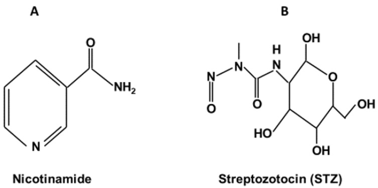
In this brief review, I focus on one particular type 2 diabetes animal model, which is created by using nicotinamide (NA, Figure 1A) and streptozotocin (STZ, Figure 1B) [21,22,23]. This non-obese type 2 diabetes animal model was initially established in rats [24] but has since been extended to include mice with modifications of the original protocol. In comparison with the high-fat-diet (HFD)-STZ-induced type 2 diabetes animal models [25,26,27,28] and genetically engineered diabetic animal models, such as db/db mice [29] and zsf1 obese rats [30], the NA/STZ model is time-saving and less expensive. Therefore, this model can be equally used as a platform for not only exploring the pathogenesis of DN but also screening and testing potential antidiabetic agents [31] or renoprotective compounds for their therapeutic effects [21].
Figure 1.
Chemical structures of nicotinamide and streptozotocin. (A): nicotinamide; (B): streptozotocin.
2. Mechanisms Underlying NA/STZ Diabetes Induction
Masiello P. et al. initially developed this non-obese type 2 diabetes in rats in 1998 [24]. Since then, this model has been widely used to test a variety of antidiabetic materials for their beneficial effects on diabetes and diabetic complications, including DN. The establishment of this model takes advantage of the contradictory effects of the two chemicals on β cells as STZ is β cell cytotoxic while NAD is globally cytoprotective. Therefore, STZ-induced β cell damage can be blunted by nicotinamide [23]. Consequently, a certain percentage of β cells are viable and respond to glucose stimulation to release insulin [24]. It should be noted that the percentage of β cells that can survive really depends on the doses of the two chemicals. For a fixed dose of STZ, if the NA is too low, there will be no blunting effect from the NA and all β cells can be destroyed by STZ. On the other hand, if the NA is too high, the blunting or protective effects of the NA could be too high. In fact, the blunting or protective effects of the NA could reach 100% and no diabetes would be induced. For NA to play a protective role, NA is often given before STZ administration. Nonetheless, that NA is given shortly after STZ ingestion has also been reported in the literature [32,33,34,35]. In these cases, however, whether there are any differences between NA being given first or STZ being given first in diabetes induction and the severity of kidney injury has not been investigated. Notwithstanding, based on the observation that NA given immediately after STZ is equally protective [36], any difference should be minimal when the NA is administered right after STZ administration.
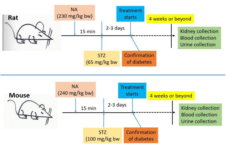
STZ is a nitrosourea compound that has a component similar to glucose (Figure 1B) [37]. Hence, STZ is also known as a glucose analog [37]. Because of this structural similarity to glucose, the STZ enters into β cells via the glucose transporter-2 (Glut2) [37] that is abundantly expressed on the β cell surface [38]. Once inside the β cells, the nitrosoamide moiety of STZ can attack DNA and causes DNA alkylation and is thus responsible for STZ genotoxicity and cytotoxicity [23]. STZ-caused DNA damage can activate poly (ADP-ribose) polymerase-1 (PARP-1) that can then repair damaged DNA using NAD+ as a substrate [39]. As a result, NAD+ could be potentially depleted by the activated PARP-1 [23], thereby leading to cell death. When NA is administered prior to STZ administration, the damaging effect of STZ is greatly mitigated. This mitigating effect is thought to be due to two establishments. One is that NA is a direct inhibitor of PARP-1 [23], the other is that NA is a precursor of NAD+ [23]. Hence, the STZ cytotoxic effects can be greatly blunted by NA and the blunting magnitude is known to be NA-concentration-dependent [23]. In rats, although different investigators would use a different dosage combination of NA and STZ, the initially established dosages of NA and STZ (230 mg/kg and 65 mg/kg, respectively) still seem to prevail in the literature (Figure 2), though the application of lower concentrations of NA and STZ has been reported. In mice, the ingested concentrations of the two chemicals also vary widely. Nevertheless, it should be noted that the concentration of STZ for mice can be higher than that for rats. It appears that the use of 240 mg/kg NA and 100 mg/kg STZ in a mouse model is a prevailing approach in the literature [40]. It should also be noted that when a mouse is used as a model, multiple daily injections of NA and STZ (up to a week) may be conducted [41]. Certain investigators have also reported using high-fat-diet (HFD) feeding followed by NA and STZ ingestions [40]. Regardless of whether rats or mice are being modeled, the key point is that a given investigator should stick to their own protocol of NA and STZ administrations, such as the dosages and routes of chemical ingestions [21,23], so that a reproducibility and data comparison could potentially be achieved. It should also be noted that the severity of the diabetic disorders depends on how long the animals are kept after diabetes induction by the two chemicals. In the absence of any interventions, diabetic disorders will progress, mimicking various stages of clinical practice in humans.
Figure 2.
Diagrams showing representative flow charts of non-obese type 2 diabetes animal models induced by nicotinamide and streptozotocin. Renoprotective materials can be tested for their beneficial effects by this model, which is also outlined in the diagram. Note that for mice to be used as a model, more than one NA and STZ injection may be performed. Depending on the objective of a given study, the mouse model of NA-STZ diabetes induction may also involve HFD feeding for weeks before NA and STZ administrations (please see the text for a detailed discussion).
Additionally, based on a modified mouse model of type 2 diabetes induced by combining the HFD-NA-STZ treatments [42], investigations of DN created by HFD feeding in conjunction with NA and STZ administrations have also been reported [43,44]. It should be noted that when an HFD is involved, the creation of such a model would take longer than when only NA and STZ are used.
3. Renal Pathophysiology in this NA/STZ Animal Model
It has been reported that when diabetes was induced in mice by injection of 230 mg/kg NAD along with 50 mg/kg or 65 mg/kg STZ, the kidney organ index (kidney weight vs. body weight) for both STZ doses showed an increase when compared with that of the controls [45]. For a six-week duration of testing, urinary and serum parameters, such as creatinine, urea, and uric acid, were enhanced in the NA-STZ diabetic animals. In the presence of NAD, mice lived longer than those that received only STZ administration [45]. Such a result further demonstrates the blunting effects of NA on STZ cytotoxicity.
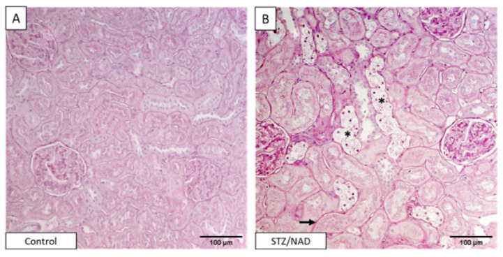
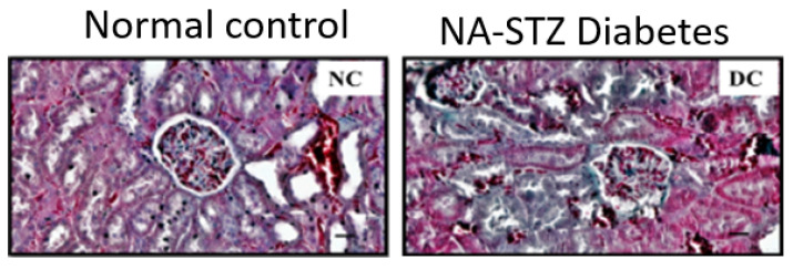
It should be noted that while DN can be classified into five stages (Table 1), none of the NA-STZ-involved animal studies published so far have systematically addressed the five stages of diabetic kidney injury. Therefore, future studies on the progression of DN from stage 1 to stage 5 in the NA-STZ animal model need to be conducted. Moreover, numerous diabetic kidney injury biomarkers, such as those recently reported by Pelle et al. [46] and Natesan et al. [11], have also not been systematically and comprehensively evaluated in this NA-STZ animal model. Most studies use popular kidney injury parameters [47] such as blood urea nitrogen (BUN), serum cystatin C, creatinine, uric acid, or/and estimated glomerular flow rate (eGFR) for the evaluation of diabetic kidney injury after NA-STZ injections. The histopathological staining of kidney is also frequently used for the analysis of kidney injury in this NA-STZ animal model. Figure 3 and Figure 4 [48,49] show a typical staining of the kidney tissues by the periodic acid–Schiff and Masson trichrome, respectively. As can be observed from these histochemical stainings, the pathophysiological changes are obvious in the NA-STZ diabetic kidneys. Table 2 summarizes the renal pathophysiological measurements in the NA-STZ diabetic animal models in the absence of any interventions.
Table 1.
Pathophysiological stages of diabetic nephropathy.
| Stage 1: Glomerular basement membrane thickening, normal GFR *, no urinary albumin; high blood pressure is often observed |
| Stage 2: Mild to severe mesangial expansion, increased mesangial matrix, normal GFR |
| Stage 3: Damaged glomerular and increased albuminuria can be observed. This stage is also known as nodular sclerosis |
| Stage 4: Advanced stage of glomerulosclerosis |
| Stage 5: Complete kidney failure, GFR is well below 15 mL/min/1.73 m2 |
Figure 3.
Histopathology staining of DN. Periodic acid–Schiff (PAS)-stained renal sections of a non-diabetic control rat (A) and STZ/NAD (B) diabetic rat at week 12. Indicated are tubular epithelial cell necrosis (asterisk), thickening of tubular basement membrane (arrow). This figure was reproduced from Corremans et al. [48].
Figure 4.
NA-STZ diabetic kidney histopathology stained by Masson trichrome. Kidney tissues were collected and processed for staining after 28 days of diabetes induction. This figure was reproduced from Arigela et al. [49].
Table 2.
Renal pathophysiology in the NA-STZ rodent model.
| Rodent Model (NA/STZ, mg/kg) | Analysis Time Point after NA/STZ Injections | Measured Renal Pathophysiology | Reference |
|---|---|---|---|
| Rat (200/55) | 2 months | Increased serum Cre and proteinuria, advanced glomerulosclerosis | [51] |
| Rat (110/65) | 4 weeks | Increased kidney index and BUN, decreased NAD and ATP contents in renal cells, increased oxidative damage | [52] |
| Rat (110/65) | 6 weeks | Increased renal triglycerides, enlarged Bowman’s capsule, Congested glomeruli, elevated serum Cre and BUN | [53] |
| Rat (200/65) | 2 weeks | Increased renal vitamin A and C | [54] |
| Rat (110/55) | 4 weeks | Increased BUN and serum creatinine, increased renal Oxidative stress, and decreased renal antioxidants | [49] |
| Rat (230/65) | 30 days | Increased levels of serum urea, uric acid, creatinine, and BUN | [55] |
| Rat (200/55) | 21 days | Increased urinary α1-macroglobulin excretion, increased serum uric acid and BUN, and enlarged glomerular diameter | [56] |
| Rat (120/50) | 4 weeks | Elevated serum fructosamine, increased serum creatinine and urea | [57] |
| Rat (100/50) | 8 weeks | Increased urinary N-acetyl-β-D-glucosaminidase, urea uric acid, and Cre | [58] |
| Rat (110/65) | 6 weeks | Increased serum Cre, BUN, and uric acid | [59] |
| Rat (110/65) | 6 weeks | Increased kidney–body index, elevated levels of serum Cre, BUN, uric acid, and urinary protein | [60] |
| Mouse (180/60/HFD) | 8 weeks | Increased serum Cre and kidney–body index | [61] |
| Rat (110/65) | 4 weeks | Increased serum Cre and BUN | [62] |
| Rat (230/65) | 45 days | Elevated levels of BUN, serum uric acid, and Cre | [63] |
| Rat (230/65) | 8 weeks | Increased ratio of urinary albumin to urinary Cre | [64] |
| Rat (230/65) | 12 weeks | Increased albuminuria and increased serum Cre | [65] |
| Rat (110/65) | 35 days | Increase in histological tubular injury | [66] |
| Rat (230/65) | 30 days | Increased levels of serum urea, uric acid, Cre, and BUN | [67] |
| Rat (110/45) | 45 days | Multiple foci of hemorrhage, necrosis, and swelling tubules | [68] |
| Rat (120/40) | 4 weeks | Increased BUN and serum Cre, increased kidney index and glomerular size | [69] |
| Mouse (120/60) | 5 weeks | Increased levels of BUN, serum Cre, uric acid, and urea, elevated levels of urine protein | [70] |
| Rat (110/65) | 40 days | Increased levels of urea, uric acid, and Cre in the sera | [71] |
| Rat (120/60) | 5 weeks | Increased renal tubular vacuolation and tubular degeneration | [72] |
| Rat (120/60) | 45 days | Increased levels of serum urea, uric acid, and Cre | [73] |
| Rat (85/65) | 8 weeks | Increased serum glucose, urea, and Cre with albuminuria | [74] |
| Rat (110/55) | 6 weeks | Increase in: Kim-1, serum Cre, BUN, uric acid, and urine albumin/Cre ratio | [75] |
| Rat (110/55) | 6 weeks | Glomerular and tubular injuries observed histochemically | [76] |
| Rat (230/65) | 12 weeks | Increased serum Cre and albumin to Cre ratio, glomerular and tubular injury observed histochemically | [48] |
| Rat (230/55) | 6 weeks | Increased serum Cre and BUN with decreased urine Cre | [77] |
| Mouse (240/100/HFD) | 8 weeks | Increased microphage infiltration in the kidney | [78] |
| Rat (230/65) | 45 days | Increased levels of blood urea, uric acid, BUN, and Cre | [79] |
| Rat (110/55) | 28 days | Decreased Cre clearance, increased BUN and uric acid, increased urine protein contents | [80] |
| Rat (110/65) | 21 days | Decrased renal antioxidant power with increased renal oxidative damage | [81] |
| Rat (230/65) | 12 weeks | Increased hemorrhage and neutrophils gathering in the kidney | [82] |
| Rat (110/50) | 30 days | Decrease in Cre clearance, tubular lumen dilation, swelling of proximal tubular cells with tubular cell necrosis and intraluminal casts | [83] |
| Rat (100/60) | 4 weeks | Increased kidney index, increased urine albumin, thickening of the basement membrane of renal tubule | [84] |
| Rat (110/45) | 45 days | Increased levels of Cre and proteinuria, podocyte hypertrophy | [85] |
| Mouse (120/100) | 4 weeks | Increased fibrotic deposition in the kidney | [86] |
| Rat (120/60) | 60 days | Increased urine volume and urine albumin, increased serum uric acid | [87] |
| Rat (110/65) | 4 weeks | Tubules with vacuolated cells, glomerulai exhibiting mesangial thickening | [88] |
| Rat (120/60) | 28 days | Increased blood urea, glomerular enlargement, and sclerosis | [89] |
| Rat (110/65) | 45 days | Increased fatty acid contents in the kidney | [90] |
| Rat (210/55) | 8 weeks | Increased BUN and serum Cre with elevated proteinuria | [19] |
| Rat (110/50) | 6 weeks | Increased serum Cre, uric acid, and proteinuria, decrease in creatinine clearance | [91] |
| Rat (110/55) | 28 days | Increased BUN, serum creatinine, and uric acid with proteinuria | [92] |
| Rat (100/55) | 28 days | Increased serum Cre and urea, glomerular architecture deranged | [93] |
Abbreviations: BUN, blood urea nitrogen; Cre, creatinine; HFD, high fat diet.
4. Application of this Model in DN Research
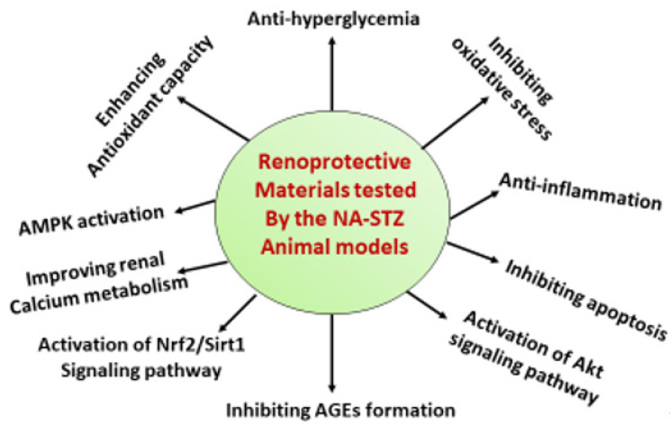
As mentioned above, this NA-STZ diabetes animal model is a non-obese type 2 diabetes model [23]. The pathogenesis of diabetes in this model may be different from that of HFD/STZ or genetically engineered models, such as the db/db mouse model and the zsf1 obese rat model [30,94,95,96]. Nevertheless, the NA-STZ model may provide a unique platform for the study of non-obese diabetes and diabetic complications [43,97]. With respect to DN research, this model has been widely used for testing the therapeutic effects of numerous antidiabetic or renoprotective materials (Table 3). Most of these materials are natural products derived from plants, such as herbs, trees, teas, and vegetables. Table 3 lists the representative materials that exhibit renoprotective effects in DN in the NA-STZ rodent model of type 2 diabetes. It should be noted that as oxidative stress has been established as one of the major mechanisms underlying DN, many of the listed materials in Table 3 thus have antioxidant properties. Figure 5 summarizes the major mechanisms of the renoprotective materials tested by this NA-STZ animal model. It should also be noted that the renoprotective effects of many of the tested materials are in a dose-dependent manner, such as reported in reference [98] and others.
Table 3.
Renoprotective materials tested by the NA-STZ type 2 diabetes animal models.
| Renoprotective Materials |
Rodent Model (NA/STZ, mg/kg) |
Mechanism | Reference |
|---|---|---|---|
| 1,8 Cineole | Rat (200/55) | Glyoxalase-I induction | [51] |
| Abroma augusta L leaf | Rat (110/65) | Inhibiting oxidative stress | [52] |
| Acetate | Rat (110/65) | Suppressing xanthine oxidase activity | [53] |
| Abrus precatorius leaf | Rat (110/60) | Total antioxidant increase in kidney | [98] |
| Ascomycetes | Rat (200/65) | Inhibiting oxidative stress | [54] |
| Betanin | Rat (110/45) | Antioxidative damage | [99] |
| Bitter Gourd Honey | Rat (110/55) | Antioxidation, anti-inflammation | [49] |
| Bocopa monnieri | Rat (230/65) | Inhibiting AGEs formation | [55] |
| Brucea javanica seeds | Rat (100/60) | Inhibiting alpha-glucosidase | [100] |
| Cichorium intybus L seed | Rat (200/55) | Improving blood and urine parameters | [56] |
| Citrus reticulate fruit peel | Rat (120/50) | Antioxidative stress | [57] |
| Combretum micranthum | Rat (100/50) | Elevating SOD and catalase activities | [58] |
| CoQ-10/metformin | Rat (110/65) | Inhibiting oxidative stress | [59] |
| CoQ-10/sitagliptin | Rat (110/65) | Enhancing antioxidant system | [60] |
| Cordyceps militaris | Mouse (180/60) | Decreasing serum creatinine levels | [61] |
| Crocin | Rat (110/65) | Antioxidation | [62] |
| Curculigo orchiodies | Rat (230/65) | Antioxidation, anti-hyperlipidemia | [63] |
| Dapagliflozin | Rat (230/65) | Normalizing renal corpuscles histology | [64] |
| Dapagliflozin/irbesartan | Rat (230/65) | Inhibiting AGEs formation | [65] |
| Dietary flaxseed | Rat (110/65) | Antioxidative stress | [66] |
| Dillenia Indica L | Rat (230/65) | Inhibiting AGEs formation | [67] |
| Diosmin | Rat (110/45) | Inhibiting oxidative stress | [68] |
| Ellagic acid/pioglitazone | Rat (175/65) | Improving kidney function markers | [101] |
| Empagliflozin | Rat (120/40) | Decreasing BUN, creatinine, and oxidative stress | [69] |
| Eysenhardtia polystachya | Mouse (120/60) | Inhibiting glycation | [70] |
| Garlic extract | Rat (110/65) | Inhibiting oxidative stress | [71] |
| Grain amaranth | Rat (120/60) | Improving renal calcium metabolism | [72] |
| Glycosin | Rat (120/60) | Decreasing blood urea and creatinine | [73] |
| Hypericum perforatum | Rat (85/65) | Antioxidative stress | [74] |
| Lipoic acid | Rat (110/55) | Activating CSE/H2S pathway | [75,76] |
| L-NAME | Rat (230/65) | Increasing blood glucose | [48] |
| Manilkara zapota extract | Rat (120/60) | Reversing glomerulosclerosis | [102] |
| Metformin | Rat (230/55) | Decreasing BUN and serum creatinine | [77] |
| Myrciaria cauliflora | Mouse (240/100) | Inhibiting oxidative stress | [78] |
| Naringenin | Rat (120/60) | TRB3-FoxO1 downregulation | [97] |
| Oligo-fucoidan | HFD-Mouse (200/50) | Activation of Nrf2 and Sirt1 | [41] |
| Paeonia emodi | Rat (230/65) | Inhibiting glycation end products | [79] |
| Phyllanthus niruri leaves | Rat (110/55) | Antioxidative stress | [80] |
| Pioglitazone | Rat (110/65) | Antioxidation | [81] |
| Pomegranate | Rat (120/60) | Antioxidative stress | [103] |
| Quercetin | Rat (230/65) | Anti-apoptosis | [82] |
| Resveratrol | Rat (110/50) | Attenuating oxidative stress | [83,104] |
| Rhinacanthins | Rat (100/60) | Inhibiting oxidative stress | [84] |
| S-allylcysteine | Rat (110/45) | Attenuating oxidative stress | [85] |
| SGLT2 inhibitors | Mouse (120/100) | AMPK activation | [86] |
| Silymarin | Rat (120/60) | Lowering serum creatinine and uric acid | [87] |
| Strawberry | Rat (110/65) | Enhancing kidney antioxidant defense | [88] |
| Syzygium calophyllifolium Rat | (120/60) | Enhancing kidney antioxidant defense | [89] |
| Tetrahydrocurcumin | Rat (110/65) | Preventing fatty acid changes in the kidney | [90] |
| Tetramethylpyrazine | Rat (210/55) | Akt signaling pathway activation | [19] |
| Vanillic acid | Rat (110/50) | Attenuating oxidative stress | [91] |
| Tilianin | Rat (110/55) | Nrf2 signaling pathway activation | [92] |
| Zanthoxylum | |||
| Zanthoxyloides extract | Rat (100/55) | Improved kidney histology and biomarkers | [93] |
Note: This table is not meant to be exhaustive and only shows the materials tested for their renoprotective effects. Therefore, antidiabetic materials screened using this model but not focusing on diabetic nephropathy are not included in this table. AGEs = advanced glycation end products.
Figure 5.
Diagram summarizing the representative renoprotective mechanisms of the materials listed in Table 3, using the NA-STZ non-obese type 2 diabetes animal models. AGEs stands for “advanced glycation end products”.
5. Redox-Related Mechanisms That Remain to Be Elucidated in this NA-STZ Model
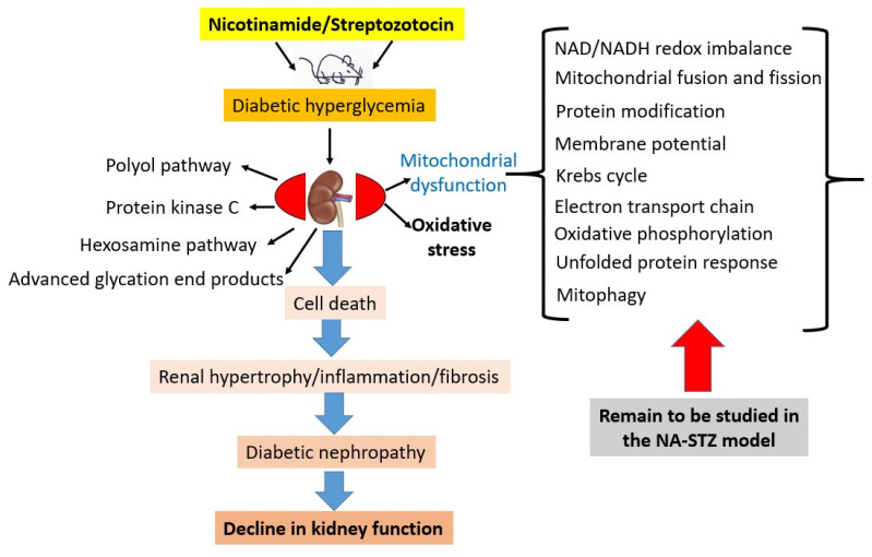
The non-tissue-specific mechanisms involved in cellular injury are thought to be implicated in the development of diabetic nephropathy [5,50]. These mechanisms, as shown in Figure 6, include the activation of the polyol pathway [105] and protein kinase C signaling, the hexosamine pathway, and the increased formation of the advanced glycation products [5,106]. However, what has been lacking is the underlying pathological mechanisms of DN in this unique NA-STZ model, in particular, redox signaling and the mitochondrial mechanisms of NA-STZ-induced DN. In fact, numerous aspects remain to be investigated in detail. These include mitochondrial redox imbalance [39]; sources of mitochondrial reactive oxygen species [107]; proteomics of mitochondrial protein oxidation [108,109]; mitochondrial abnormalities such as the derangement of mitochondrial metabolic pathways, including the Krebs cycle and electron transport chain [29]; fatty acid oxidation [110,111]; mitochondrial fusion and fission [112,113]; and mitophagy and the mitochondrial unfolded protein response [114,115,116,117,118,119] (Figure 6). The changes in redox signaling during the progression of DN in this animal model also remain to be comprehensively studied. Nephron segment-specific investigations of targeted genes [120,121] as well as the role of epigenetics [122,123] in this DN model also remain to be fully conducted. Delineating the mechanisms of these biological processes in the diabetic kidney may provide comprehensive insights into the underpinnings of DN. Additionally, this model could also provide a platform for testing the therapeutic effects of stem cells and gene therapy on DN [11]. For studying multiple kidney disease-causing risk factors, this model could also be combined with other kidney disease animal models, such as those induced by folic acid [47,124,125,126], cisplatin [127,128], cadmium [129,130,131], lipopolysaccharide [132,133], and hypoxia or ischemia reperfusion [134,135,136,137,138,139,140,141].
Figure 6.
Potential mechanisms underlying diabetic nephropathy in the NA-STZ animal model. While common deleterious mechanisms operate in the kidney upon hyperglycemic challenge, those potential mitochondrial mechanisms underlying kidney injury remain to be elucidated (right side of the figure). These deleterious mechanisms would eventually converge on renal hypertrophy and renal fibrosis, leading to phenotype of diabetic nephropathy and kidney functional decline.
Finally, this NA-STZ diabetes animal model may also be used to evaluate any potential renoprotective effects of caloric restriction [142,143,144], intermittent caloric restriction [145,146], exercise [147,148,149,150], and ketone bodies [151,152,153,154], which all have been demonstrated to provide beneficial effects on the kidney in a variety of pathological conditions [155,156]. Indeed, the underlying mechanisms of renoprotection conferred by these approaches in this non-obese type 2 diabetes model remain to be comprehensively elucidated.
6. Summary
The NA-STZ induction of a type 2 diabetic animal model is a useful tool for both studying the mechanisms of DN and screening renoprotective materials for diabetic kidney disease. The model is less time-consuming and less expensive than that created by genetic engineering or high-fat-diet feeding. The establishment of this model is based on the fact that NA can partially protect pancreatic β cells against STZ cytotoxicity, leading to the incomplete destruction of β cells and thus development of non-insulin-dependent type 2 diabetes mellitus [21,23,24]. This unique animal model should continue to serve as a utility for studying the non-obese type 2 diabetes that is highly prevalent in East Asian diabetic patients [157].
Institutional Review Board Statement
Not applicable.
Informed Consent Statement
Not applicable.
Data Availability Statement
Not applicable.
Conflicts of Interest
The author declares no conflict of interest.
Funding Statement
This research received no external funding.
Footnotes
Publisher’s Note: MDPI stays neutral with regard to jurisdictional claims in published maps and institutional affiliations.
References
- 1.Lodhi A.H., Ahmad F.-U., Furwa K., Madni A. Role of oxidative stress and reduced endogenous hydrogen sulfide in diabetic nephropathy. Drug Des. Dev. Ther. 2021;15:1031–1043. doi: 10.2147/DDDT.S291591. [DOI] [PMC free article] [PubMed] [Google Scholar]
- 2.Ji J., Tao P., Wang Q., Li L., Xu Y. Sirt1: Mechanism and protective effect in diabetic nephropathy. Endocr. Metab. Immune Disord.-Drug Targets. 2021;21:835–842. doi: 10.2174/1871530320666201029143606. [DOI] [PubMed] [Google Scholar]
- 3.Zoja C., Xinaris C., Macconi D. Diabetic nephropathy: Novel molecular mechanisms and therapeutic targets. Front. Pharmacol. 2020;11:586892. doi: 10.3389/fphar.2020.586892. [DOI] [PMC free article] [PubMed] [Google Scholar]
- 4.Chowdhury T.A., Ali O. Diabetes and the kidney. Clin. Med. Lond. 2021;21:e318–e322. doi: 10.7861/clinmed.2021-0144. [DOI] [PMC free article] [PubMed] [Google Scholar]
- 5.Yan L.J. Nadh/nad (+) redox imbalance and diabetic kidney disease. Biomolecules. 2021;11:730. doi: 10.3390/biom11050730. [DOI] [PMC free article] [PubMed] [Google Scholar]
- 6.Nakhoul F., Abassi Z., Morgan M., Sussan S., Mirsky N. Inhibition of diabetic nephropathy in rats by an oral antidiabetic material extracted from yeast. J. Am. Soc. Nephrol. 2006;17:S127–S131. doi: 10.1681/ASN.2005121333. [DOI] [PubMed] [Google Scholar]
- 7.Machado D.I., Silva E.D.O., Ventura S., Vattimo M.D.F.F. The effect of curcumin on renal ischemia/reperfusion injury in diabetic rats. Nutrients. 2022;14:2798. doi: 10.3390/nu14142798. [DOI] [PMC free article] [PubMed] [Google Scholar]
- 8.Hernandez L.F., Eguchi N., Whaley D., Alexander M., Tantisattamo E., Ichii H. Anti-Oxidative therapy in diabetic nephropathy. Front. Biosci. 2022;14:14. doi: 10.31083/j.fbs1402014. [DOI] [PubMed] [Google Scholar]
- 9.Eboh C., Chowdhury T.A. Management of diabetic renal disease. Ann. Transl. Med. 2015;3:154. doi: 10.3978/j.issn.2305-5839.2015.06.25. [DOI] [PMC free article] [PubMed] [Google Scholar]
- 10.Sheng X., Dong Y., Cheng D., Wang N., Guo Y. Efficacy and safety of bailing capsules in the treatment of type 2 diabetic nephropathy: A meta-Analysis. Ann. Palliat. Med. 2020;9:3885–3898. doi: 10.21037/apm-20-1799. [DOI] [PubMed] [Google Scholar]
- 11.Natesan V., Kim S.J. Diabetic nephropathy—A review of risk factors, progression, mechanism, and dietary management. Biomol. Ther. 2021;29:365–372. doi: 10.4062/biomolther.2020.204. [DOI] [PMC free article] [PubMed] [Google Scholar]
- 12.Fabris B., Candido R., Armini L., Fischetti F., Calci M., Bardelli M., Fazio M., Campanacci L., Carretta R. Control of glomerular hyperfiltration and renal hypertrophy by an angiotensin converting enzyme inhibitor prevents the progression of renal damage in hypertensive diabetic rats. J. Hypertens. 1999;17:1925–1931. doi: 10.1097/00004872-199917121-00023. [DOI] [PubMed] [Google Scholar]
- 13.Li Z., Guo H., Li J., Ma T., Zhou S., Zhang Z., Miao L., Cai L. Sulforaphane prevents type 2 diabetes-Induced nephropathy via ampk-Mediated activation of lipid metabolic pathways and nrf2 antioxidative function. Clin. Sci. 2020;134:2469–2487. doi: 10.1042/CS20191088. [DOI] [PubMed] [Google Scholar]
- 14.Palygin O., Spires D., Levchenko V., Bohovyk R., Fedoriuk M., Klemens C.A., Sykes O., Bukowy J.D., Cowley A.W., Lazar J., et al. Progression of diabetic kidney disease in t2dn rats. Am. J. Physiol. Ren. Physiol. 2019;317:F1450–F1461. doi: 10.1152/ajprenal.00246.2019. [DOI] [PMC free article] [PubMed] [Google Scholar]
- 15.Thomas H.Y., Versypt A.N.F. Pathophysiology of mesangial expansion in diabetic nephropathy: Mesangial structure, glomerular biomechanics, and biochemical signaling and regulation. J. Biol. Eng. 2022;16:19. doi: 10.1186/s13036-022-00299-4. [DOI] [PMC free article] [PubMed] [Google Scholar]
- 16.Ma J., Zhao N., Du L., Wang Y. Downregulation of lncrna neat1 inhibits mouse mesangial cell proliferation, fibrosis, and inflammation but promotes apoptosis in diabetic nephropathy. Int. J. Clin. Exp. Pathol. 2019;12:1174–1183. [PMC free article] [PubMed] [Google Scholar]
- 17.Zang X.J., Li L., Du X., Yang B., Mei C.L. Lncrna tug1 inhibits the proliferation and fibrosis of mesangial cells in diabetic nephropathy via inhibiting the pi3k/akt pathway. Eur. Rev. Med. Pharm. Sci. 2019;23:7519–7525. doi: 10.26355/eurrev_201909_18867. [DOI] [PubMed] [Google Scholar]
- 18.Zheng S., Powell D.W., Zheng F., Kantharidis P., Gnudi L. Diabetic nephropathy: Proteinuria, inflammation, and fibrosis. J. Diabetes Res. 2016;2016:5241549. doi: 10.1155/2016/5241549. [DOI] [PMC free article] [PubMed] [Google Scholar]
- 19.Rai U., Kosuru R., Prakash S., Tiwari V., Singh S. Tetramethylpyrazine alleviates diabetic nephropathy through the activation of akt signalling pathway in rats. Eur. J. Pharmacol. 2019;865:172763. doi: 10.1016/j.ejphar.2019.172763. [DOI] [PubMed] [Google Scholar]
- 20.Joost H.-G., Al-Hasani H., Schurmann A. Animal Models in Diabetes Research. Volume 933. Humana Press; New York, NY, USA: 2012. p. 325. [Google Scholar]
- 21.Ghasemi A., Khalifi S., Jedi S. Streptozotocin-Nicotinamide-Induced rat model of type 2 diabetes (review) Acta Physiol. Hung. 2014;101:408–420. doi: 10.1556/APhysiol.101.2014.4.2. [DOI] [PubMed] [Google Scholar]
- 22.Sathaye S., Kaikini A.A., Dhodi D., Muke S., Peshattiwar V., Bagle S., Korde A., Sarnaik J., Kadwad V., Sachdev S. Standardization of type 1 and type 2 diabetic nephropathy models in rats: Assessment and characterization of metabolic features and renal injury. J. Pharm. Bioallied Sci. 2020;12:295–307. doi: 10.4103/jpbs.JPBS_239_19. [DOI] [PMC free article] [PubMed] [Google Scholar]
- 23.Szkudelski T. Streptozotocin-Nicotinamide-Induced diabetes in the rat. Characteristics of the experimental model. Exp. Biol. Med. 2012;237:481–490. doi: 10.1258/ebm.2012.011372. [DOI] [PubMed] [Google Scholar]
- 24.Masiello P., Broca C., Gross R., Roye M., Manteghetti M., Hillaire-Buys D., Novelli M., Ribes G. Experimental niddm: Development of a new model in adult rats administered streptozotocin and nicotinamide. Diabetes. 1998;47:224–229. doi: 10.2337/diab.47.2.224. [DOI] [PubMed] [Google Scholar]
- 25.Gheibi S., Jeddi S., Carlstrom M., Kashfi K., Ghasemi A. Hydrogen sulfide potentiates the favorable metabolic effects of inorganic nitrite in type 2 diabetic rats. Nitric Oxide. 2019;92:60–72. doi: 10.1016/j.niox.2019.08.006. [DOI] [PubMed] [Google Scholar]
- 26.Jeddi S., Gheibi S., Kashfi K., Ghasemi A. Sodium hydrosulfide has no additive effects on nitrite-Inhibited renal gluconeogenesis in type 2 diabetic rats. Life Sci. 2021;283:119870. doi: 10.1016/j.lfs.2021.119870. [DOI] [PubMed] [Google Scholar]
- 27.Javrushyan H., Nadiryan E., Grigoryan A., Avtandilyan N., Maloyan A. Antihyperglycemic activity of l-Norvaline and l-Arginine in high-Fat diet and streptozotocin-Treated male rats. Exp. Mol. Pathol. 2022;126:104763. doi: 10.1016/j.yexmp.2022.104763. [DOI] [PubMed] [Google Scholar]
- 28.Liu P., Zhang Z., Li Y. Relevance of the pyroptosis-Related inflammasome pathway in the pathogenesis of diabetic kidney disease. Front. Immunol. 2021;12:603416. doi: 10.3389/fimmu.2021.603416. [DOI] [PMC free article] [PubMed] [Google Scholar]
- 29.Wu J., Luo X., Thangthaeng N., Sumien N., Chen Z., Rutledge M.A., Jing S., Forster M.J., Yan L.J. Pancreatic mitochondrial complex i exhibits aberrant hyperactivity in diabetes. Biochem. Biophys. Rep. 2017;11:119–129. doi: 10.1016/j.bbrep.2017.07.007. [DOI] [PMC free article] [PubMed] [Google Scholar]
- 30.Li C.Y., Ma W.X., Yan L.J. 5-Methoxyindole-2-Carboxylic acid (mica) fails to retard development and progression of type ii diabetes in zsf1 diabetic rats. React. Oxyg. Species Apex NC. 2020;9:144–147. [PMC free article] [PubMed] [Google Scholar]
- 31.Dugbartey G.J., Wonje Q.L., Alornyo K.K., Adams I., Diaba D.E. Alpha-Lipoic acid treatment improves adverse cardiac remodelling in the diabetic heart—The role of cardiac hydrogen sulfide-Synthesizing enzymes. Biochem. Pharmacol. 2022;203:115179. doi: 10.1016/j.bcp.2022.115179. [DOI] [PubMed] [Google Scholar]
- 32.Qasem M.A., Noordin M.I., Arya A., Alsalahi A., Jayash S.N. Evaluation of the glycemic effect of ceratonia siliqua pods (carob) on a streptozotocin-Nicotinamide induced diabetic rat model. PeerJ. 2018;6:e4788. doi: 10.7717/peerj.4788. [DOI] [PMC free article] [PubMed] [Google Scholar]
- 33.Patra S., Bhattacharya S., Bala A., Haldar P.K. Antidiabetic effect of drymaria cordata leaf against streptozotocin-Nicotinamide-Induced diabetic albino rats. J. Adv. Pharm. Technol. Res. 2020;11:44–52. doi: 10.4103/japtr.JAPTR_98_19. [DOI] [PMC free article] [PubMed] [Google Scholar]
- 34.Kumar E.K., Janardhana G.R. Antidiabetic activity of alcoholic stem extract of nervilia plicata in streptozotocin-Nicotinamide induced type 2 diabetic rats. J. Ethnopharmacol. 2011;133:480–483. doi: 10.1016/j.jep.2010.10.025. [DOI] [PubMed] [Google Scholar]
- 35.Balaji P., Madhanraj R., Rameshkumar K., Veeramanikandan V., Eyini M., Arun A., Thulasinathan B., Al Farraj D., Elshikh M., Alokda A., et al. Evaluation of antidiabetic activity of pleurotus pulmonarius against streptozotocin-Nicotinamide induced diabetic wistar albino rats. Saudi J. Biol. Sci. 2020;27:913–924. doi: 10.1016/j.sjbs.2020.01.027. [DOI] [PMC free article] [PubMed] [Google Scholar]
- 36.Szkudelski T. The mechanism of alloxan and streptozotocin action in b cells of the rat pancreas. Physiol. Res. 2001;50:537–546. [PubMed] [Google Scholar]
- 37.Wu J., Yan L.J. Streptozotocin-Induced type 1 diabetes in rodents as a model for studying mitochondrial mechanisms of diabetic beta cell glucotoxicity. Diabetes Metab. Syndr. Obes. Targets Ther. 2015;8:181–188. doi: 10.2147/DMSO.S82272. [DOI] [PMC free article] [PubMed] [Google Scholar]
- 38.Lenzen S. The mechanisms of alloxan- and streptozotocin-Induced diabetes. Diabetologia. 2008;51:216–226. doi: 10.1007/s00125-007-0886-7. [DOI] [PubMed] [Google Scholar]
- 39.Wu J., Jin Z., Zheng H., Yan L.J. Sources and implications of nadh/nad (+) redox imbalance in diabetes and its complications. Diabetes Metab. Syndr. Obes. 2016;9:145–153. doi: 10.2147/DMSO.S106087. [DOI] [PMC free article] [PubMed] [Google Scholar]
- 40.Wu C.C., Hung C.N., Shin Y.C., Wang C.J., Huang H.P. Myrciaria cauliflora extracts attenuate diabetic nephropathy involving the ras signaling pathway in streptozotocin/nicotinamide mice on a high fat diet. J. Food Drug Anal. 2016;24:136–146. doi: 10.1016/j.jfda.2015.10.001. [DOI] [PMC free article] [PubMed] [Google Scholar]
- 41.Yu W.C., Huang R.Y., Chou T.C. Oligo-Fucoidan improves diabetes-Induced renal fibrosis via activation of sirt-1, glp-1r, and nrf2/ho-1: An in vitro and in vivo study. Nutrients. 2020;12:3068. doi: 10.3390/nu12103068. [DOI] [PMC free article] [PubMed] [Google Scholar]
- 42.Nakamura T., Terajima T., Ogata T., Ueno K., Hashimoto N., Ono K., Yano S. Establishment and pathophysiological characterization of type 2 diabetic mouse model produced by streptozotocin and nicotinamide. Biol. Pharm. Bull. 2006;29:1167–1174. doi: 10.1248/bpb.29.1167. [DOI] [PubMed] [Google Scholar]
- 43.Weng Y., Yu L., Cui J., Zhu Y.-R., Guo C., Wei G., Duan J.-L., Yin Y., Guan Y., Wang Y.-H., et al. Antihyperglycemic, hypolipidemic and antioxidant activities of total saponins extracted from aralia taibaiensis in experimental type 2 diabetic rats. J. Ethnopharmacol. 2014;152:553–560. doi: 10.1016/j.jep.2014.02.001. [DOI] [PubMed] [Google Scholar]
- 44.Bayrasheva V.K., Babenko A.Y., Dobronravov V.A., Dmitriev Y.V., Chefu S.G., Pchelin I.Y., Ivanova A.N., Bairamov A.A., Alexeyeva N.P., Shatalov I.S., et al. Uninephrectomized high-Fat-Fed nicotinamide-Streptozotocin-Induced diabetic rats: A model for the investigation of diabetic nephropathy in type 2 diabetes. J. Diabetes Res. 2016;2016:8317850. doi: 10.1155/2016/8317850. [DOI] [PMC free article] [PubMed] [Google Scholar]
- 45.Sasongko H., Nurrochmad A., Rohman A., Nugroho A.E. Characteristic of Streptozotocin-Nicotinamide-Induced Inflammation in A Rat Model of Diabetes-Associated Renal Injury. Open Access Maced. J. Med. Sci. 2022;10:16–22. [Google Scholar]
- 46.Pelle M.C., Provenzano M., Busutti M., Porcu C.V., Zaffina I., Stanga L., Arturi F. Up-Date on diabetic nephropathy. Life. 2022;12:1202. doi: 10.3390/life12081202. [DOI] [PMC free article] [PubMed] [Google Scholar]
- 47.Yan L.J. Folic acid-Induced animal model of kidney disease. Animal. Model. Exp. Med. 2021;4:329–342. doi: 10.1002/ame2.12194. [DOI] [PMC free article] [PubMed] [Google Scholar]
- 48.Corremans R., D’Haese P.C., Vervaet B.A., Verhulst A. L-Name administration enhances diabetic kidney disease development in an stz/nad rat model. Int. J. Mol. Sci. 2021;22:12767. doi: 10.3390/ijms222312767. [DOI] [PMC free article] [PubMed] [Google Scholar]
- 49.Arigela C.S., Nelli G., Gan S.H., Sirajudeen K.N.S., Krishnan K., Abdul Rahman N., Pasupuleti V.R. Bitter gourd honey ameliorates hepatic and renal diabetic complications on type 2 diabetes rat models by antioxidant, anti-Inflammatory, and anti-Apoptotic mechanisms. Foods. 2021;10:2872. doi: 10.3390/foods10112872. [DOI] [PMC free article] [PubMed] [Google Scholar]
- 50.Agarwal R. Chronic Kidney Disease and Type 2 Diabetes. American Diabetes Association; Arlington, VA, USA: 2021. Pathogenesis of diabetic nephropathy; pp. 2–7. [Google Scholar]
- 51.Mahdavifard S., Nakhjavani M. 1,8 cineole protects type 2 diabetic rats against diabetic nephropathy via inducing the activity of glyoxalase-I and lowering the level of transforming growth factor-1beta. J. Diabetes Metab. Disord. 2022;21:567–572. doi: 10.1007/s40200-022-01014-2. [DOI] [PMC free article] [PubMed] [Google Scholar]
- 52.Khanra R., Dewanjee S., Dua T.K., Sahu R., Gangopadhyay M., De Feo V., Zia-Ul-Haq M. Abroma augusta l. (malvaceae) leaf extract attenuates diabetes induced nephropathy and cardiomyopathy via inhibition of oxidative stress and inflammatory response. J. Transl. Med. 2015;13:6. doi: 10.1186/s12967-014-0364-1. [DOI] [PMC free article] [PubMed] [Google Scholar]
- 53.Olaniyi K.S., Amusa O.A., Akinnagbe N.T., Ajadi I.O., Ajadi M.B., Agunbiade T.B., Michael O.S. Acetate ameliorates nephrotoxicity in streptozotocin-Nicotinamide-Induced diabetic rats: Involvement of xanthine oxidase activity. Cytokine. 2021;142:155501. doi: 10.1016/j.cyto.2021.155501. [DOI] [PubMed] [Google Scholar]
- 54.Wu W.T., Hsu T.H., Lee C.H., Lo H.C. Fruiting bodies of chinese caterpillar mushroom, ophiocordyceps sinensis (ascomycetes) alleviate diabetes-Associated oxidative stress. Int. J. Med. Mushrooms. 2020;22:15–29. doi: 10.1615/IntJMedMushrooms.2019033275. [DOI] [PubMed] [Google Scholar]
- 55.Kishore L., Kaur N., Singh R. Renoprotective effect of bacopa monnieri via inhibition of advanced glycation end products and oxidative stress in stz-Nicotinamide-Induced diabetic nephropathy. Ren. Fail. 2016;38:1528–1544. doi: 10.1080/0886022X.2016.1227920. [DOI] [PubMed] [Google Scholar]
- 56.Pourfarjam Y., Rezagholizadeh L., Nowrouzi A., Meysamie A., Ghaseminejad S., Ziamajidi N., Norouzi D. Effect of cichorium intybus l. Seed extract on renal parameters in experimentally induced early and late diabetes type 2 in rats. Ren. Fail. 2017;39:211–221. doi: 10.1080/0886022X.2016.1256317. [DOI] [PMC free article] [PubMed] [Google Scholar]
- 57.Ali A.M., Gabbar M.A., Abdel-Twab S.M., Fahmy E.M., Ebaid H., Alhazza I.M., Ahmed O.M. Antidiabetic potency, antioxidant effects, and mode of actions of citrus reticulata fruit peel hydroethanolic extract, hesperidin, and quercetin in nicotinamide/streptozotocin-Induced wistar diabetic rats. Oxidative Med. Cell. Longev. 2020;2020:1730492. doi: 10.1155/2020/1730492. [DOI] [PMC free article] [PubMed] [Google Scholar]
- 58.Kpemissi M., Potârniche A.-V., Lawson-Evi P., Metowogo K., Melila M., Dramane P., Taulescu M., Chandramohan V., Suhas D.S., Puneeth T.A., et al. Nephroprotective effect of combretum micranthum g. Don in nicotinamide-Streptozotocin induced diabetic nephropathy in rats: In-Vivo and in-Silico experiments. J. Ethnopharmacol. 2020;261:113133. doi: 10.1016/j.jep.2020.113133. [DOI] [PubMed] [Google Scholar]
- 59.Maheshwari R.A., Balaraman R., Sen A.K., Seth A.K. Effect of coenzyme q10 alone and its combination with metformin on streptozotocin-Nicotinamide-Induced diabetic nephropathy in rats. Indian J. Pharmacol. 2014;46:627–632. doi: 10.4103/0253-7613.144924. [DOI] [PMC free article] [PubMed] [Google Scholar]
- 60.Maheshwari R., Balaraman R., Sen A.K., Shukla D., Seth A. Effect of concomitant administration of coenzyme q10 with sitagliptin on experimentally induced diabetic nephropathy in rats. Ren. Fail. 2017;39:130–139. doi: 10.1080/0886022X.2016.1254659. [DOI] [PMC free article] [PubMed] [Google Scholar]
- 61.Yu S.H., Dubey N.K., Li W.S., Liu M.C., Chiang H.S., Leu S.J., Shieh Y.H., Tsai F.C., Deng W.P. Cordyceps militaris treatment preserves renal function in type 2 diabetic nephropathy mice. PLoS ONE. 2016;11:e0166342. doi: 10.1371/journal.pone.0166342. [DOI] [PMC free article] [PubMed] [Google Scholar]
- 62.Margaritis I., Angelopoulou K., Lavrentiadou S., Mavrovouniotis I.C., Tsantarliotou M., Taitzoglou I., Theodoridis A., Veskoukis A., Kerasioti E., Kouretas D., et al. Effect of crocin on antioxidant gene expression, fibrinolytic parameters, redox status and blood biochemistry in nicotinamide-Streptozotocin-Induced diabetic rats. J. Biol. Res. 2020;27:4. doi: 10.1186/s40709-020-00114-5. [DOI] [PMC free article] [PubMed] [Google Scholar]
- 63.Singla K., Singh R. Nephroprotective effect of curculigo orchiodies in streptozotocin-Nicotinamide induced diabetic nephropathy in wistar rats. J. Ayurveda Integr. Med. 2020;11:399–404. doi: 10.1016/j.jaim.2020.05.006. [DOI] [PMC free article] [PubMed] [Google Scholar]
- 64.El Medany A.M.H., Hammadi S.H.M., Khalifa H.M., Ghazala R.A., Mohammed H.S.Z. The vascular impact of dapagliflozin, liraglutide, and atorvastatin alone or in combinations in type 2 diabetic rat model. Fundam. Clin. Pharmacol. 2022;36:731–741. doi: 10.1111/fcp.12765. [DOI] [PubMed] [Google Scholar]
- 65.Abdel-Wahab A.F., Bamagous G.A., Al-Harizy R.M., ElSawy N.A., Shahzad N., Ibrahim I.A., Ghamdi S.S.A. Renal protective effect of sglt2 inhibitor dapagliflozin alone and in combination with irbesartan in a rat model of diabetic nephropathy. Biomed. Pharm. 2018;103:59–66. doi: 10.1016/j.biopha.2018.03.176. [DOI] [PubMed] [Google Scholar]
- 66.Jangale N.M., Devarshi P.P., Bansode S.B., Kulkarni M.J., Harsulkar A.M. Dietary flaxseed oil and fish oil ameliorates renal oxidative stress, protein glycation, and inflammation in streptozotocin-Nicotinamide-Induced diabetic rats. J. Physiol. Biochem. 2016;72:327–336. doi: 10.1007/s13105-016-0482-8. [DOI] [PubMed] [Google Scholar]
- 67.Kaur N., Kishore L., Singh R. Dillenia indica l. Attenuates diabetic nephropathy via inhibition of advanced glycation end products accumulation in stz-Nicotinamide induced diabetic rats. J. Tradit. Complement Med. 2018;8:226–238. doi: 10.1016/j.jtcme.2017.06.004. [DOI] [PMC free article] [PubMed] [Google Scholar]
- 68.Srinivasan S., Pari L. Ameliorative effect of diosmin, a citrus flavonoid against streptozotocin-Nicotinamide generated oxidative stress induced diabetic rats. Chem.-Biol. Interact. 2012;195:43–51. doi: 10.1016/j.cbi.2011.10.003. [DOI] [PubMed] [Google Scholar]
- 69.El-Kader M.A., Hashish H.A. Potential role of empagliflozin in prevention of nephropathy in streptozotocin-Nicotinamideinduced type 2 diabetes: An ultrastructural study. Anatomy. 2019;13:137–148. [Google Scholar]
- 70.Gutierrez R.M.P., Campoy A.H.G., Carrera S.P.P., Ramirez A.M., Flores J.M.M., Valle S.O.F. 3′-O-Beta-D-Glucopyranosyl-Alpha,4,2′,4′,6′-Pentahydroxy-Dihydrochalcone, from bark of eysenhardtia polystachya prevents diabetic nephropathy via inhibiting protein glycation in stz-Nicotinamide induced diabetic mice. Molecules. 2019;24:1214. doi: 10.3390/molecules24071214. [DOI] [PMC free article] [PubMed] [Google Scholar]
- 71.Ziamajidi N., Nasiri A., Abbasalipourkabir R., Sadeghi Moheb S. Effects of garlic extract on tnf-Alpha expression and oxidative stress status in the kidneys of rats with stz + nicotinamide-Induced diabetes. Pharm. Biol. 2017;55:526–531. doi: 10.1080/13880209.2016.1255978. [DOI] [PMC free article] [PubMed] [Google Scholar]
- 72.Kasozi K.I., Namubiru S., Safiriyu A.A., Ninsiima H.I., Nakimbugwe D., Namayanja M., Valladares M.B. Grain amaranth is associated with improved hepatic and renal calcium metabolism in type 2 diabetes mellitus of male wistar rats. Evid.-Based Complement. Altern. Med. 2018;2018:4098942. doi: 10.1155/2018/4098942. [DOI] [PMC free article] [PubMed] [Google Scholar]
- 73.Selvaraj G., Kaliamurthi S., Thirugnasambandan R. Effect of glycosin alkaloid from rhizophora apiculata in non-Insulin dependent diabetic rats and its mechanism of action: In vivo and in silico studies. Phytomedicine. 2016;23:632–640. doi: 10.1016/j.phymed.2016.03.004. [DOI] [PubMed] [Google Scholar]
- 74.Abd El Motteleb D.M., Abd El Aleem D.I. Renoprotective effect of hypericum perforatum against diabetic nephropathy in rats: Insights in the underlying mechanisms. Clin. Exp. Pharmacol. Physiol. 2017;44:509–521. doi: 10.1111/1440-1681.12729. [DOI] [PubMed] [Google Scholar]
- 75.Dugbartey G.J., Alornyo K.K., N’Guessan B.B., Atule S., Mensah S.D., Adjei S. Supplementation of conventional anti-Diabetic therapy with alpha-Lipoic acid prevents early development and progression of diabetic nephropathy. Biomed. Pharmacother. 2022;149:112818. doi: 10.1016/j.biopha.2022.112818. [DOI] [PubMed] [Google Scholar]
- 76.Dugbartey G.J., Alornyo K.K., Diaba D.E., Adams I. Activation of renal cse/h2s pathway by alpha-Lipoic acid protects against histological and functional changes in the diabetic kidney. Biomed. Pharmacother. 2022;153:113386. doi: 10.1016/j.biopha.2022.113386. [DOI] [PubMed] [Google Scholar]
- 77.Deshmukh A., Manjalkar P. Synergistic effect of micronutrients and metformin in alleviating diabetic nephropathy and cardiovascular dysfunctioning in diabetic rat. J. Diabetes Metab. Disord. 2021;20:533–541. doi: 10.1007/s40200-021-00776-5. [DOI] [PMC free article] [PubMed] [Google Scholar]
- 78.Hsu J.D., Wu C.C., Hung C.N., Wang C.J., Huang H.P. Myrciaria cauliflora extract improves diabetic nephropathy via suppression of oxidative stress and inflammation in streptozotocin-Nicotinamide mice. J. Food Drug Anal. 2016;24:730–737. doi: 10.1016/j.jfda.2016.03.009. [DOI] [PMC free article] [PubMed] [Google Scholar]
- 79.Kishore L., Kaur N., Singh R. Nephroprotective effect of paeonia emodi via inhibition of advanced glycation end products and oxidative stress in streptozotocin-Nicotinamide induced diabetic nephropathy. J. Food Drug Anal. 2017;25:576–588. doi: 10.1016/j.jfda.2016.08.009. [DOI] [PMC free article] [PubMed] [Google Scholar]
- 80.Giribabu N., Karim K., Kilari E.K., Salleh N. Phyllanthus niruri leaves aqueous extract improves kidney functions, ameliorates kidney oxidative stress, inflammation, fibrosis and apoptosis and enhances kidney cell proliferation in adult male rats with diabetes mellitus. J. Ethnopharmacol. 2017;205:123–137. doi: 10.1016/j.jep.2017.05.002. [DOI] [PubMed] [Google Scholar]
- 81.Afzal H.R., Khan N.U.H., Sultana K., Mobashar A., Lareb A., Khan A., Gull A., Afzaal H., Khan M.T., Rizwan M., et al. Schiff bases of pioglitazone provide better antidiabetic and potent antioxidant effect in a streptozotocin-Nicotinamide-Induced diabetic rodent model. ACS Omega. 2021;6:4470–4479. doi: 10.1021/acsomega.0c06064. [DOI] [PMC free article] [PubMed] [Google Scholar]
- 82.Lin C.F., Kuo Y.T., Chen T.Y., Chien C.T. Quercetin-Rich guava (psidium guajava) juice in combination with trehalose reduces autophagy, apoptosis and pyroptosis formation in the kidney and pancreas of type ii diabetic rats. Molecules. 2016;21:334. doi: 10.3390/molecules21030334. [DOI] [PMC free article] [PubMed] [Google Scholar]
- 83.Palsamy P., Subramanian S. Resveratrol protects diabetic kidney by attenuating hyperglycemia-Mediated oxidative stress and renal inflammatory cytokines via nrf2-Keap1 signaling. Biochim. Biophys. Acta-Mol. Basis Dis. 2011;1812:719–731. doi: 10.1016/j.bbadis.2011.03.008. [DOI] [PubMed] [Google Scholar]
- 84.Zhao L.L., Makinde E.A., Shah M.A., Olatunji O.J., Panichayupakaranant P. Rhinacanthins-Rich extract and rhinacanthin c ameliorate oxidative stress and inflammation in streptozotocin-Nicotinamide-Induced diabetic nephropathy. J. Food Biochem. 2019;43:e12812. doi: 10.1111/jfbc.12812. [DOI] [PubMed] [Google Scholar]
- 85.Uddandrao V.V.S., Brahmanaidu P., Ravindarnaik R., Suresh P., Vadivukkarasi S., Saravanan G. Restorative potentiality of s-Allylcysteine against diabetic nephropathy through attenuation of oxidative stress and inflammation in streptozotocin-Nicotinamide-Induced diabetic rats. Eur. J. Nutr. 2019;58:2425–2437. doi: 10.1007/s00394-018-1795-x. [DOI] [PubMed] [Google Scholar]
- 86.Inoue M.-K., Matsunaga Y., Nakatsu Y., Yamamotoya T., Ueda K., Kushiyama A., Sakoda H., Fujishiro M., Ono H., Iwashita M., et al. Possible involvement of normalized pin1 expression level and ampk activation in the molecular mechanisms underlying renal protective effects of sglt2 inhibitors in mice. Diabetol. Metab. Syndr. 2019;11:57. doi: 10.1186/s13098-019-0454-6. [DOI] [PMC free article] [PubMed] [Google Scholar]
- 87.Sheela N., Jose M.A., Sathyamurthy D., Kumar B.N. Effect of silymarin on streptozotocin-Nicotinamide-Induced type 2 diabetic nephropathy in rats. Iran. J. Kidney Dis. 2013;7:117–123. [PubMed] [Google Scholar]
- 88.Mandave P., Khadke S., Karandikar M., Pandit V., Ranjekar P., Kuvalekar A., Mantri N. Antidiabetic, lipid normalizing, and nephroprotective actions of the strawberry: A potent supplementary fruit. Int. J. Mol. Sci. 2017;18:124. doi: 10.3390/ijms18010124. [DOI] [PMC free article] [PubMed] [Google Scholar]
- 89.Chandran R., Parimelazhagan T., Shanmugam S., Thankarajan S. Antidiabetic activity of syzygium calophyllifolium in streptozotocin-Nicotinamide induced type-2 diabetic rats. Biomed. Pharmacother. 2016;82:547–554. doi: 10.1016/j.biopha.2016.05.036. [DOI] [PubMed] [Google Scholar]
- 90.Murugan P., Pari L. Protective role of tetrahydrocurcumin on changes in the fatty acid composition in streptozotocin-Nicotinamide induced type 2 diabetic rats. J. Appl. Biomed. 2007;5:31–38. [Google Scholar]
- 91.Singh B., Kumar A., Singh H., Kaur S., Arora S., Singh B. Protective effect of vanillic acid against diabetes and diabetic nephropathy by attenuating oxidative stress and upregulation of nf-Kappab, tnf-Alpha and cox-2 proteins in rats. Phytother. Res. 2022;36:1338–1352. doi: 10.1002/ptr.7392. [DOI] [PubMed] [Google Scholar]
- 92.Zhang R., Lu M., Zhang S., Liu J. Renoprotective effects of tilianin in diabetic rats through modulation of oxidative stress via nrf2-Keap1 pathway and inflammation via tlr4/mapk/nf-Kappab pathways. Int. Immunopharmacol. 2020;88:106967. doi: 10.1016/j.intimp.2020.106967. [DOI] [PubMed] [Google Scholar]
- 93.Kyei-Barffour I., Kwarkoh R.K.B., Arthur O.D., Akwetey S.A., Acheampong D.O., Aboagye B., Brah A.S., Amponsah I.K., Adokoh C.K. Alkaloidal extract from zanthoxylum zanthoxyloides stimulates insulin secretion in normoglycemic and nicotinamide/streptozotocin-Induced diabetic rats. Heliyon. 2021;7:e07452. doi: 10.1016/j.heliyon.2021.e07452. [DOI] [PMC free article] [PubMed] [Google Scholar]
- 94.Homer B.L., Dower K. 41-Week study of progressive diabetic nephropathy in the zsf1 fa/fa(cp) rat model. Toxicol. Pathol. 2018;46:976–977. doi: 10.1177/0192623318803278. [DOI] [PubMed] [Google Scholar]
- 95.Zhao Y., Yan T., Xiong C., Chang M., Gao Q., Yao S., Wu W., Yi X., Xu G. Overexpression of lipoic acid synthase gene alleviates diabetic nephropathy of lepr(db/db) mice. BMJ Open Diabetes Res. Care. 2021;9:e002260. doi: 10.1136/bmjdrc-2021-002260. [DOI] [PMC free article] [PubMed] [Google Scholar]
- 96.Zhang B., Zhang X., Zhang C., Sun G., Sun X. Berberine improves the protective effects of metformin on diabetic nephropathy in db/db mice through trib1-Dependent inhibiting inflammation. Pharm. Res. 2021;38:1807–1820. doi: 10.1007/s11095-021-03104-x. [DOI] [PubMed] [Google Scholar]
- 97.Khan M.F., Mathur A., Pandey V.K., Kakkar P. Endoplasmic reticulum stress-Dependent activation of trb3-Foxo1 signaling pathway exacerbates hyperglycemic nephrotoxicity: Protection accorded by naringenin. Eur. J. Pharmacol. 2022;917:174745. doi: 10.1016/j.ejphar.2022.174745. [DOI] [PubMed] [Google Scholar]
- 98.Boye A., Acheampong D.O., Gyamerah E.O., Asiamah E.A., Addo J.K., Mensah D.A., Brah A.S., Ayiku P.J. Glucose lowering and pancreato-Protective effects of abrus precatorius (l.) leaf extract in normoglycemic and stz/nicotinamide-Induced diabetic rats. J. Ethnopharmacol. 2020;258:112918. doi: 10.1016/j.jep.2020.112918. [DOI] [PubMed] [Google Scholar]
- 99.Indumathi D., Sujithra K., Srinivasan S., Vinothkumar V. Protective effect of betanin against streptozotocin-Nicotinamide induced liver, kidney and pancreas damage by attenuating lipid byproducts and improving renal biomarkers in wistar rats. Int. J. Adv. Res. Biol. Sci. 2017;4:160–170. [Google Scholar]
- 100.Ablat A., Halabi M.F., Mohamad J., Hasnan M.H., Hazni H., Teh S.H., Shilpi J.A., Mohamed Z., Awang K. Antidiabetic effects of brucea javanica seeds in type 2 diabetic rats. BMC Complement. Altern. Med. 2017;17:94. doi: 10.1186/s12906-017-1610-x. [DOI] [PMC free article] [PubMed] [Google Scholar]
- 101.Nankar R.P., Doble M. Hybrid drug combination: Anti-Diabetic treatment of type 2 diabetic wistar rats with combination of ellagic acid and pioglitazone. Phytomedicine. 2017;37:4–9. doi: 10.1016/j.phymed.2017.10.014. [DOI] [PubMed] [Google Scholar]
- 102.Karle P.P., Dhawale S.C., Navghare V.V. Amelioration of diabetes and its complications by manilkara zapota (l) p. Royen fruit peel extract and its fractions in alloxan and stz-Na induced diabetes in wistar rats. J. Diabetes Metab. Disord. 2022;21:493–510. doi: 10.1007/s40200-022-01000-8. [DOI] [PMC free article] [PubMed] [Google Scholar]
- 103.Aboonabi A., Rahmat A., Othman F. Antioxidant effect of pomegranate against streptozotocin-Nicotinamide generated oxidative stress induced diabetic rats. Toxicol. Rep. 2014;1:915–922. doi: 10.1016/j.toxrep.2014.10.022. [DOI] [PMC free article] [PubMed] [Google Scholar]
- 104.Soufi F.G., Vardyani M., Sheervalilou R., Mohammadi M., Somi M.H. Long-Term treatment with resveratrol attenuates oxidative stress pro-Inflammatory mediators and apoptosis in streptozotocin-Nicotinamide-Induced diabetic rats. Gen. Physiol. Biophys. 2012;31:431–438. doi: 10.4149/gpb_2012_039. [DOI] [PubMed] [Google Scholar]
- 105.Yan L.J. Redox imbalance stress in diabetes mellitus: Role of the polyol pathway. Animal Model. Exp. Med. 2018;1:7–13. doi: 10.1002/ame2.12001. [DOI] [PMC free article] [PubMed] [Google Scholar]
- 106.Luo X., Wu J., Jing S., Yan L.J. Hyperglycemic stress and carbon stress in diabetic glucotoxicity. Aging Dis. 2016;7:90–110. doi: 10.14336/AD.2015.0702. [DOI] [PMC free article] [PubMed] [Google Scholar]
- 107.Yan L.J., Sumien N., Thangthaeng N., Forster M.J. Reversible inactivation of dihydrolipoamide dehydrogenase by mitochondrial hydrogen peroxide. Free Radic. Res. 2013;47:123–133. doi: 10.3109/10715762.2012.752078. [DOI] [PMC free article] [PubMed] [Google Scholar]
- 108.Wu J., Luo X., Jing S., Yan L.J. Two-Dimensional gel electrophoretic detection of protein carbonyls derivatized with biotin-Hydrazide. J. Chromatogr. B. 2016;1019:128–131. doi: 10.1016/j.jchromb.2015.11.003. [DOI] [PMC free article] [PubMed] [Google Scholar]
- 109.Zheng H., Wu J., Jin Z., Yan L.J. Protein modifications as manifestations of hyperglycemic glucotoxicity in diabetes and its complications. Biochem. Insights. 2016;9:BCI-S36141. doi: 10.4137/BCI.S36141. [DOI] [PMC free article] [PubMed] [Google Scholar]
- 110.Murea M., Freedman B.I., Parks J.S., Antinozzi P.A., Elbein S.C., Ma L. Lipotoxicity in diabetic nephropathy: The potential role of fatty acid oxidation. Clin. J. Am. Soc. Nephrol. 2010;5:2373–2379. doi: 10.2215/CJN.08160910. [DOI] [PubMed] [Google Scholar]
- 111.Jang H.S., Noh M.R., Kim J., Padanilam B.J. Defective mitochondrial fatty acid oxidation and lipotoxicity in kidney diseases. Front. Med. 2020;7:65. doi: 10.3389/fmed.2020.00065. [DOI] [PMC free article] [PubMed] [Google Scholar]
- 112.Yang S.-K., Li A.-M., Han Y.-C., Peng C.-H., Song N., Yang M., Zhan M., Zeng L.-F., Song P.-A., Zhang W., et al. Mitochondria-Targeted peptide ss31 attenuates renal tubulointerstitial injury via inhibiting mitochondrial fission in diabetic mice. Oxidative Med. Cell. Longev. 2019;2019:2346580. doi: 10.1155/2019/2346580. [DOI] [PMC free article] [PubMed] [Google Scholar]
- 113.Ma Y., Chen Z., Tao Y., Zhu J., Yang H., Liang W., Ding G. Increased mitochondrial fission of glomerular podocytes in diabetic nephropathy. Endocr. Connect. 2019;8:1206–1212. doi: 10.1530/EC-19-0234. [DOI] [PMC free article] [PubMed] [Google Scholar]
- 114.Higgins G.C., Coughlan M.T. Mitochondrial dysfunction and mitophagy: The beginning and end to diabetic nephropathy? Br. J. Pharmacol. 2014;171:1917–1942. doi: 10.1111/bph.12503. [DOI] [PMC free article] [PubMed] [Google Scholar]
- 115.Sun J., Zhu H., Wang X., Gao Q., Li Z., Huang H. Coq10 ameliorates mitochondrial dysfunction in diabetic nephropathy through mitophagy. J. Endocrinol. 2019;240:445–465. doi: 10.1530/JOE-18-0578. [DOI] [PubMed] [Google Scholar]
- 116.Cybulsky A.V. Endoplasmic reticulum stress, the unfolded protein response and autophagy in kidney diseases. Nat. Rev. Nephrol. 2017;13:681–696. doi: 10.1038/nrneph.2017.129. [DOI] [PubMed] [Google Scholar]
- 117.Wang H., Karnati S., Madhusudhan T. Regulation of the homeostatic unfolded protein response in diabetic nephropathy. Pharmaceuticals. 2022;15:401. doi: 10.3390/ph15040401. [DOI] [PMC free article] [PubMed] [Google Scholar]
- 118.Lu C., Wu B., Liao Z., Xue M., Zou Z., Feng J., Sheng J. Dusp1 overexpression attenuates renal tubular mitochondrial dysfunction by restoring parkin-Mediated mitophagy in diabetic nephropathy. Biochem. Biophys. Res. Commun. 2021;559:141–147. doi: 10.1016/j.bbrc.2021.04.032. [DOI] [PubMed] [Google Scholar]
- 119.Sherkhane B., Kalvala A.K., Arruri V.K., Khatri D.K., Singh S.B. Renoprotective potential of myo-Inositol on diabetic kidney disease: Focus on the role of the pink1/parkin pathway and mitophagy receptors. J. Biochem. Mol. Toxicol. 2022;36:e23032. doi: 10.1002/jbt.23032. [DOI] [PubMed] [Google Scholar]
- 120.Asico L.D., Cuevas S., Ma X., Jose P.A., Armando I., Konkalmatt P.R. Nephron segment-Specific gene expression using aav vectors. Biochem. Biophys. Res. Commun. 2018;497:19–24. doi: 10.1016/j.bbrc.2018.01.169. [DOI] [PMC free article] [PubMed] [Google Scholar]
- 121.Thallas-Bonke V., Tan S.M., Lindblom R.S., Snelson M., Granata C., Jha J.C., Sourris K.C., Laskowski A., Watson A., Tauc M., et al. Targeted deletion of nicotinamide adenine dinucleotide phosphate oxidase 4 from proximal tubules is dispensable for diabetic kidney disease development. Nephrol. Dial. Transpl. 2021;36:988–997. doi: 10.1093/ndt/gfaa376. [DOI] [PubMed] [Google Scholar]
- 122.Shao B.Y., Zhang S.F., Li H.D., Meng X.M., Chen H.Y. Epigenetics and inflammation in diabetic nephropathy. Front. Physiol. 2021;12:649587. doi: 10.3389/fphys.2021.649587. [DOI] [PMC free article] [PubMed] [Google Scholar]
- 123.Li X., Lu L., Hou W., Huang T., Chen X., Qi J., Zhao Y., Zhu M. Epigenetics in the pathogenesis of diabetic nephropathy. Acta Biochim. Biophys. Sin. 2022;54:163–172. doi: 10.3724/abbs.2021016. [DOI] [PMC free article] [PubMed] [Google Scholar]
- 124.Aparicio-Trejo O.E., Reyes-Fermin L.M., Briones-Herrera A., Tapia E., Leon-Contreras J.C., Hernandez-Pando R., Sanchez-Lozada L.G., Pedraza-Chaverri J. Protective effects of n-Acetyl-Cysteine in mitochondria bioenergetics, oxidative stress, dynamics and s-Glutathionylation alterations in acute kidney damage induced by folic acid. Free Radic. Biol. Med. 2019;130:379–396. doi: 10.1016/j.freeradbiomed.2018.11.005. [DOI] [PubMed] [Google Scholar]
- 125.Aparicio-Trejo O.E., Avila-Rojas S.H., Tapia E., Rojas-Morales P., Leon-Contreras J.C., Martinez-Klimova E., Hernandez-Pando R., Sanchez-Lozada L.G., Pedraza-Chaverri J. Chronic impairment of mitochondrial bioenergetics and beta-Oxidation promotes experimental aki-To-Ckd transition induced by folic acid. Free Radic. Biol. Med. 2020;154:18–32. doi: 10.1016/j.freeradbiomed.2020.04.016. [DOI] [PubMed] [Google Scholar]
- 126.Gupta A., Puri V., Sharma R., Puri S. Folic acid induces acute renal failure (arf) by enhancing renal prooxidant state. Exp. Toxicol. Pathol. 2012;64:225–232. doi: 10.1016/j.etp.2010.08.010. [DOI] [PubMed] [Google Scholar]
- 127.Perse M., Veceric-Haler Z. Cisplatin-Induced rodent model of kidney injury: Characteristics and challenges. Biomed. Res. Int. 2018;2018:1462802. doi: 10.1155/2018/1462802. [DOI] [PMC free article] [PubMed] [Google Scholar]
- 128.Oh G.S., Kim H.J., Shen A., Lee S.B., Yang S.H., Shim H., Cho E.Y., Kwon K.B., Kwak T.H., So H.S. New therapeutic concept of nad redox balance for cisplatin nephrotoxicity. Biomed. Res. Int. 2016;2016:4048390. doi: 10.1155/2016/4048390. [DOI] [PMC free article] [PubMed] [Google Scholar]
- 129.Prozialeck W.C., Vaidya V.S., Liu J., Waalkes M.P., Edwards J.R., Lamar P.C., Bernard A.M., Dumont X., Bonventre J.V. Kidney injury molecule-1 is an early biomarker of cadmium nephrotoxicity. Kidney Int. 2007;72:985–993. doi: 10.1038/sj.ki.5002467. [DOI] [PMC free article] [PubMed] [Google Scholar]
- 130.Prozialeck W.C., Edwards J.R. Mechanisms of cadmium-Induced proximal tubule injury: New insights with implications for biomonitoring and therapeutic interventions. J. Pharmacol. Exp. Ther. 2012;343:2–12. doi: 10.1124/jpet.110.166769. [DOI] [PMC free article] [PubMed] [Google Scholar]
- 131.Yan L.J., Allen D.C. Cadmium-Induced kidney injury: Oxidative damage as a unifying mechanism. Biomolecules. 2021;11:1575. doi: 10.3390/biom11111575. [DOI] [PMC free article] [PubMed] [Google Scholar]
- 132.Chi P.J., Lee C.J., Hsieh Y.J., Lu C.W., Hsu B.G. Dapagliflozin ameliorates lipopolysaccharide related acute kidney injury in mice with streptozotocin-Induced diabetes mellitus. Int. J. Med. Sci. 2022;19:729–739. doi: 10.7150/ijms.69031. [DOI] [PMC free article] [PubMed] [Google Scholar]
- 133.Tang J.L., Xin M., Zhang L.C. Protective effect of astragalus membranaceus and astragaloside iv in sepsis-Induced acute kidney injury. Aging. 2022;14:5855. doi: 10.18632/aging.204189. [DOI] [PMC free article] [PubMed] [Google Scholar]
- 134.Pabla N., Bajwa A. Role of mitochondrial therapy for ischemic-Reperfusion injury and acute kidney injury. Nephron. 2022;146:253–258. doi: 10.1159/000520698. [DOI] [PMC free article] [PubMed] [Google Scholar]
- 135.Hesp A.C., Schaub J.A., Prasad P.V., Vallon V., Laverman G.D., Bjornstad P., van Raalte D.H. The role of renal hypoxia in the pathogenesis of diabetic kidney disease: A promising target for newer renoprotective agents including sglt2 inhibitors? Kidney Int. 2020;98:579–589. doi: 10.1016/j.kint.2020.02.041. [DOI] [PMC free article] [PubMed] [Google Scholar]
- 136.Ahmadi F., Hajihashemi S., Rahbari A., Ghanbari F. Effects of nitroglycerine on renal ischemia-Reperfusion injury in adult male rats. Drug Res. 2019;69:612–620. doi: 10.1055/a-0958-1987. [DOI] [PubMed] [Google Scholar]
- 137.Khbouz B., Lallemand F., Cirillo A., Rowart P., Legouis D., Sounni N.E., Noel A., De Tullio P., de Seigneux S., Jouret F. Kidney-Targeted irradiation triggers renal ischemic preconditioning in mice. Am. J. Physiol.-Ren. Physiol. 2022;323:F198–F211. doi: 10.1152/ajprenal.00005.2022. [DOI] [PubMed] [Google Scholar]
- 138.Khbouz B., Rowart P., Poma L., Dahlke E., Bottner M., Stokes M., Bolen G., Rahmouni S., Theilig F., Jouret F. The genetic deletion of the dual specificity phosphatase 3 (dusp3) attenuates kidney damage and inflammation following ischaemia/reperfusion injury in mouse. Acta Physiol. 2022;234:e13735. doi: 10.1111/apha.13735. [DOI] [PubMed] [Google Scholar]
- 139.Yoo Y.C., Yoo K.J., Lim B.J., Jun J.H., Shim J.K., Kwak Y.L. Propofol attenuates renal ischemia-Reperfusion injury aggravated by hyperglycemia. J. Surg. Res. 2013;183:783–791. doi: 10.1016/j.jss.2013.02.017. [DOI] [PubMed] [Google Scholar]
- 140.Sehirli O., Sener E., Cetinel S., Yuksel M., Gedik N., Sener G. Alpha-Lipoic acid protects against renal ischaemia-reperfusion injury in rats. Clin. Exp. Pharmacol. Physiol. 2008;35:249–255. doi: 10.1111/j.1440-1681.2007.04810.x. [DOI] [PubMed] [Google Scholar]
- 141.Gang G.-T., Hwang J.H., Kim Y.-H., Noh J.-R., Kim K.-S., Jeong J.Y., Choi D.E., Lee K.W., Jung J.-Y., Shong M., et al. Protection of nad (p) h:Quinone oxidoreductase 1 against renal ischemia/reperfusion injury in mice. Free Radic. Biol. Med. 2014;67:139–149. doi: 10.1016/j.freeradbiomed.2013.10.817. [DOI] [PubMed] [Google Scholar]
- 142.Xu X.M., Cai G.Y., Bu R., Wang W.J., Bai X.Y., Sun X.F., Chen X.M. Beneficial effects of caloric restriction on chronic kidney disease in rodent models: A meta-Analysis and systematic review. PLoS ONE. 2015;10:e0144442. doi: 10.1371/journal.pone.0144442. [DOI] [PMC free article] [PubMed] [Google Scholar]
- 143.Koehler F.C., Spath M.R., Hoyer-Allo K.J.R., Muller R.U. Mechanisms of caloric restriction-Mediated stress-Resistance in acute kidney injury. Nephron. 2022;146:234–238. doi: 10.1159/000517733. [DOI] [PMC free article] [PubMed] [Google Scholar]
- 144.Serna J.D.C., Amaral A.G., Caldeira da Silva C.C., Munhoz A.C., Vilas-Boas E.A., Menezes-Filho S.L., Kowaltowski A.J. Regulation of kidney mitochondrial function by caloric restriction. Am. J. Physiol. Renal. Physiol. 2022;323:F92–F106. doi: 10.1152/ajprenal.00461.2021. [DOI] [PubMed] [Google Scholar]
- 145.Bai M., Wang Y., Han R., Xu L., Huang M., Zhao J., Lin Y., Song S., Chen Y. Intermittent caloric restriction with a modified fasting-Mimicking diet ameliorates autoimmunity and promotes recovery in a mouse model of multiple sclerosis. J. Nutr. Biochem. 2021;87:108493. doi: 10.1016/j.jnutbio.2020.108493. [DOI] [PubMed] [Google Scholar]
- 146.Bai M., Cao P., Lin Y., Yu P., Song S., Chen L., Wang L., Chen Y. Intermittent caloric restriction promotes erythroid development and ameliorates phenylhydrazine-Induced anemia in mice. Front. Nutr. 2022;9:892435. doi: 10.3389/fnut.2022.892435. [DOI] [PMC free article] [PubMed] [Google Scholar]
- 147.Yang L., Li D.X., Cao B.Q., Liu S.J., Xu D.H., Zhu X.Y., Liu Y.J. Exercise training ameliorates early diabetic kidney injury by regulating the h2 s/sirt1/p53 pathway. FASEB J. 2021;35:e21823. doi: 10.1096/fj.202100219R. [DOI] [PubMed] [Google Scholar]
- 148.Monno I., Ogura Y., Xu J., Koya D., Kitada M. Exercise ameliorates diabetic kidney disease in type 2 diabetic fatty rats. Antioxidants. 2021;10:1754. doi: 10.3390/antiox10111754. [DOI] [PMC free article] [PubMed] [Google Scholar]
- 149.Sabet N., Soltani Z., Khaksari M. The effects of exercise on kidney injury: The role of sirt1. Mol. Biol. Rep. 2022;49:4025–4038. doi: 10.1007/s11033-022-07122-9. [DOI] [PubMed] [Google Scholar]
- 150.Gajos-Draus A., Duda M., Beresewicz A. Exercise and nitrite prevent and nomega-Nitrol-L-Arginine methyl ester reproduces imbalance in the nuclear factor-Kappab/nadph oxidase 2 and nuclear factor erythroid 2-Related factor 2/nadph oxidase 4/endothelial nitric oxide synthase systems in diabetes. J. Physiol. Pharmacol. 2021;72 doi: 10.26402/jpp.2021.5.03. [DOI] [PubMed] [Google Scholar]
- 151.Rojas-Morales P., León-Contreras J.C., Sánchez-Tapia M., Silva-Palacios A., Cano-Martínez A., González-Reyes S., Jiménez-Osorio A.S., Hernández-Pando R., Osorio-Alonso H., Sánchez-Lozada L.G., et al. A ketogenic diet attenuates acute and chronic ischemic kidney injury and reduces markers of oxidative stress and inflammation. Life Sci. 2022;289:120227. doi: 10.1016/j.lfs.2021.120227. [DOI] [PubMed] [Google Scholar]
- 152.Han Y.M., Ramprasath T., Zou M.H. Beta-Hydroxybutyrate and its metabolic effects on age-Associated pathology. Exp. Mol. Med. 2020;52:548–555. doi: 10.1038/s12276-020-0415-z. [DOI] [PMC free article] [PubMed] [Google Scholar]
- 153.Kolb H., Kempf K., Rohling M., Lenzen-Schulte M., Schloot N.C., Martin S. Ketone bodies: From enemy to friend and guardian angel. BMC Med. 2021;19:313. doi: 10.1186/s12916-021-02185-0. [DOI] [PMC free article] [PubMed] [Google Scholar]
- 154.Wei T., Tian W., Liu F., Xie G. Protective effects of exogenous beta-Hydroxybutyrate on paraquat toxicity in rat kidney. Biochem. Biophys. Res. Commun. 2014;447:666–671. doi: 10.1016/j.bbrc.2014.04.074. [DOI] [PubMed] [Google Scholar]
- 155.Rojas-Morales P., Pedraza-Chaverri J., Tapia E. Ketone bodies for kidney injury and disease. Adv. Redox Res. 2021;2:100009. [Google Scholar]
- 156.Rojas-Morales P., Pedraza-Chaverri J., Tapia E. Ketone bodies, stress response, and redox homeostasis. Redox Biol. 2020;29:101395. doi: 10.1016/j.redox.2019.101395. [DOI] [PMC free article] [PubMed] [Google Scholar]
- 157.Islam M.S., Wilson R.D. Experimentally induced rodent models of type 2 diabetes. Methods Mol. Biol. 2012;933:161–174. doi: 10.1007/978-1-62703-068-7_10. [DOI] [PubMed] [Google Scholar]
Associated Data
This section collects any data citations, data availability statements, or supplementary materials included in this article.
Data Availability Statement
Not applicable.


