Abstract
Fusarium head blight (FHB), caused by the fungal pathogen Fusarium graminearum, is a destructive disease worldwide. Ascospores are the primary inoculum of F. graminearum, and sexual reproduction is a critical step in its infection cycle. In this study, we characterized the functions of FgCsn12. Although the ortholog of FgCsn12 in budding yeast was reported to have a direct interaction with Csn5, which served as the core subunit of the COP9 signalosome, the interaction between FgCsn12 and FgCsn5 was not detected through the yeast two-hybrid assay. The deletion of FgCSN12 resulted in slight defects in the growth rate, conidial morphology, and pathogenicity. Instead of forming four-celled, uninucleate ascospores, the Fgcsn12 deletion mutant produced oval ascospores with only one or two cells and was significantly defective in ascospore discharge. The 3′UTR of FgCsn12 was dispensable for vegetative growth but essential for sexual reproductive functions. Compared with those of the wild type, 1204 genes and 2240 genes were up- and downregulated over twofold, respectively, in the Fgcsn12 mutant. Taken together, FgCsn12 demonstrated an important function in the regulation of ascosporogenesis in F. graminearum.
Keywords: crop disease, phytopathogenic fungus, Fusarium graminearum, COP9 signalosome complex, sexual reproduction, ascospore
1. Introduction
Fusarium head blight (FHB) caused by F. graminearum (teleomorph Gibberella zeae) is a destructive disease of wheat and barley worldwide [1]. In addition to causing severe yield losses [2,3], the pathogen is a producer of the trichothecene mycotoxin deoxynivalenol (DON) and estrogenic zearalenone (ZEN) in infested grains, which are harmful to human and animal health [4]. F. graminearum overwinters on infected maize and rice residues, and ascospores are forcibly discharged into the air to infect the wheat head. Ascospores are the main primary infection sources of wheat scab; therefore, sexual reproduction is important for the infection cycle of F. graminearum [5,6].
F. graminearum is a homothallic ascomycete with high homologous recombination frequency and fertility [7,8,9]. Over the past twenty years, protein kinase, G-protein-coupled receptor (GPCR), phosphatase, and transcription factor genes that are important for sexual reproduction have been reported [10,11,12,13]. Although a large number of genes are required for asexual function, some genes have a sexual stage-specific role in F. graminearum, such as AMD1 [14,15] and AMA1 [16,17], which are critical for ascospore release and morphology, respectively.
In addition, although the highly similar cyclin-dependent kinase (CDK) and beta-tubulin genes functionally overlap during the asexual stage, only CDC2A and TUB1 play an important role during ascosporogenesis [18,19,20]; therefore, there are differences in the regulation of the cell cycle and microtubule cytoskeleton between asexual development and sexual reproduction.
As a multiprotein complex, the COP9 signalosome (CSN) is involved in the regulation of sexual fruiting body formation and secondary metabolism in Aspergillus nidulans [21,22]. In A. nidulans, a total of eight subunits of COP9 signalosome including CsnA, CsnB, CsnC, CsnD, CsnE, CsnF, CsnG, and CsnH, are identified [23]. Csn12 acts as a regulator of the ubiquitin conjugation pathway and mating pheromone response in Saccharomyces cerevisiae [24]; however, it was not considered as subunit of the COP9 complex in A. nidulans, and its functions in other fungi are still unknown.
In this study, we characterized FgCsn12, which is important for sexual reproduction in F. graminearum. Although the ortholog of FgCsn12 in budding yeast interacts with Csn5 (the core subunit of the COP9 signalosome complex carrying the metalloprotease catalytic center) and is important for maintaining the integrity of the complex [25], the interaction between FgCsn12 and FgCsn5 was not detected with a yeast two-hybrid assay. The Fgcsn12 deletion mutant showed severe defects in ascospore morphology and discharge but was only slightly defective in fungal growth and pathogenicity. Indeed, the role of Csn12 orthologs in ascosporogenesis has not been reported in fungi before. In addition, RNA-seq analysis revealed that FgCsn12 regulated the expression of genes related to sexual development. Overall, FgCsn12 is involved in the regulation of ascosporogenesis independent of the COP9 signalosome complex.
2. Results
2.1. FgCsn12 Is Not Directly Associated with the COP9 Complex in F. graminearum
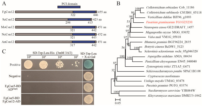
In the genome sequence of F. graminearum strain PH-1 (YL1), the predicted gene FG1G23230 encodes a protein with a C-terminal PCI domain that is orthologous to yeast ScCsn12 with 26.33% amino-acid identity [26]. We, therefore, named this gene FgCSN12 (Figure 1A). Phylogenetic analysis revealed that FgCsn12 orthologs widely exist in filamentous ascomycetes and yeasts (Figure 1B). FgCSN12 was expressed in hyphae, infected wheat heads, and perithecia (Supplementary Figure S1).
Figure 1.
Phylogenetic analysis of FgCSN12 and its interaction with FgCSN5. (A) Schematic drawing of the conserved domains of FgCsn12, ScCsn12, MoCsn12, NcCsn12, and AnCsn12 in F. graminearum, S. cerevisiae, Magnaporthe oryzae, Neurospora crassa, and Aspergillus nidulans. The PCI domain is labeled with a blue box. (B) Phylogenetic analysis of full-length amino acid sequences of FgCsn12 and its orthologs from S. cerevisiae, Schizosaccharomyces pombe, M. oryzae, N. crassa, A. nidulans, Botrytis cinerea, Sclerotinia sclerotiorum, Cryptococcus neoformans, Zymoseptoria tritici, Colletotrichum orbiculare, Colletotrichum sublineola, Verticillium dahliae, Valsa mali, Blumeria graminis, Penicillium chrysogenum, Ustilago maydis, Puccinia graminis, and Kluyveromyces marxianus. The phylogenetic tree was constructed by the neighbor-joining method using MEGA5 software [27]. The bootstrap values shown were estimated based on 1000 replications. The red font represents FgCsn12 in F. graminearum. (C) Different concentrations of yeast cells (cells/mL) of the transformants expressing the indicated bait and prey constructs (left) were assayed for growth on SD-Trp-Leu-His plates and LacZ activities.
However, in comparison with the other two stages, a relatively lower transcription level of FgCSN12 in infected wheat heads was detected by quantitative reverse transcription (qRT)-PCR assays (Supplementary Figure S1). In the genome of F. graminearum, a total of 7 COP9 signalosome subunits were identified. Whereas the PCI (proteasome, COP9 signalosome, initiation factor 3) domain existed in FgCsn1 (FG1G37750), FgCsn2 (FG1G02730), FgCsn4 (FG1G09260), and FgCsn7 (FG1G23010), we found that FgCsn5 (FG1G38480) and FgCsn6 (FG4G27310) had a conserved MPN (Mpr-Pad1-N-terminal) domain in their N-terminal region, and FgCsn3 (FG1G46710) had no conserved domain (Supplementary Figure S2).
To determine the association between FgCsn12 and the COP9 complex in F. graminearum, we cloned full-length FgCSN5 (FG1G38480) and FgCSN12 into Matchmaker vectors as the prey and bait constructs, respectively. The yeast transformants carrying the FgCsn5 bait and FgCsn12 prey constructs were unable to grow on SD-Trp-Leu-His medium and lacked LacZ activity (Figure 1C), indicating no physical interaction between these two proteins. Therefore, FgCSN12 is not directly associated with the Cop9 complex in F. graminearum and likely functions differently from its orthologs in yeast.
2.2. The Fgcsn12 Deletion Mutant Is Slightly Defective in Vegetative Growth, Conidial Morphology, and Plant Infection
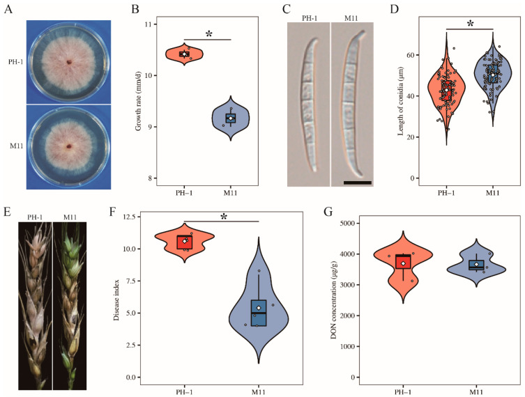
To determine the function of FgCSN12 in F. graminearum, we generated the Fgcsn12 mutant M11 in the wild-type strain PH-1 with the split-marker approach (Table 1 and Supplementary Figures S3–S5) [28]. Compared with that of the wild type, the Fgcsn12 deletion mutant M11 (Table 1) showed a 12% reduction in the growth rate (Figure 2A,B and Table 2). In 5-day-old CMC cultures, the Fgcsn12 mutant produced similar amount of conidia as the wild type (Table 2); however, conidia produced by the Fgcsn12 mutant were longer than those of the wild type (Figure 2C,D and Table 2).
Table 1.
The wild type and transformants of Fusarium graminearum used in this study.
| Strain | Brief Description | Reference |
|---|---|---|
| PH-1 | Wild-type | [28] |
| M11 | Fgcsn12 deletion mutant of PH-1 | This study |
| CG1 | Fgcsn12/FgCSN12-GFP transformant of M11 | This study |
| CU3 | Fgcsn12/FgCSN12UTR transformant of M11 | This study |
| M11R | Fgcsn12/H1-RFP transformant of M11 | This study |
| S406G | Fgcsn12/FgCSN12S406G transformant of M11 | This study |
| S406S | Fgcsn12/FgCSN12S406S transformant of M11 | This study |
Figure 2.
Defects of the Fgcsn12 mutant in vegetative growth, conidial morphology, and pathogenicity. (A) Three-day-old PDA cultures of the wild type (PH-1) and Fgcsn12 mutant (M11). (B) Growth rate of the indicated strains based on data from three biological replicates. (C) Conidial morphology of the indicated strains. Bar = 10 μm. (D) The average conidia length was calculated with data from 100 conidia. (E) Representative images of wheat heads infected with the indicated strains were photographed at 14 dpi. Black dots mark the inoculated spikelet. (F) The disease index of the indicated strains was estimated with data from five independent biological replicates. (G) DON levels in diseased wheat spikelets inoculated with the indicated strains based on data from three biological replicates. * Indicates significant differences based on t-test analysis followed by Duncan’s multiple range test (p = 0.05).
Table 2.
The growth rate, conidiation, length of conidia, and virulence of the Fgcsn12 mutant.
| Strains | Growth Rate (mm/day) a |
Conidiation (104/mL) b |
Length of Conidia (μm) |
Disease Index c | DON Concentration (μg/g) |
|---|---|---|---|---|---|
| PH-1 (wild type) | 10.42 ± 0.05 *A | 40.00 ± 2.25 A | 42.89 ± 0.72 A | 10.60 ± 0.24 A | 3692.86 ± 285.63 A |
| M11 (Fgcsn12 mutant) | 9.17 ± 0.10 B | 40.17 ± 1.64 A | 50.24 ± 0.65 B | 5.4 ± 0.75 B | 3664.83 ± 179.56 A |
| CU3 (Fgcsn12/FgCSN12UTR) | 10.56 ± 0.03 A | 40.50 ± 1.61 A | 42.81 ± 0.65 A | 10.20 ± 0.20 A | 3698.33 ± 161.06 A |
a Average daily extension in colony radius on PDA plates. b Conidiation in 5-day-old CMC cultures. c The number of diseased spikelets on each inoculated wheat head at 14 dpi. * The mean and standard deviation were calculated with results from at least three replicates. The data were analyzed with Duncan’s pairwise comparison. Different uppercase letters indicate significant differences (p = 0.05).
Moreover, the conidial germination rate (80.73%) of the Fgcsn12 mutant was lower than that of the wild type (98.08%). In infection assays with wheat heads, the Fgcsn12 deletion mutant was reduced in virulence (Figure 2E). The average disease index (diseased spikelets per head) was 5.4 for the Fgcsn12 mutant and 10.6 for the wild type (Figure 2F and Table 2). In F. graminearum, DON is considered to be an important virulence factor [29]. Therefore, we assayed DON production in inoculated wheat kernels; however, no defects of the Fgcsn12 mutant in DON biosynthesis were found (Figure 2G and Table 2). These results suggested that FgCsn12 plays a minor role in the regulation of vegetative growth, conidial morphology, and pathogenicity.
2.3. FgCsn12 Is Important for Ascosporogenesis
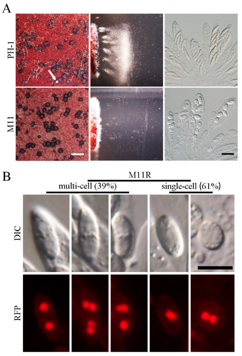
On the self-mating plates, the Fgcsn12 mutant formed a great number of melanized perithecia of normal size and morphology at 7 days postfertilization (dpf). However, ascospore cirrhi were rarely observed on the mutant’s perithecia (Figure 3A), indicating a severe defect in ascospore release. We then assayed the ascospore discharge to confirm this observation as previously described by Luo et al. [30]. After 16 h of incubation, abundant ascospores were forcibly discharged in the wild type. In contrast, only a few ascospores were discharged from the Fgcsn12 mutant under the same conditions (Figure 3A). Therefore, FgCsn12 is required for the forcible discharge of ascospores from perithecia in F. graminearum.
Figure 3.
Assays for the defects of the Fgcsn12 mutant in sexual reproduction. (A) Mating cultures of the wild type (PH-1) and Fgcsn12 mutant (M11) were examined for perithecium formation (left), ascospore discharge (middle), and asci with ascospores (right) 8 days postfertilization (dpf). Ascospore cirrhi are indicated by white arrows. White bar = 1 mm; black bar = 20 μm. (B) Ascospores of M11 expressing H1-RFP (M11R) were examined by differential interference contrast (DIC) or epifluorescence microscopy (RFP). The number of cells was randomly calculated from 600 ascospores. Bar = 10 μm.
Perithecia formed by both the wild-type and the Fgcsn12 mutant contained rosettes of asci with similar sizes. However, in the Fgcsn12 mutant, the number of ascospores in most asci was less than eight (Figure 3A and Supplementary Figure S6), suggesting a potential role of FgCSN12 in the first-round of postmeiotic mitosis. Elongated ascospores which had four compartments were observed in the wild-type, while the Fgcsn12 mutant produced only oval ascospores, with one or two nuclei (indicated by H1-RFP) in each compartment (Figure 3B and Supplementary Figure S7).
These results indicated that deletion of FgCSN12 blocked septation after ascospore delimitation and second-round postmeiotic mitosis in developing ascospores (Supplementary Figure S8). In addition, the ascospores formed by Fgcsn12 mutant had a 9.48% reduction in germination rates when compared with the wild type. Therefore, FgCSN12 plays a more critical role in sexual reproduction than in vegetative growth, asexual reproduction, and pathogenesis.
2.4. The 3′-UTR Sequence of FgCSN12 Is Required for its Function in Sexual Reproduction
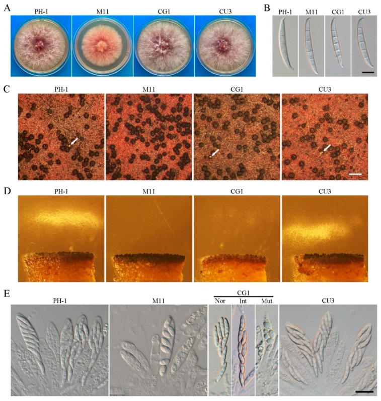
For complementation assays, FgCSN12-GFP fusion constructs were generated and introduced into Fgcsn12 deletion mutants. The resulting Fgcsn12/FgCSN12-GFP transformant CG1 (Table 1) was normal in vegetative growth, conidial morphology, and perithecia formation (Figure 4A–C). However, the ascospore discharge of transformant CG1 was partially resumed (Figure 4D).
Figure 4.
The expression of FgCSN12-GFP failed to rescue the defects of the Fgcsn12 mutant in ascosporogenesis. (A) Three-day-old PDA cultures of the wild type (PH-1), Fgcsn12 mutant (M11), Fgcsn12/FgCSN12-GFP transformant (CG1), and Fgcsn12/FgCSN12UTR transformant (CU3). (B) Conidial morphology of the same set of strains. Bar = 10 μm. (C) Mating cultures of the same set of strains were examined at 8 dpf. Arrows point to cirrhi. Bar = 1 mm. (D) Ascospore discharge was assayed with 7 dpf perithecia of the same set of strains. (E) The same set of strains was examined for asci and ascospores in 8 dpf perithecia. Nor, Int, and Mut represent normal, intermediate, and mutant ascospores from transformant CG1, respectively. Bar = 20 μm.
Normal ascospores, elongated ascospores (intermediate type), and mutant ascospores were produced in the asci of transformant CG1 (Figure 4E), indicating that the FgCsn12-GFP fusion construct failed to rescue the defect of the Fgcsn12 mutant in ascospore morphology. Since the FgCsn12-GFP fusion construct was generated in the coding region of FgCSN12, the defects of CG1 in sexual reproduction are likely due to the GFP tag or the lack of a 3′-UTR sequence. We, therefore, constructed a FgCSN12 complementation construct with an 855-bp 3′-UTR sequence based on published RNA-seq data [16] and transformed it into the Fgcsn12 mutant.
The resulting Fgcsn12/FgCSN12UTR transformant CU3 (Table 1) was normal in vegetative growth, conidial morphology, perithecia formation, asci development, and ascospore discharge (Figure 4A–E), indicating that the 3′-UTR sequence of FgCSN12 is able to rescue sexual reproduction defects. Thus, the 3′-UTR sequence of FgCSN12 is not essential for its functions in vegetative growth and asexual reproduction; however, it is important for sexual reproduction in F. graminearum.
2.5. FgCsn12-GFP Localizes to the Nucleus
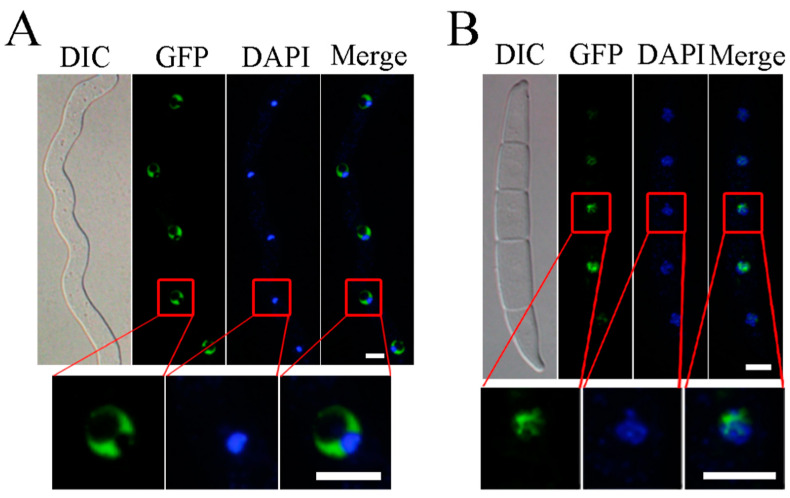
In the Fgcsn12/FgCSN12-GFP transformants CG1, GFP signals accumulated but were unevenly distributed in the nuclei of germlings and conidia (Figure 5A,B). The germlings and conidia were further stained with DAPI, and the fluorescent signals of DAPI and FgCsn12-GFP rarely overlapped with each other (Figure 5A,B). When analyzed by cNLS Mapper (http://nls-mapper.iab.keio.ac.jp/cgi-bin/NLS_Mapper_form.cgi#opennewwindow, accessed on 23 June 2009), a predicted 10-amino-acid nuclear localization signal (TAHKRKLDHD, 60 to 69 aa) was identified in the N-terminal region of FgCsn12. Previous studies have reported that regions stained with DAPI usually correspond to centromeric heterochromatin [31], therefore, FgCsn12 is likely enriched in euchromatin.
Figure 5.
The subcellular localization of the FgCsn12-GFP fusion protein. (A) Germlings of transformant CG1 were stained with DAPI and examined by DIC or epifluorescence microscopy (GFP). Bar = 5 μm. (B) Conidia of transformant CG1 were stained with DAPI and examined by DIC or epifluorescence microscopy. Bar = 10 μm.
2.6. Deletion of FgCSN12 Affects the Expression of More Than 3000 Genes
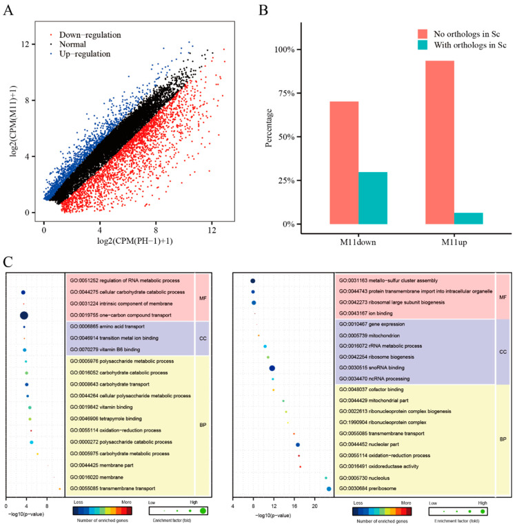
To identify genes affected by the deletion of FgCSN12, RNA-seq analysis was performed with RNA isolated from perithecia sampled at 7 dpf. In the Fgcsn12 mutant, 1204 genes and 2240 genes were upregulated and downregulated, respectively, over two-fold more than that of the wild type (Figure 6A and Supplementary Table S1). To verify the RNA-seq data, we selected five differentially expressed gene (DEGs) in the Fgcsn12 mutant for qRT-PCR assays. All of them had similar changes in their expression levels in the RNA-seq data and qRT-PCR results (Supplementary Figure S9).
Figure 6.
Assay for the role of FgCsn12 in transcriptional regulation. (A) Genes that were significantly increased (blue dots) and decreased (red dots) over two-fold in the Fgcsn12 mutant in comparison with the wild type. The x-axis and y-axis are the logarithms of CPM (wild-type PH-1) + 1 and CPM (M11) + 1, respectively. (B) The proportion of downregulated genes and upregulated genes with or without orthologs in budding yeast. (C) GO enrichment analysis of the upregulated and downregulated genes in M11. BP, MF, and CC represent biological process, molecular function, and cellular component, respectively.
More than 90% of the upregulated genes and approximately 70% of the downregulated genes had no homologs in S. cerevisiae, indicating that those genes appear to be unique to F. graminearum and other filamentous fungi (Figure 6B). Gene Ontology (GO) enrichment analysis showed that genes upregulated in the Fgcsn12 mutant were related to transmembrane transport, membrane parts, the carbohydrate metabolic process, and the polysaccharide catabolic process and were significantly enriched (Figure 6C).
A number of genes related to sexual reproduction had increased transcription levels in the Fgcsn12 mutant, such as the L-type calcium ion channel gene CCH1 (FG1G20950) [32] and peptide transporter gene FgPTR2D (FG2G00070) [33]. In contrast, genes downregulated in the Fgcsn12 mutant were enriched for preribosome, nucleolus, oxidoreductase activity, nucleolar part, ribonucleoprotein complex biogenesis, ncRNA processing, ribosome biogenesis, and gene expression (Figure 6C).
Some of the genes downregulated in the Fgcsn12 mutant are known to be related to asci and ascospore development, such as the pyruvate decarboxylase gene PDC1 (FG4G23780) [34], protein kinase gene FgFPK1 (FG2G38150) [10], and Rho family small GTPase gene FgRHO2 (FG2G33460) [35]. Reducing the transcription level of these genes may be responsible for the defects of the Fgcsn12 mutant in ascosporogenesis.
2.7. Both Editable and Noneditable FgCSN12 Alleles Complement the Defects of the Fgcsn12 Mutant in Sexual Development
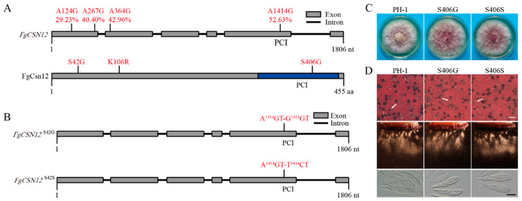
FgCSN12 transcripts had three A-to-I RNA editing sites during sexual reproduction (Liu et al., 2016). These three editing events caused an amino acid change in S42G, K106R, and S406G, which had editing levels of 29.23%, 42.96%, and 52.63%, respectively, in perithecia harvested at 8 days postfertilization (Figure 7A). Among them, S406G had an editing level higher than 50% and occurred in the C-terminal PCI domain (Figure 7A).
Figure 7.
Effects of expressing edited and uneditable alleles of FgCSN12 on sexual reproduction. (A) Schematic drawing of FgCSN12 and its protein. The A-to-I RNA editing sites and efficiency are marked in red. The amino acid changes caused by A-to-I RNA editing are also marked in red. The PCI domain is labeled with a blue box. (B) Schematic drawing of the FgCSN12S406G and FgCSN12S406S mutants. The FgCSN12S406G mutant had the A1414GT to G1414GT mutation. The FgCSN12S406S mutant had the A1414GT to T1414CT mutation. (C) Three-day-old PDA cultures of the wild type (PH-1) and transformants of the Fgcsn12 mutant expressing the FgCSN12S406G (S406G) and FgCSN12S406S (S406S) alleles. Photographs were taken after 3 days of incubation. (D) Mating cultures of PH-1, S406G, and S406S were examined for perithecium formation (upper), ascospore discharge (middle), and asci with ascospores (lower) in 8 dpf perithecia. White arrows point to cirrhi. White bar = 1 mm; black bar = 20 μm.
To determine whether this S406G editing event is associated with the role of FgCsn12 in sexual development, we introduced A1414GT (S) to T1414CT (S) and G1414GT (G) mutations into the complementation construct to generate noneditable and edited alleles (Figure 7B). All the resulting transformants expressing these mutant alleles of FgCsn12 had no obvious defects in vegetative growth (Figure 7C) or sexual development (Figure 7D), suggesting that the editing event that occurred in S406 had no significant effect on FgCsn12 functions in F. graminearum.
3. Discussion
Csn5 is a well-conserved subunit that acts as the catalytic center for the COP9 signalosome complex in fungi [36]. The incorporation of Csn5 into the COP9 signalosome is dependent on Csn12 in budding yeast [37], indicating that Csn12 is associated with the COP9 signalosome complex for its organization and stabilization [24]. However, a direct interaction between FgCsn5 and FgCsn12 was not detected in F. graminearum, suggesting that FgCsn12 was not directly associated with the COP9 signalosome complex.
In other words, the COP9 complexes in F. graminearum and S. cerevisiae were organized differently. Indeed, the subunit composition of the COP9 signalosome in fungi is rather divergent. Protein purification of the COP9 signalosome revealed that Neurospora crassa lacks the Csn8 subunit [38], whereas, in Schizosaccharomyces pombe, both Csn6 and Csn8 were not detected in this complex [39]. As FgCsn12 was not directly associated with FgCsn5, it might have some functions independent of the COP9 signalosome. The deletion of FgCSN12 resulted in the formation of oval ascospores, which were rarely discharged from the perithecia, suggesting FgCsn12 has an important role in ascosporogenesis.
Round or oval ascospores were also observed in Gzsnf1- and Fgama1-deletion mutants in F. graminearum [17,40]. GzSNF1 encodes a serine/threonine protein kinase, and its orthologs in Arabidopsis and wheat are involved in binding SCF ubiquitin ligase [11,41]. FgAMA1, a gene specifically expressed during sexual reproduction, encodes a meiosis-specific activator of APC/C. APC/C and SCF are evolutionarily related ubiquitin ligase complexes that control the sequential degradation of cell-cycle-progression proteins during cell division.
Therefore, both GzSNF1 and FgAMA1 also appeared to be related to SCF ubiquitin ligase based on their function during ascosporogenesis. Interestingly, although a direct relationship between Csn12 and SCF ubiquitin ligase was not reported, Csn12 is known to be associated with Dss1, which functions as a ubiquitin receptor [42]. It is likely that FgCsn12, FgAma1, and GzSnf1 regulate ascosporogenesis in the same manner, potentially through SCF ubiquitin ligase-mediated protein degradation.
In previous studies, many genes, including FNG1, AMD1, and PAL1, could fully complement their corresponding deletion mutants with GFP fusion constructs in F. graminearum [14,43,44]. In this study, FgCSN12-GFP complemented the defects of the Fgcsn12 mutant in vegetative growth and asexual reproduction but not during the sexual development process.
However, with the addition of the 3′-UTR, FgCSN12 successfully complemented the defects in ascosporogenesis, indicating that the function of FgCSN12 3′UTR sequences is only required during sexual reproduction. A similar phenomenon was also observed in complementation assays for Fgama1 and Fgtub1 mutants [17,20]. These genes may have stage-specific transcription termination or alternative polyadenylation sites (APAs) in ascogenous tissues.
Adenosine to inosine (A-to-I) RNA editing is the most prevalent type of RNA editing in mammals; however, most editing sites are in the noncoding regions [45]. The A-to-I editing in fungi specifically occurred in the sexual development stage, and the majority of the editing sites resulted in changes of protein recoding [46]. In F. graminearum, the PUK1, AMD1, and AMA1 genes require A-to-I RNA editing during sexual reproduction to encode a full-length protein [14,16,17]. In this study, several nonsynonymous editing sites were identified in FgCSN12, introducing amino acid sequence variations in F. graminearum. Although the exact roles of editing sites in FgCSN12 are not known yet, nonsynonymous editing events were generally beneficial and favored by positive selection during evolution in N. crassa [46]
4. Materials and Methods
4.1. Strains and Culture Conditions
The F. graminearum wild-type strain PH-1 and all the transformants generated in this study were cultured on potato dextrose agar (PDA) plates at 25 °C. Conidiation in liquid carboxymethyl cellulose (CMC) medium and growth rate in PDA medium were assayed as described by Zhou et al. [47,48]. For sexual reproduction, aerial hyphae of 7-day-old carrot agar cultures were pressed down with sterile 0.1% Tween 20 as described by Zheng et al. [9,49]. The perithecia, cirrhi, asci, and ascospore discharge were examined as described by Cavinder et al. [50].
Protoplast preparation and polyethylene glycol (PEG)-mediated transformation were performed as described by Hou et al. [47]. Hygromycin B (CalBiochem, La Jolla, CA, USA), geneticin (Sigma–Aldrich, St. Louis, MO, USA), and zeocin (Invitrogen, Carlsbad, CA, USA) were added to final concentrations of 300, 400, and 450 μg/mL, respectively, for transformant selection.
4.2. Quantitative Reverse Transcription (qRT) PCR Assays
RNA samples of the wild type from vegetative hyphae harvested from 24 h liquid YEPD cultures, Inoculated spikelets of flowering wheat heads of cultivar Xiaoyan22 collected 3 days after inoculation, perithecia from carrot agar plates at 4 and 7 dpf, and Fgcsn12 mutant from carrot agar plates at 7 dpf were isolated with the Eastep Super Total RNA Extraction Kit (Promega, Madison, WI, USA). The FastKing RT Kit (TIANGEN, Beijing, China) was used to synthesize cDNA, and qRT-PCR assays were performed with the CFX96 Real-Time System (Bio-RAD, Hercules, CA, USA) [51]. The comparative 2−ΔΔCt method was used to calculate the relative fold changes in the expression of FgCSN12 from the samples collected. The relative expression levels of target genes were assayed using qRT-PCR with the primers listed in Supplementary Table S2 using the F. graminearum actin gene FG4G14550 as the internal control [52].
4.3. Yeast Two-Hybrid Assays
The interaction of FgCsn12 with FgCsn5 was assayed with the Matchmaker yeast two-hybrid system (Clontech, Mountain View, CA, USA). The ORF of FgCSN12 was amplified from cDNA of PH-1 synthesized as described by Zhou et al. [53] with the primers FgCSN12AD/F-FgCSN12AD/R (Supplementary Table S2) and cloned into pGADT7 as the prey construct. The ORF of FgCSN5 was amplified with the primers FgCSN5BD/F-FgCSN5BD/R (Supplementary Table S2) and cloned into pGBKT7 as the bait construct.
The resulting bait and prey vectors were cotransformed in pairs into yeast strain AH109. To check for autoactivation, the bait construct of FgCSN5 was cotransformed with an empty pGADT7 vector. The resulting transformants were then assayed for growth on a synthetic dropout (SD) medium lacking tryptophan, leucine, and histidine (SD-Trp-Leu-His) and β-galactosidase activities as described by Zhou et al. [54].
4.4. Generation of the Fgcsn12 Deletion Mutant
To generate the gene replacement construct for the FgCSN12 gene using the split marker approach, the 0.7 kb upstream and 0.7 kb downstream flanking sequences were amplified by polymerase chain reaction (PCR) from wild-type genomic DNA. The resulting PCR products were connected to the hygromycin phosphotransferase (hph) resistance gene cassette by overlapping PCR and transformed into wild-type protoplasts as described by Xu et al. [55]. Hygromycin-resistant transformants were screened for Fgcsn12 deletion mutants by PCR.
4.5. Generation of the FgCSN12UTR and FgCSN12-GFP Transformants
For complementation assays, the FgCSN12 gene, including its 754-bp promoter region and 855-bp 3′-end sequence, was amplified with the primer pair FgCSN12N/F- FgCSN12U/R (Supplementary Table S2) and cotransformed with XhoI-digested pFL2 (carrying the geneticin resistance marker) into yeast strain XK1-25 by the yeast gap repair approach [56] to generate the FgCSN12UTR construct. The same approach was used to generate the FgCSN12-GFP construct with the primer pair FgCSN12N/F-FgCSN12G/R (Supplementary Table S2) [56]. The resulting fusion constructs rescued from Trp+ yeast transformants were confirmed by sequencing analysis and transformed into the Fgcsn12 mutant M11 (Table 1). Transformants resistant to both hygromycin and geneticin were screened by PCR.
4.6. Plant Infection and DON Production Assays
For infection assays, conidia of PH-1 and mutant strains were obtained by filtration from CMC cultures and resuspended to 105 spores/mL in sterile distilled water [57,58]. The spikelet of each wheat head (cultivar Xiaoyan 22) was inoculated with 10 μL of conidial suspension as described by Jiang et al. [59]. All of the inoculated wheat heads were examined at 14 days postinfection (dpi) to estimate the disease index [29]. Inoculated wheat kernels were collected and assayed for DON production as described by Bluhm et al. [51,60].
4.7. Generation of the FgCSN12S406G and FgCSN12S406S Transformants
The S406G mutation was introduced into FgCSN12 by overlapping PCR with fragments amplified with the primer pairs FgCSN12N/F-FgCSN12S406G/R and FgCSN12S406G/F–FgCSN12U/R (Supplementary Table S2) (primers FgCSN12S406G/R and FgCSN12S406G/F carrying the mutations). FgCSN12S406G (edited) was cloned into pFL2 by yeast gap repair [56] to generate the FgCSN12S406G fusion construct.
The same approach was used to generate the FgCSN12S406S (unedited) construct with the primer pairs FgCSN12N/F-FgCSN12S406S/R and FgCSN12S406S/F–FgCSN12U/R (Supplementary Table S2) (primers FgCSN12S406S/R and FgCSN12S406S/F carrying the mutations). The FgCSN12S406G and FgCSN12S406S constructs were confirmed by sequencing analysis and transformed into the Fgcsn12 mutant M11 (Table 1). Transformants resistant to both hygromycin and geneticin were screened using PCR.
4.8. RNA-Seq Analysis
Perithecia of PH-1 and Fgcsn12 mutants were harvested at 7 dpf from carrot agar cultures and used for RNA extraction with TRIzol (Invitrogen, USA). RNA was isolated from two independent biological replicates for each strain. Strand-specific RNA-seq libraries were prepared with the NEBNext Ultra Directional RNA Library Prep Kit (NEB, Ipswich, MA, USA) following the instructions provided by the manufacturer and sequenced with an Illumina HiSeq 2500 system using the 2 × 150 bp paired-end read model at the Novogene Bioinformatics Institute (Beijing, China). For each replicate, at least 24 Mb paired-end reads were obtained.
The resulting RNA-seq reads were mapped onto the reference genome of F. graminearum strain PH-1 [28,61] by HISAT2 [62]. The number of reads (counts) mapped to each gene was calculated using featureCounts [63]. Differential expression analysis of all genes was performed using the edgeRun package [64] with the exactTest function. Genes with a log2FC (log2fold change) above 1 and FDR (false discovery rate) below 0.05 were considered to be differentially expressed genes. GO enrichment analysis was performed with Blast2GO [65], and the p values were adjusted with the Benjamin–Hochberg procedure by controlling the false discovery rate (FDR) to 0.05 as previously described [66].
5. Conclusions
In summary, we functionally characterized the FgCSN12 gene, which is important for sexual reproduction in F. graminearum. The Fgcsn12 deletion mutant has slight defects in vegetative growth, conidiation, and plant infection. However, the deletion of FgCSN12 resulted in severe defects in ascospore morphology and discharge. Instead of forming four-celled, uninucleate ascospores, the Fgcsn12 mutant produced oval, single- or two-celled ascospores that were significantly defective in ascospore discharge. FgCsn12-GFP localizes to the nucleus in the hyphae and conidia.
RNA-seq analysis revealed that more than 3000 differentially expressed genes were identified in the Fgcsn12 mutant, including a variety of genes related to sexual reproduction. Although the ortholog of FgCsn12 in budding yeast had a direct interaction with Csn5, the core subunit of the COP9 signalosome, FgCsn12, did not interact with FgCsn5 in the yeast two-hybrid assay. Taken together, FgCsn12 is involved in the regulation of ascosporogenesis independent of the COP9 signalosome complex.
Acknowledgments
We thank Qinhu Wang, Guanghui Wang, Xue Zhang, and Ming Xu for the fruitful discussions.
Supplementary Materials
The following supporting information can be downloaded at: https://www.mdpi.com/article/10.3390/ijms231810445/s1.
Author Contributions
Methodology, H.J., Y.Z., W.W. and H.X.; Data Curation, X.C., H.L. and J.Q.; Writing—Original Draft Preparation, H.J.; Writing—Review and Editing, C.J.; Supervision, C.W.; Funding Acquisition, C.J. and H.J. All authors have read and agreed to the published version of the manuscript.
Institutional Review Board Statement
Not applicable.
Informed Consent Statement
Not applicable.
Data Availability Statement
RNA-seq data were deposited in the NCBI SRA database under accession number PRJNA655824 accessed on 7 August 2020 at https://www.ncbi.nlm.nih.gov/.
Conflicts of Interest
The authors declare no conflict of interest. The funders had no role in the study design, data collection and analysis, decision to publish, or preparation of the manuscript.
Funding Statement
This research was funded by the National Natural Science Foundation of China (grant number 32102181) and Shaanxi Science Fund for Distinguished Young Scholars (grant number 2022JC-14).
Footnotes
Publisher’s Note: MDPI stays neutral with regard to jurisdictional claims in published maps and institutional affiliations.
References
- 1.Xu X., Nicholson P. Community ecology of fungal pathogens causing wheat head blight. Annu. Rev. Phytopathol. 2009;47:83–103. doi: 10.1146/annurev-phyto-080508-081737. [DOI] [PubMed] [Google Scholar]
- 2.Goswami R.S., Kistler H.C. Heading for disaster: Fusarium graminearum on cereal crops. Mol. Plant Pathol. 2004;5:515–525. doi: 10.1111/j.1364-3703.2004.00252.x. [DOI] [PubMed] [Google Scholar]
- 3.Bai G., Shaner G. Management and resistance in wheat and barley to fusarium head blight. Annu. Rev. Phytopathol. 2004;42:135–161. doi: 10.1146/annurev.phyto.42.040803.140340. [DOI] [PubMed] [Google Scholar]
- 4.Audenaert K., Vanheule A., Höfte M., Haesaert G. Deoxynivalenol: A major player in the multifaceted response of Fusarium to its environment. Toxins. 2013;6:1–19. doi: 10.3390/toxins6010001. [DOI] [PMC free article] [PubMed] [Google Scholar]
- 5.Bennett R.J., Turgeon B.G. Fungal sex: The ascomycota. Microbiol Spectr. 2016;4:FUNK-0005-2016. doi: 10.1128/microbiolspec.FUNK-0005-2016. [DOI] [PubMed] [Google Scholar]
- 6.Trail F., Gaffoor I., Vogel S. Ejection mechanics and trajectory of the ascospores of Gibberella zeae (anamorph Fuarium graminearum) Fungal Genet. Biol. 2005;42:528–533. doi: 10.1016/j.fgb.2005.03.008. [DOI] [PubMed] [Google Scholar]
- 7.Kim H.K., Cho E.J., Lee S., Lee Y.S., Yun S.H. Functional analyses of individual mating-type transcripts at MAT loci in Fusarium graminearum and Fusarium asiaticum. FEMS Microbiol. Lett. 2012;337:89–96. doi: 10.1111/1574-6968.12012. [DOI] [PubMed] [Google Scholar]
- 8.Kim H.K., Jo S.M., Kim G.Y., Kim D.W., Kim Y.K., Yun S.H. A large-scale functional analysis of putative target genes of mating-type loci provides insight into the regulation of sexual development of the cereal pathogen Fusarium graminearum. PLoS Genet. 2015;11:e1005486. doi: 10.1371/journal.pgen.1005486. [DOI] [PMC free article] [PubMed] [Google Scholar]
- 9.Zheng Q., Hou R., Zhang J., Ma J., Wu Z., Wang G., Wang C., Xu J.-R. The MAT locus genes play different roles in sexual reproduction and pathogenesis in Fusarium graminearum. PLoS ONE. 2013;8:e66980. doi: 10.1371/journal.pone.0066980. [DOI] [PMC free article] [PubMed] [Google Scholar]
- 10.Wang C., Zhang S., Hou R., Zhao Z., Zheng Q., Xu Q., Zheng D., Wang G., Liu H., Gao X., et al. Functional analysis of the kinome of the wheat scab fungus Fusarium graminearum. PLoS Pathog. 2011;7:e1002460. doi: 10.1371/journal.ppat.1002460. [DOI] [PMC free article] [PubMed] [Google Scholar]
- 11.Jiang C., Cao S., Wang Z., Xu H., Liang J., Liu H., Wang G., Ding M., Wang Q., Gong C., et al. An expanded subfamily of G-protein-coupled receptor genes in Fusarium graminearum required for wheat infection. Nat. Microbiol. 2019;4:1582–1591. doi: 10.1038/s41564-019-0468-8. [DOI] [PubMed] [Google Scholar]
- 12.Yun Y., Liu Z., Yin Y., Jiang J., Chen Y., Xu J.-R., Ma Z. Functional analysis of the Fusarium graminearum phosphatome. New Phytol. 2015;207:119–134. doi: 10.1111/nph.13374. [DOI] [PubMed] [Google Scholar]
- 13.Son H., Seo Y.S., Min K., Park A.R., Lee J., Jin J.M., Lin Y., Cao P., Hong S.Y., Kim E.K., et al. A phenome-based functional analysis of transcription factors in the cereal head blight fungus, Fusarium graminearum. PLoS Pathog. 2011;7:e1002310. doi: 10.1371/journal.ppat.1002310. [DOI] [PMC free article] [PubMed] [Google Scholar]
- 14.Cao S., He Y., Hao C., Xu Y., Zhang H., Wang C., Liu H., Xu J.-R. RNA editing of the AMD1 gene is important for ascus maturation and ascospore discharge in Fusarium graminearum. Sci. Rep. 2017;7:4617. doi: 10.1038/s41598-017-04960-7. [DOI] [PMC free article] [PubMed] [Google Scholar]
- 15.Son H., Lee J., Lee Y.W. A novel gene, GEA1, is required for ascus cell-wall development in the ascomycete fungus Fusarium graminearum. Microbiology. 2013;159:1077–1085. doi: 10.1099/mic.0.064287-0. [DOI] [PubMed] [Google Scholar]
- 16.Liu H., Wang Q., He Y., Chen L., Hao C., Jiang C., Li Y., Dai Y., Kang Z., Xu J.-R. Genome-wide A-to-I RNA editing in fungi independent of ADAR enzymes. Genome Res. 2016;26:499–509. doi: 10.1101/gr.199877.115. [DOI] [PMC free article] [PubMed] [Google Scholar]
- 17.Hao C., Yin J., Sun M., Wang Q., Liang J., Bian Z., Liu H., Xu J.-R. The meiosis-specific APC activator FgAMA1 is dispensable for meiosis but important for ascosporogenesis in Fusarium graminearum. Mol. Microbiol. 2019;111:1245–1262. doi: 10.1111/mmi.14219. [DOI] [PubMed] [Google Scholar]
- 18.Zhao Z., Liu H., Luo Y., Zhou S., An L., Wang C., Jin Q., Zhou M., Xu J.R. Molecular evolution and functional divergence of tubulin superfamily in the fungal tree of life. Sci. Rep. 2014;4:6746. doi: 10.1038/srep06746. [DOI] [PMC free article] [PubMed] [Google Scholar]
- 19.Liu H., Zhang S., Ma J., Dai Y., Li C., Lyu X., Wang C., Xu J.R. Two Cdc2 kinase genes with distinct functions in vegetative and infectious hyphae in Fusarium graminearum. PLoS Pathog. 2015;11:e1004913. doi: 10.1371/journal.ppat.1004913. [DOI] [PMC free article] [PubMed] [Google Scholar]
- 20.Chen D., Wu C., Hao C., Huang P., Liu H., Bian Z., Xu J.R. Sexual specific functions of Tub1 beta-tubulins require stage-specific RNA processing and expression in Fusarium graminearum. Environ. Microbiol. 2018;20:4009–4021. doi: 10.1111/1462-2920.14441. [DOI] [PubMed] [Google Scholar]
- 21.Qin N., Xu D., Li J., Deng X.W. COP9 signalosome: Discovery, conservation, activity, and function. J. Integr. Plant Biol. 2020;62:90–103. doi: 10.1111/jipb.12903. [DOI] [PubMed] [Google Scholar]
- 22.Beckmann E.A., Köhler A.M., Meister C., Christmann M., Draht O.W., Rakebrandt N., Valerius O., Braus G.H. Integration of the catalytic subunit activates deneddylase activity in vivo as final step in fungal COP9 signalosome assembly. Mol. Microbiol. 2015;97:110–124. doi: 10.1111/mmi.13017. [DOI] [PubMed] [Google Scholar]
- 23.Busch S., Schwier E.U., Nahlik K., Bayram O., Helmstaedt K., Draht O.W., Krappmann S., Valerius O., Lipscomb W.N., Braus G.H. An eight-subunit COP9 signalosome with an intact JAMM motif is required for fungal fruit body formation. Proc. Natl. Acad. Sci. USA. 2007;104:8089–8094. doi: 10.1073/pnas.0702108104. [DOI] [PMC free article] [PubMed] [Google Scholar]
- 24.Maytal-Kivity V., Piran R., Pick E., Hofmann K., Glickman M.H. COP9 signalosome components play a role in the mating pheromone response of S. cerevisiae. EMBO Rep. 2002;3:1215–1221. doi: 10.1093/embo-reports/kvf235. [DOI] [PMC free article] [PubMed] [Google Scholar]
- 25.Jin D., Li B., Deng X.W., Wei N. Plant COP9 signalosome subunit 5, CSN5. Plant Sci. 2014;224:54–61. doi: 10.1016/j.plantsci.2014.04.001. [DOI] [PubMed] [Google Scholar]
- 26.Lu P., Chen D., Qi Z., Wang H., Chen Y., Wang Q., Jiang C., Xu J.R., Liu H. Landscape and regulation of alternative splicing and alternative polyadenylation in a plant pathogenic fungus. New Phytol. 2022;235:674–689. doi: 10.1111/nph.18164. [DOI] [PubMed] [Google Scholar]
- 27.Tamura K., Peterson D., Peterson N., Stecher G., Nei M., Kumar S. MEGA5: Molecular evolutionary genetics analysis using maximum likelihood, evolutionary distance, and maximum parsimony methods. Mol. Biol. Evol. 2011;28:2731–2739. doi: 10.1093/molbev/msr121. [DOI] [PMC free article] [PubMed] [Google Scholar]
- 28.Cuomo C.A., Güldener U., Xu J.-R., Trail F., Turgeon B.G., Di Pietro A., Walton J.D., Ma L.-J., Baker S.E., Rep M., et al. The Fusarium graminearum genome reveals a link between localized polymorphism and pathogen specialization. Science. 2007;317:1400–1402. doi: 10.1126/science.1143708. [DOI] [PubMed] [Google Scholar]
- 29.Jiang C., Zhang S., Zhang Q., Tao Y., Wang C., Xu J.R. FgSKN7 and FgATF1 have overlapping functions in ascosporogenesis, pathogenesis and stress responses in Fusarium graminearum. Environ. Microbiol. 2015;17:1245–1260. doi: 10.1111/1462-2920.12561. [DOI] [PubMed] [Google Scholar]
- 30.Luo Y., Zhang H., Qi L., Zhang S., Zhou X., Zhang Y., Xu J.R. FgKin1 kinase localizes to the septal pore and plays a role in hyphal growth, ascospore germination, pathogenesis, and localization of Tub1 beta-tubulins in Fusarium graminearum. New Phytol. 2014;204:943–954. doi: 10.1111/nph.12953. [DOI] [PubMed] [Google Scholar]
- 31.Rodgers W., Byrum J.N., Simpson D.A., Hoolehan W., Rodgers K.K. RAG2 localization and dynamics in the pre-B cell nucleus. PLoS ONE. 2019;14:e0216137. doi: 10.1371/journal.pone.0216137. [DOI] [PMC free article] [PubMed] [Google Scholar]
- 32.Hallen H.E., Trail F. The L-type calcium ion channel Cch1 affects ascospore discharge and mycelial growth in the filamentous fungus Gibberella zeae (anamorph Fusarium graminearum) Eukaryot. Cell. 2008;7:415–424. doi: 10.1128/EC.00248-07. [DOI] [PMC free article] [PubMed] [Google Scholar]
- 33.Droce A., Sørensen J.L., Sondergaard T.E., Rasmussen J.J., Lysøe E., Giese H. PTR2 peptide transporters in Fusarium graminearum influence secondary metabolite production and sexual development. Fungal Biol. 2017;121:515–527. doi: 10.1016/j.funbio.2017.02.003. [DOI] [PubMed] [Google Scholar]
- 34.Son H., Min K., Lee J., Choi G.J., Kim J.C., Lee Y.W. Differential roles of pyruvate decarboxylase in aerial and embedded mycelia of the ascomycete Gibberella zeae. FEMS Microbiol. Lett. 2012;329:123–130. doi: 10.1111/j.1574-6968.2012.02511.x. [DOI] [PubMed] [Google Scholar]
- 35.Zhang C., Wang Y., Wang J., Zhai Z., Zhang L., Zheng W., Zheng W., Yu W., Zhou J., Lu G., et al. Functional characterization of Rho family small GTPases in Fusarium graminearum. Fungal Genet. Biol. 2013;61:90–99. doi: 10.1016/j.fgb.2013.09.001. [DOI] [PubMed] [Google Scholar]
- 36.Wee S., Hetfeld B., Dubiel W., Wolf D.A. Conservation of the COP9/signalosome in budding yeast. BMC Genet. 2002;3:15. doi: 10.1186/1471-2156-3-41. [DOI] [PMC free article] [PubMed] [Google Scholar]
- 37.Maytal-Kivity V., Pick E., Piran R., Hofmann K., Glickman M.H. The COP9 signalosome-like complex in S. cerevisiae and links to other PCI complexes. Int. J. Biochem. Cell Biol. 2003;35:706–715. doi: 10.1016/S1357-2725(02)00378-3. [DOI] [PubMed] [Google Scholar]
- 38.He Q., Cheng P., He Q., Liu Y. The COP9 signalosome regulates the Neurospora circadian clock by controlling the stability of the SCFFWD-1 complex. Genes Dev. 2005;19:1518–1531. doi: 10.1101/gad.1322205. [DOI] [PMC free article] [PubMed] [Google Scholar]
- 39.Liu C., Powell K.A., Mundt K., Wu L., Carr A.M., Caspari T. Cop9/signalosome subunits and Pcu4 regulate ribonucleotide reductase by both checkpoint-dependent and -independent mechanisms. Genes Dev. 2003;17:1130–1140. doi: 10.1101/gad.1090803. [DOI] [PMC free article] [PubMed] [Google Scholar]
- 40.Lee S.H., Lee J., Lee S., Park E.H., Kim K.W., Kim M.D., Yun S.H., Lee Y.W. GzSNF1 is required for normal sexual and asexual development in the ascomycete Gibberella zeae. Eukaryot. Cell. 2009;8:116–127. doi: 10.1128/EC.00176-08. [DOI] [PMC free article] [PubMed] [Google Scholar]
- 41.Farrás R., Ferrando A., Jásik J., Kleinow T., Okrész L., Tiburcio A., Salchert K., del Pozo C., Schell J., Koncz C. SKP1-SnRK protein kinase interactions mediate proteasomal binding of a plant SCF ubiquitin ligase. EMBO J. 2001;20:2742–2756. doi: 10.1093/emboj/20.11.2742. [DOI] [PMC free article] [PubMed] [Google Scholar]
- 42.Kragelund B.B., Schenstrøm S.M., Rebula C.A., Panse V.G., Hartmann-Petersen R. DSS1/Sem1, a multifunctional and intrinsically disordered protein. Trends Biochem. Sci. 2016;41:446–459. doi: 10.1016/j.tibs.2016.02.004. [DOI] [PubMed] [Google Scholar]
- 43.Jiang H., Xia A., Ye M., Ren J., Li D., Liu H., Wang Q., Lu P., Wu C., Xu J.R., et al. Opposing functions of Fng1 and the Rpd3 HDAC complex in H4 acetylation in Fusarium graminearum. PLoS Genet. 2020;16:e1009185. doi: 10.1371/journal.pgen.1009185. [DOI] [PMC free article] [PubMed] [Google Scholar]
- 44.Yin J., Hao C., Niu G., Wang W., Wang G., Xiang P., Xu J.R., Zhang X. FgPal1 regulates morphogenesis and pathogenesis in Fusarium graminearum. Environ. Microbiol. 2020;22:5373–5386. doi: 10.1111/1462-2920.15266. [DOI] [PubMed] [Google Scholar]
- 45.Wang C., Xu J.R., Liu H. A-to-I RNA editing independent of ADARs in filamentous fungi. RNA Biol. 2016;13:940–945. doi: 10.1080/15476286.2016.1215796. [DOI] [PMC free article] [PubMed] [Google Scholar]
- 46.Liu H., Li Y., Chen D., Qi Z., Wang Q., Wang J., Jiang C., Xu J.R. A-to-I RNA editing is developmentally regulated and generally adaptive for sexual reproduction in Neurospora crassa. Proc. Natl. Acad. Sci. USA. 2017;114:E7756–E7765. doi: 10.1073/pnas.1702591114. [DOI] [PMC free article] [PubMed] [Google Scholar]
- 47.Hou Z., Xue C., Peng Y., Katan T., Kistler H.C., Xu J.-R. A mitogen-activated protein kinase gene (MGV1) in Fusarium graminearum is required for female fertility, heterokaryon formation, and plant infection. Mol. Plant. Microbe Interact. 2002;15:1119–1127. doi: 10.1094/MPMI.2002.15.11.1119. [DOI] [PubMed] [Google Scholar]
- 48.Zhou X., Heyer C., Choi Y.E., Mehrabi R., Xu J.R. The CID1 cyclin C-like gene is important for plant infection in Fusarium graminearum. Fungal Genet. Biol. 2010;47:143–151. doi: 10.1016/j.fgb.2009.11.001. [DOI] [PubMed] [Google Scholar]
- 49.Gao X., Jin Q., Jiang C., Li Y., Li C., Liu H., Kang Z., Xu J.-R. FgPrp4 kinase is important for spliceosome B-complex activation and splicing efficiency in Fusarium graminearum. PLoS Genet. 2016;12:e1005973. doi: 10.1371/journal.pgen.1005973. [DOI] [PMC free article] [PubMed] [Google Scholar]
- 50.Cavinder B., Sikhakolli U., Fellows K.M., Trail F. Sexual development and ascospore discharge in Fusarium graminearum. J. Vis. Exp. 2012;61:e3895. doi: 10.3791/3895. [DOI] [PMC free article] [PubMed] [Google Scholar]
- 51.Jiang C., Zhang C., Wu C., Sun P., Hou R., Liu H., Wang C., Xu J.-R. TRI6 and TRI10 play different roles in the regulation of deoxynivalenol (DON) production by cAMP signalling in Fusarium graminearum. Environ. Microbiol. 2016;18:3689–3701. doi: 10.1111/1462-2920.13279. [DOI] [PubMed] [Google Scholar]
- 52.Livak K.J., Schmittgen T.D. Analysis of relative gene expression data using real-time quantitative PCR and the 2−ΔΔCT method. Methods. 2001;25:402–408. doi: 10.1006/meth.2001.1262. [DOI] [PubMed] [Google Scholar]
- 53.Zhou X., Zhang H., Li G., Shaw B., Xu J.-R. The Cyclase-associated protein Cap1 is important for proper regulation of infection-related morphogenesis in Magnaporthe oryzae. PLoS Pathog. 2012;8:e1002911. doi: 10.1371/journal.ppat.1002911. [DOI] [PMC free article] [PubMed] [Google Scholar]
- 54.Zhou X., Liu W., Wang C., Xu Q., Wang Y., Ding S., Xu J.R. A MADS-box transcription factor MoMcm1 is required for male fertility, microconidium production and virulence in Magnaporthe oryzae. Mol. Microbiol. 2011;80:33–53. doi: 10.1111/j.1365-2958.2011.07556.x. [DOI] [PubMed] [Google Scholar]
- 55.Xu H., Ye M., Xia A., Jiang H., Huang P., Liu H., Hou R., Wang Q., Li D., Xu J.R., et al. The Fng3 ING protein regulates H3 acetylation and H4 deacetylation by interacting with two distinct histone-modifying complexes. New Phytol. 2022;235:2350–2364. doi: 10.1111/nph.18294. [DOI] [PubMed] [Google Scholar]
- 56.Zhou X., Li G., Xu J.-R. Efficient approaches for generating GFP fusion and epitope-tagging constructs in filamentous fungi. Methods Mol. Biol. 2011;722:199–212. doi: 10.1007/978-1-61779-040-9_15. [DOI] [PubMed] [Google Scholar]
- 57.Yin T., Zhang Q., Wang J., Liu H., Wang C., Xu J.-R., Jiang C. The cyclase-associated protein FgCap1 has both protein kinase A-dependent and -independent functions during deoxynivalenol production and plant infection in Fusarium graminearum. Mol. Plant Pathol. 2018;19:552–563. doi: 10.1111/mpp.12540. [DOI] [PMC free article] [PubMed] [Google Scholar]
- 58.Ren J., Zhang Y., Wang Y., Li C., Bian Z., Zhang X., Liu H., Xu J.-R., Jiang C. Deletion of all three MAP kinase genes results in severe defects in stress responses and pathogenesis in Fusarium graminearum. Stress Biol. 2022;2:6. doi: 10.1007/s44154-021-00025-y. [DOI] [PMC free article] [PubMed] [Google Scholar]
- 59.Jiang C., Hei R., Yang Y., Zhang S., Wang Q., Wang W., Zhang Q., Yan M., Zhu G., Huang P., et al. An orphan protein of Fusarium graminearum modulates host immunity by mediating proteasomal degradation of TaSnRK1α. Nat. Commun. 2020;11:4382. doi: 10.1038/s41467-020-18240-y. [DOI] [PMC free article] [PubMed] [Google Scholar]
- 60.Bluhm B.H., Zhao X., Flaherty J.E., Xu J.R., Dunkle L.D. RAS2 regulates growth and pathogenesis in Fusarium graminearum. Mol. Plant. Microbe Interact. 2007;20:627–636. doi: 10.1094/MPMI-20-6-0627. [DOI] [PubMed] [Google Scholar]
- 61.King R., Urban M., Hammond-Kosack M.C.U., Hassani-Pak K., Hammond-Kosack K.E. The completed genome sequence of the pathogenic ascomycete fungus Fusarium graminearum. BMC Genom. 2015;16:544. doi: 10.1186/s12864-015-1756-1. [DOI] [PMC free article] [PubMed] [Google Scholar]
- 62.Kim D., Langmead B., Salzberg S.L. HISAT: A fast spliced aligner with low memory requirements. Nat. Methods. 2015;12:357–360. doi: 10.1038/nmeth.3317. [DOI] [PMC free article] [PubMed] [Google Scholar]
- 63.Liao Y., Smyth G.K., Shi W. featureCounts: An efficient general purpose program for assigning sequence reads to genomic features. Bioinformatics. 2014;30:923–930. doi: 10.1093/bioinformatics/btt656. [DOI] [PubMed] [Google Scholar]
- 64.Dimont E., Shi J., Kirchner R., Hide W. EdgeRun: An R package for sensitive, functionally relevant differential expression discovery using an unconditional exact test. Bioinformatics. 2015;31:2589–2590. doi: 10.1093/bioinformatics/btv209. [DOI] [PMC free article] [PubMed] [Google Scholar]
- 65.Conesa A., Götz S., García-Gómez J.M., Terol J., Talón M., Robles M. Blast2GO: A universal tool for annotation, visualization and analysis in functional genomics research. Bioinformatics. 2005;21:3674–3676. doi: 10.1093/bioinformatics/bti610. [DOI] [PubMed] [Google Scholar]
- 66.Wang Q., Jiang C., Wang C., Chen C., Xu J.-R., Liu H. Characterization of the two-speed subgenomes of Fusarium graminearum reveals the fast-speed subgenome specialized for adaption and infection. Front. Plant Sci. 2017;8:140. doi: 10.3389/fpls.2017.00140. [DOI] [PMC free article] [PubMed] [Google Scholar]
Associated Data
This section collects any data citations, data availability statements, or supplementary materials included in this article.
Supplementary Materials
Data Availability Statement
RNA-seq data were deposited in the NCBI SRA database under accession number PRJNA655824 accessed on 7 August 2020 at https://www.ncbi.nlm.nih.gov/.


