Abstract
The pigmentation of the fungiform papillae of the tongue is a rare idiopathic condition in which only the fungiform papillae appear hyperpigmented. In the absence of any reviews on the subject, we conducted a systematic review of the aetiopathogenesis and pathophysiology of pigmented fungiform papillae (PFP) of the tongue, including its demographic and histopathological features, trying to outline a possible aetiology. The preferred reporting items for systematic reviews and meta-analyses (PRISMA) was performed using PubMed, Scopus, EMBASE databases and manual searches, for publications between January 1974 and July 2022. Inclusion criteria were case reports defining patients’ characteristics, their general medical and dental conditions, histopathological and/or immunohistochemical findings, all with a final definitive diagnosis of PFP. Overall, 51 studies comprising 69 cases of PFP which included histopathological descriptions were reviewed. Prominent features consisted of hyperpigmentation of melanocytes, melanophages, chromatophores, and a lymphocytic infiltrate in the subepidermal area of the fungiform papillae. On special staining, PFP contained melanin, not iron or hemosiderin. On immunohistochemistry, immune-reactive CD3+ T lymphocytes, S-100 and Sox10, but non-immune-reactive melan-A intraepithelial melanocytes were noted in some studies. The presence of hyperpigmented melanocytes and melanophages, with non-immune-reactive melan-A, suggests that PFP are a benign and physiological form of pigmentation. The inflammatory infiltrates described in some papillary lesions could possibly be due to traumatic events during mastication. Nevertheless, the true reasons for the hyperpigmentation of the fungiform papillae are as of yet elusive, and remain to be determined.
Keywords: tongue, papillae, fungiform, pigmentation, melanocytes
1. Introduction
Oral mucosal pigmentation can be induced by either intrinsic or extrinsic causes [1]. The causes can be categorised as physiological [2], or pathological. The associated pathological causes include systemic infections such as the human immunodeficiency virus (HIV) infection [3,4,5,6,7,8], malignancies [9], inflammatory conditions [10,11], iatrogenic and drug-induced hyperpigmentations [12,13,14,15,16,17,18], tobacco-related [19], or idiopathic aetiology [20,21,22]. Overwhelmingly, most oral mucosal hyperpigmentations are innocuous and appear as a normal variant of the mucous colouration, which does not require further intervention (e.g., racial hyperpigmentation and amalgam tattoos), except when patients are deeply concerned.
Other well-recognised conditions that affect lingual mucosa are fissured tongue (also known as scrotal tongue or lingua plicata) [23,24], geographic tongue [25], coated tongue, hairy tongue [26], ankyloglossia [27], crenated tongue, and lingual varices [28]. Some rare conditions/syndromes, such as Laugier–Hunziker syndrome [3,29] and Dowling–Degos disease (DDS) [30], may present with PFP of the tongue.
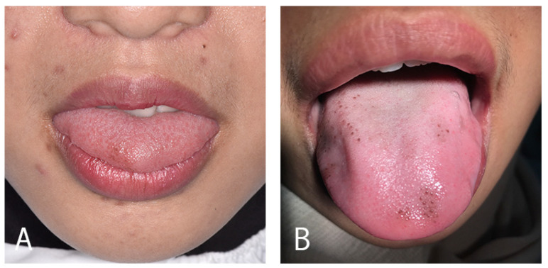
Fungiform papillae are located on the tip and lateral portions of the tongue; infrequently, they may become hyperpigmented solely, appearing as mushroom-like structures with a brown or dark colour [31] (Figure 1).
Figure 1.
Clinical appearance of PFP, showing the Holzwanger Type 1 (A) and Type 2 (B) variants.
Clinically, PFP can be easily diagnosed by careful naked eye examination, with or without dermoscopy [32]. The latter provides a much clearer visualisation of the lesions, which usually have a cobblestone or rose-petal appearance [32]. Some authors have used tissue biopsy as a diagnostic tool to rule out underlying malignancies.
In the absence of any reviews on the subject in the literature to our knowledge, we conducted the current systematic review on PFP, its possible aetiopathogenesis, and pathophysiology.
2. Materials and Methods
A systematic search of the literature, including publications from January 1974 to July 2022, was conducted using the databases PubMed, Scopus, and EMBASE, as well as a manual search utilizing the MESH terms and keywords “pigmented fungiform papillae”, “oral”, and “tongue”.
The PFP have been classified into three types by Holzwanger [33] (Figure 1). In type 1, the pigmentation affects a well circumscribed area on the anterolateral sides or towards the tip of the tongue; in type 2, the pigmentation affects a few fungiform papillae of the dorsum of the tongue; while in type 3, the pigmentation involves all of the dorsum fungiform papillae.
From all databases, a total of 82 articles were identified, and those with duplicate titles/resources were excluded. The remaining total of 51 articles was then screened based on the availability of the abstracts and full texts. Further filtering of these yielded 51 articles for final review. The 51 articles were divided into two categories, 21 articles, encompassing 36 cases that presented or discussed a PFP case by including histopathological findings accompanied by another examination. The other 30 articles encompassed 33 cases that presented or discussed a PFP case.
3. Results
3.1. Cumulative Data
The review yielded 51 studies comprising 69 cases of PFP, and all of them were case reports. Of these, approximately three-quarters of the cases were reported in females (55/69; 79.71%), the youngest being 7 years old and the oldest 65 years old. Most patients were from Asia, Caucasian (n = 1), Indonesia (Javanese) (n = 2), Japanese (n = 3), Taiwanese (n = 1), Indian (n = 2), Korean (n = 3), and Vietnamese (n = 1) origin, while the remainder were Europeans (Hispanic; n = 3 and Italian; n = 2), African (n = 13), American (n = 4), or mixed ethnicity (n = 15) origin. The ethnicity or nationality was not reported in 15 cases (Table 1).
Table 1.
Characteristics of PFP of the tongue as reported in selected studies.
| Gender | Age | Ethnicity | Location of Affected Papillae | Clinical Appearance | Dermoscopy | Type | Ref. |
|---|---|---|---|---|---|---|---|
| Female | 7 | Ethiopian | Antero-dorsal | Multiple brown pigmented spots | RP | Type 2 | [63] |
| Female | 8 | African | Antero-lateral | Multiple dark pinhead papules | RP | Type 2 | [57] |
| Female | 9 | Japanese | Anterial and dorsal | Multiple pigmentation | CS | Type 1 | [64] |
| Female | 9 | NR | Lateral | Multiple pigmentation | NR | Type 1 | [43] |
| Female | 10 | Indian | Dorsal and lateral | Multiple sharply bordered macules | NR | Type 2 | [55] |
| Female | 12 | Moroccan | Anterial | Multiple hyperpigmented papillae in a diffuse and symmetrical pattern | RP | Type 1 | [46] |
| Female | 12 | African | Anterial, lateral, and dorsal | Multiple hyperpigmented papillae presenting as dark patches | RP | Type 2 | [36] |
| Female | 12 | NR | Anterial and dorsal | Multiple discrete tan-brown pinhead papules | RP | Type 2 | [58] |
| Female | 12 | NR | Dorsal and lateral | Brown pigmentations | RP CS |
Type 1 | [47] |
| Female | 12 | Asian | Anterial | Tiny pigmented macules | CS | Type 2 | [65] |
| Female | 13 | South Asian | Anterial | Light to dark brown pigmentation, round or polygonal in shape, and circumscribed | CS | Type 1 | [66] |
| Female | 13 | Mexican | NR | Multiple hyperchromic macules, light brown, mottled, 1 mm in diameter | RP | Type 2 | [48] |
| Female | 15 | NR | Anterial, lateral, and dorsal | Asymptomatic and multiple brown pigmentations | CS RP |
Type 1 Type 2 |
[49] |
| Female | 15 | NR | Dorsal | Multiple brown macules | NR | Type 2 | [31] |
| Female | 18 | North African | Anterial | Multiple small erythematous and hyperpigmented papules | RP | Type 1 | [50] |
| Female | 18 | Black | Dorsal | Bluish-black to black macular hyperpigmentation measuring 30–70 mm | NR | Type 1 | [33] |
| Female | 18 | NR | Dorsal | Dark spots | NR | NCP | [51] |
| Female | 20 | NR | Antero-lateral and dorsal | Multiple brown macules | RP | Type 2 | [67] |
| Female | 20 | Moroccan | Dorsal | NR | NR | Type 2 | [68] |
| Female | 21 | NR | Antero-lateral | Irregularly distributed pigmentation | RP | Type 2 | [42] |
| Female | 23 | NR | Antero-lateral | Multiple hyperpigmented papillae in a diffuse pattern | NR | Type 2 | [56] |
| Female | 24 | Hispanic | Dorsal and lateral | Diffuse punctate pigmentation in a symmetrical pattern, and others grouped in a mottled pattern | RP | Type 1 | [69] |
| Female | 25 | Saudi | Dorsal | Diffuse tan, brown, patches with prominent dark papillae | NR | Type 2 | [52] |
| Female | 25 | Korean | Antero-lateral | Multiple dark brownish macules | NR | Type 1 | [70] |
| Female | 33 | Korean | Antero-lateral | Multiple dark brownish macules | NR | Type 1 | |
| Female | 26 | Black | Antero-lateral | Small reddish-brown pigmented lesions | NR | Type 1 | [71] |
| Female | 26 | Indian | Dorsal | Multiple tiny brown macules | NR | Type 1 | [37] |
| Female | 27 | Italian | Antero-lateral | Multiple blue-grey pigmentation, diffuse with a symmetrical pattern | RP | Type 2 | [72] |
| Female | 28 | Haitian | Antero-lateral | Multiple dark brown macules | NR | Type 2 | [73] |
| Female | 28 | NR | Dorsal | Multiple discrete tan-brown pin-head papules | RP | Type 2 | [53] |
| Female | 29 | NR | Dorsal | Hyperpigmentation, diffuse with a symmetrical pattern | NR | Type 2 | [61] |
| Female | 30 | NR | Dorsal | Hyperpigmented macules | RP | Type 2 | [30] |
| Female | 30 | Black | Anterial | Multiple hyperpigmented papillae | RP | Type 2 | [74] |
| Female | 32 | Caucasian | Dorsal | 6 mm oval area with brown pigmentation | NR | Type 2 | [75] |
| Female | 35 | Japanese | Antero-lateral | Hyperpigmented papillae | RP | Type 1 | [44] |
| Female | 35 | African | Dorsal | Groups of 15 to 20 papillae with a mottled appearance | NR | Type 2 | [76] |
| Female | 43 | South American | Dorsal | Diffuse and symmetrical pattern of macules | NR | Type 1 | |
| Female | 40 | Black | Anterial | Multiple hyperpigmented papillae | RP | Type 2 | [39] |
| Female | 44 | Black | Anterial | Multiple hyperpigmented papillae | RP | Type 2 | |
| Female | 44 | African | Antero-lateral | NR | NR | Type 2 | [77] |
| Female | 45 | Hispanic | Lateral | Multiple dark-brown pigmented papules | NR | Type 1 | [78] |
| Female | NR * | NR | Dorsal and lateral | Multiple hyperpigmented papillae and patches | CS | Type 2 | [38] |
| Male | 8 | Latin America | Anterial and dorsal | Multiple asymptomatic and sharply demarcated hyperpigmented pinhead papules | CS | Type 2 | [59] |
| Male | 11 | Brazilian | Anterial and lateral | Multiple tiny brown macules | NR | Type 1 | [62] |
| Male | 12 | NR | Dorsal and lateral | Multiple pigmented papule in a symmetrical pattern | NR | Type 1 | [41] |
| Male | 17 | Korean | Anterial | Well-demarcated small, black, clustered hyperpigmented papules | NR | Type 1 | [79] |
| Male | 26 | Taiwanese | Antero-lateral | Multiple tiny brown macules | CS | Type 2 | [80] |
| Male | 28 | NR | Anterial | Multiple dark-brown macules and dome shaped papules | CS | Type 1 | [34] |
| Male | 28 | Hispanic | Latero-distal | Multiple dark-brown pigmented papules | RP | NCP | [35] |
| Male | 36 | Italian | Dorsal and lateral | Multiple brown papillae | CS | Type 2 | [81] |
| Male | 42 | Japanese | Lateral | Black pigmented papillae | NR | Type 1 | [40] |
| Male | 65 | Vietnamese | Dorsal and lateral | NR | NR | Type 2 | [82] |
| Female | 21 | Javanese | Dorsal and lateral | Multiple brownish-black, diffuse, and asymptomatic macules | CS | Type 2 | [60] |
| Male | 22 | Javanese | Dorsal | Multiple macules, brownish-black and sharing a clear border | CS | Type 2 | |
| Female Female Female Female Female Female Female Female Female Female Female Female Male Male Male |
18 18 22 26 27 29 31 36 39 40 48 51 8 24 52 |
Mixed ethnicity | Not reported individually in each patient | patient | Not reported individually in each patient | Type 2 Type 2 Type 2 Type 2 Type 2 Type 2 Type 2 Type 1 Type 2 Type 2 Type 2 Type 2 Type 1 Type 2 Type 2 |
[54] |
NR: Not reported; * not mentioned specifically (mentioned as adolescent); CS: Cobblestone appearance; RP: Rose-petal appearance; NCP: No clinical picture.
3.2. Clinical Appearance of PFP
PFP were almost equally distributed over the tongue, in the following decreasing order of frequency: dorsal (13/66); antero-lateral (11/69); anterial (9/69); lateral (3/66); antero-dorsal (1/69); antero-dorsal (1/69); latero-distal (1/69); both anterial and dorsal (3/69); anterial and lateral (1/69); anterial, lateral, and dorsal (2/69); dorsal and lateral (8/69); and antero-lateral and dorsal (1/69). In 16/69 cases lesion location was not mentioned (Table 1).
The dermoscopy findings reported were: cobblestone appearance (10/69), rose-petal appearance (17/69), both cobblestone and rose-petal appearance (2/69), and non-specific and unreported (39/69). The pigmentations were classified as either Type 1 or 2, as per the Holzwanger categorisation of the affected fungiform papillae [33] (Figure 1). Accordingly, 22 of 69 (31.9%) cases could be categorised as Type 1, 43 of 69 (62.3%) as Type 2, and 1 of 69 (1.5%) as Type 1 and Type 2. The remainder (2/29) were unclassifiable due to the absence of clinical photos (Table 1).
3.3. Associated Dermatological and Other Systemic Manifestations
Twenty-six studies reported associated skin and mucosal findings in addition to PFP, such as patch stage mycosis fungoides (1/69) [34], or IA mycosis fungoides (1/69) [35], acanthosis nigricans (1/69) [36], pigmented macules on trunk (1/69) [37] or lips (1/69) [38], hand eczema (1/69) [39], asymptomatic skin hyperpigmentation (1/36) [40], yellowish hyperpigmentation of sclera and conjunctival mucosa (1/36) [41], Fitzpatrick skin type II (1/69) [42], Fitzpatrick skin type III (13/69) [43,44,45], Fitzpatrick skin type IV (11/69) [46,47,48,49,50,51,52,53,54], Dowling–Degos disease (1/69) [30], and similar PFP affects also seen in another family member, especially the mother (2/69) [55,56]. A few studies clearly stated that skin, mucosal, or nail pigmentation was not found [31,33,46,49,50,52,53,54,57,58,59,60] or that parents or family members did not present similar pigmentation of the oral mucosa [38,61,62].
3.4. Complete Blood Cell Count and Blood Chemistry Findings
The following additional investigations were reported in some studies: routine laboratory [53,58], complete blood cell count [36,37,40,41,46,47,49,62,64,69,70,71,79,81,82], blood glucose, vitamin levels, trace element (iron, zinc), serum electrolytes, and blood chemistry (urea, ferritin).
The complete blood cell count was reported in 15 studies, mostly with normal results. Only in three studies were some anomalies detected, including low hemoglobin [41], low leucocyte count [41], higher mean corpuscle volume (MCV) [82], and heterozygosity of hemoglobin [36]. The blood glucose [51,69,71,82], vitamin levels [36,49,51,52,69,82], trace element [36,46,47,62,68,82], electrolytes [37,51,69,71,81], and blood chemistry, including ferritin [49,51,52,69] and urea [37,69,70,71] were reported within normal limits, as well as metabolic panel [46,47,49,62] and some analyses stated as routine laboratory investigations [53,58]. Only one study reported a high level of iron in a Vietnamese male [82].
3.5. Liver and Kidney Function Tests
Eleven studies reported liver and renal function tests, or urinalysis [36,37,40,51,68,69,70,71,79,82]. The liver function test was reported normal as well as renal function test, creatinine levels, and urinalysis in nine studies. One study reported a high level of aspartate aminotransferase, alanine aminotransferase, and bilirubin [81], and in other study high levels of anti-smooth muscle antibodies, anti-liver antibodies, and anti-kidney microsomal antibodies were detected [82].
3.6. Infection Markers, Endocrine, Auto-Immune, Ophthalmological, and Neurological Findings
Diverse additional investigations were reported, including infection markers (hepatitis [51], syphilis [79], fungal infection [52], and HIV [76]), endocrinological tests [40,79] (synacthenor adrenal cortex function [64], hyperinsulinism [36], and thyroid [47,49,82]) and auto-immune (anti-nuclear antibodies [46,62,82]) tests, ophthalmological and neurological examinations [40,79]. All studies reported that the infection markers were negative, with the exception of one study in which the HIV test was positive [76].
3.7. Additional Findings
No antecedent of medication intake was reported in 14 studies [31,39,41,46,50,61,67,68,72,73,74,75,76,80]. Similarly, history of a relative with a similar condition [34,38,41,42,46,49,51,57,61,62,68,70,72], social habits (smoking and tobacco) [31,67,73,81], or relevant medical history [31,38,43,46,48,49,50,57,58,59,65,68,70,75,78,81] were not found in 26 studies. Only one study reported that the PFP became more noticeable during pregnancy in a Caucasian 32-year-old female [75], and one 30-year-old female patient had concomitant Dowling–Degos disease [30].
Five studies reported that PFP were found in patients with active medication. Medications included oral iron; II-glycine-sulfate [47]; tenofovir, emtricitabine, and nevirapine [76]; paucibacillary [39]; interferon and ribavirin [82]; and meloxicam [60]. Two patients were reported with higher body mass index (obesity) [36,60]. Two patients had PFP similar to their mother [55,56], the first one with Indian ethnicity and the other with no race reported. Three patients presented with iron deficiency anemia [47], condyle arthritis [60], or recurrent aphthous ulceration [82].
3.8. Histopathology
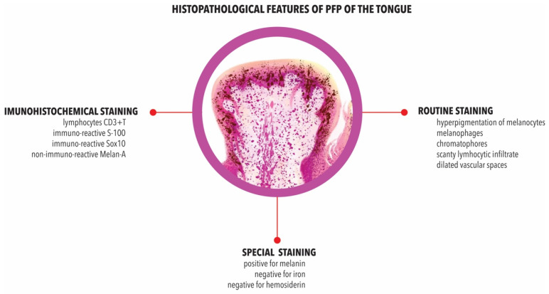
The histopathological descriptions of PFP revealed the following general characteristics: (i) hyperpigmentation of melanocytes with brown melanin of the basal cell layers of epithelium; (ii) melanophages in the lamina propria, sub-epithelial or sub-mucosal connective tissue; (iii) scant lymphocytic infiltrate in the lamina propria; (iv) chromatophores in the sub-epithelial area and surrounding the blood vessels of the fungiform papillae; and finally, (v) dilated vascular spaces (Table 2).
Table 2.
Histopathological findings in the reported cases of selected studies.
| Gender | Ages | Ethnicity | Histopathological Findings | Ref |
|---|---|---|---|---|
| Female | 9 | Japanese |
|
[64] |
| Female | 9 | NR |
|
[43] |
| Female | 12 | Asian | Melanophages in the submucosa | [65] |
| Female | 18 | Black | Chromatophores in the subepidermal area and around the blood vessels within the fungiform papillae | [33] |
| Female | 18 | NR | Melanophages were found in the lamina propria of the papillae | [51] |
| Female | 20 | NR | Melanophages in the lamina propria | [67] |
| Female | 24 | Hispanic | Melanophages in the lamina propria | [69] |
| Female | 25 | Korean | Melanophages in the upper lamina propria | [70] |
| Female | 33 | Korean | Melanophages in the upper lamina propria | |
| Female | 26 | Black |
|
[71] |
| Female | 27 | Italian |
|
[72] |
| Female | 28 | Haitian |
|
[73] |
| Female | 32 | Caucasian | Melanophages were prominent in the fungiform and adjacent filiform papillae | [75] |
| Female | 35 | Japanese |
|
[44] |
| Female | 45 | Hispanic | Melanin deposits in the epithelium | [78] |
| Male | 12 | NR | Melanophages in the subepidermal area | [41] |
| Male | 17 | Korean | Melanophages in the upper lamina propria without significant inflammation | [79] |
| Male | 26 | Taiwanese | Pigmented basal keratinocytes and melanophages in the upper lamina propria | [80] |
| Male | 28 | NR |
|
[34] |
| Male | 28 | Hispanic | Melanophages within the lamina propria | [35] |
| Male | 42 | Japanese | Melanophages in the upper lamina propria without significant inflammation | [40] |
| Female Female Female Female Female Female Female Female Female Female Female Female Male Male Male |
18 18 22 26 27 29 31 36 39 40 48 51 8 24 52 |
Mixed ethnicity |
|
[54] |
NR: not reported.
3.9. Immunohistochemistry
Immunohistochemical studies showed an immune-reactive for CD3+ T lymphocytes, S-100 and Sox10 in the intraepithelial melanocytes and non-immuno-reactive melan-A (Table 3).
Table 3.
Histological findings based on staining of PFP.
| Gender | Ages | Ethnicity | Routine Staining | Special Staining | Immunohistochemical Staining | Ref. | |||
|---|---|---|---|---|---|---|---|---|---|
| Marker | Staining | Marker | Staining | Marker | Staining | ||||
| Female | 9 | NR | Brown pigment in the basal layer of keratinocytes | HE | Iron and hemosiderin not detected | Perl’s | Immuno-reactive for Sox-10 | IHC | [43] |
| Female | 27 | Italian |
|
HE | - | - | Immuno-reactive of CD3+ T lymphocytes | IHC | [72] |
| Female | 32 | Caucasian | The stroma supporting the fungiform and adjacent filiform papillae showed prominent melanophages | HE | - | - |
|
IHC | [75] |
| Female | 35 | Japanese |
|
HE | Melanin was detected | Fontana–Masson | - | - | [44] |
| Iron and hemosiderin not detected | Berlin blue | - | - | ||||||
NR: Not reported. HE: Hematoxylin-eosin. IHC: Immunohistochemistry.
3.10. Other Features
Using specific staining, two studies showed the presence of melanin with Fontana–Masson staining. PFP were not linked with the presence of iron or hemosiderin with Berlin blue and Perl’s staining (Table 3).
4. Discussion
The tongue is the largest organ of the oral cavity, and its surface is studded with four types of papillae: filiform, fungiform, foliate, and vallate papillae. Lingual papillae are thought to increase the surface area of the tongue, serve as bearers of taste buds, and also to increase the area of contact and friction between the tongue and food [45]. The fungiform papillae are club shaped projections generally found scattered on the tip and sides of the tongue. They are, in health, pink to red in colour as the rest of the lingual mucosa, and usually do not stand out as discrete organelles. However, in some people, the fungiform papillae are pigmented and can be readily seen as discrete projections, even by naked eye examination. In most of the reports of the current review PFP appeared as dark-brown, blue-grey, or bluish-black papillae.
A number of hypotheses have been proposed for the origin of PFP. Some contend that it is due to the transfer of darker melanocytes in the basal layer of the epithelium to the superficial keratinocytes via membrane-bound organelles called melanosomes [83]. The melanin is likely to be the darker variant eumelanin found in black ethnicity, as opposed to the lighter variant pheomelanin seen in Asian ethnicity [84]. The melanin was later confirmed in PFP, and not related to the presence of iron and hemosiderin [43,44].
On histological examination of the papillary epithelium, PFP appear to be due to the hyperpigmentation of papillary keratinocytes [34,43,73,80], usually bearing a profusion of melanocytes containing melanin granules in the basal cell layer of the epithelium [64,73]. Other features include a mild to moderate lymphocytic infiltrate of the lamina propria [54,72], and chromatophores in the sub-epithelial area, and around the blood vessels of the papillae [33]. Many authors have observed melanophages containing melanin granules in the lamina propria [34,35,40,51,67,69,70,73,79], the connective tissue [64], subepithelial tissue [54,72], adjacent filiform papillae [75], and the submucosa [65,73] of the fungiform papillae (Figure 2).
Figure 2.
Schematic illustration of histopathological features of PFP of the tongue.
Melanophages—macrophages that contain melanin—are a common feature in inflammatory and non-inflammatory pigmentation [85]. Melanophages are large cells with indistinct cytoplasmic boundaries, usually located around or near superficial dermal vessels [73]. Another histological feature described by some authors in PFP is the presence of so-called ‘chromatophores’, a disputed, and confusing term used by Holzwanger [33,71] and Koplon [71] in the older literature, referring to melanocytes, or perhaps more probably to melanophages (pigment carrying cells) as mentioned by Steigmann [86]. The presence of melanin was later confirmed with Fontana–Masson staining [44].
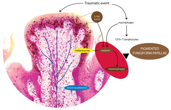
The presence of a lymphocytic infiltrate reported in some studies, could be considered as a sign of an inflammatory response associated with the genesis of PFP. In an immunohistochemical study, Ghigliotti et al. revealed that the lymphocytes in PFP were mainly CD3 + T lymphocytes [72]. The localised traumatic event or mechanical injury provokes inflammatory responses. The inflammatory responses cause inflammatory cytokine production from keratinocytes as well as from fibroblasts, which in turn stimulate melanocytes, often resulting in pigmentation. In some research, it has been mentioned that several cytokines such as interleukin, tumor necrosis factor (TNF), and prostaglandin E (PGE) modulate the proliferation and differentiation of human melanocytes to pigmentation [87]. The presence of the latter lymphocytic infiltrate, together with the melanophages, implies the possibility that the pigmentation is due to an inflammatory process, provoked most likely by a localised traumatic event (Figure 2).
Not surprisingly, some authors have investigated whether PFP is a precursor of melanoma. To our knowledge, no malignant transformation has been reported from PFP. Only in two studies were immunohistochemical staining for melanotic markers done, detecting Sox10 [43] and S-100 [75] expressions, but they were non-reactive for melan-A [75].
On further detailed review of the reports, we noted that a number of investigators have used dermoscopy to evaluate the nature of PFP. This technique increases the sensitivity and specificity of the clinical examination, and almost eliminates biopsy procedures and by extension reduces patient discomfort. Nevertheless, dermoscopy is infrequently used in dentistry [88]. A majority of clinicians in our review used dermoscopy, but in combination with biopsy examination, which seems to be a waste of resources [34,35,64,65,67,72,80].
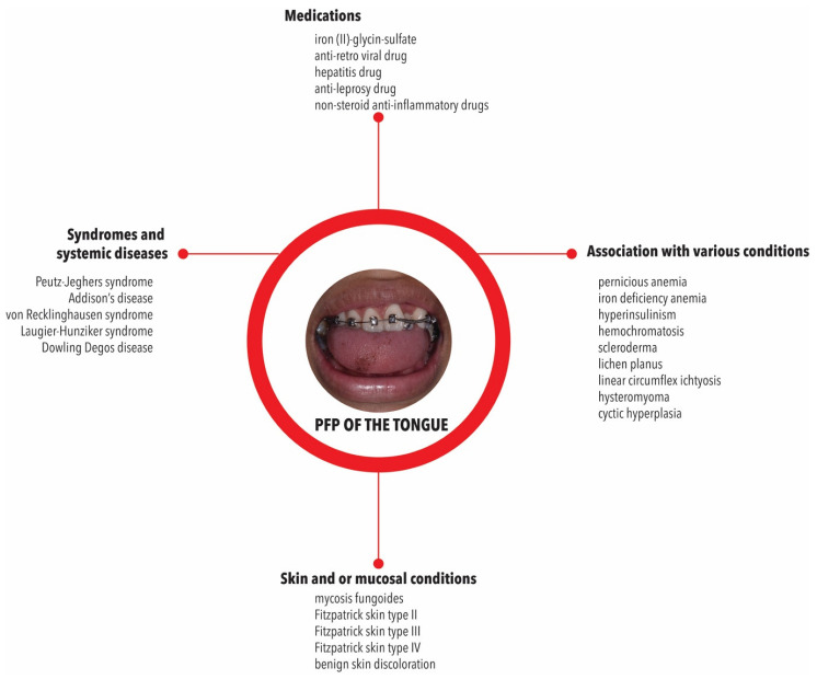
Some of the reviewed reports showed that PFP were associated with other skin and/or mucosal conditions, including mycosis fungoides [34,35], Fitzpatrick skin type II [42], Fitzpatrick skin type III [44,54], Fitzpatrick skin type IV [46,47,48,49,50,51,52,53,54], yellowish discolourations in sclera and conjunctival mucosa [41], and benign skin discolourations [36,37,38,39,40]. Nonspecific observations have been made in this context, as a few cases were associated with light brown skin—i.e., Fitzpatrick skin type IV [46,48,50]—or less frequently with fair skin and blue eyes-skin type II [42]. However, many reports did not mention the skin colour [60,79,89] (Figure 3).
Figure 3.
Associations described for PFP with syndromes, systemic conditions, medications, and other related conditions.
PFP were also described in association with other syndromes and systemic diseases including Peutz–Jeghers syndrome [35,40,51,64,67,72,73], Addison’s disease [35,40,51,64,67,72,73], von Recklinghausen syndrome [51,72], and Laugier–Hunziker syndrome [64,73,80] (Figure 3). Two cases reported that the PFP were inherited, because this condition was found in the mother and her children [55,56] as also in a case of Dowling Degos disease (EDD) [30].
The role of external factors in the origin of PFP was also analysed. Medication intake [31,39,41,46,50,61,67,68,72,73,74,75,76,80], family history [34,38,41,42,46,49,51,57,61,62,68,70,72], medical history [31,38,43,46,48,49,50,57,58,59,65,68,70,75,78,81], smoking [31,67], and tobacco use [67,73] did not appear to be related. However, in some cases, PFP were found to be related with the medication, such as iron (II)-glycinee-sulfate [47], and anti-retro viral drug [76], hepatitis drug [82], anti-leprosy drug [66], and non-steroid anti-inflammatory drugs [60]. They have also been found in HIV infected patients [76]. Specific pigmentation of the papillae related to human immunodeficiency virus (HIV) infection [8] and medication-induced [90,91] are common causes of mucosal pigmentation and are not directly involved in PFP pathogenesis.
In order to ascertain the aetiology of PFP and to rule out systemic diseases, numerous laboratory analyses have been conducted, including complete blood cell count [37,40,46,47,49,62,64,69,70,71,79,81], blood glucose [69,71,82], or fasting blood glucose level [51], liver function tests [36,37,51,69,79], renal or kidney function tests [36,68,79,82], urinalysis [40,70,71,79], adrenal cortex function (synacthen) tests [64], electrolyte levels [37,51,69,70,81], vitamin B12 [49,51,52,69], vitamin D [36], folate [82], trace element [36,46,47,62,68,82], ferritin [49,51,52], and urea [37,69,71]. The infection markers, as hepatitis markers [51], fungal [52], VDRL [79], the endocrinological [36,40,47,49,64,79,82], and anti-nuclear antibody levels [46,62,82] were assessed. The results of the foregoing tests as well as ophthalmological and neurological tests conducted by some [40,79], were negative, and did not reveal an oral-systemic connection with the origin of PFP. Only three cases showed low hemoglobin and leucocytes counts [41] and high MCV counts [82], heterozygocity of HBs [36] was noted amongst the 69 studies reviewed. These data point to the fact that PFP is a normal physiological condition with an aberration of the focal melanocyte function of the fungiform papillae.
On the other hand, PFP have been associated with various systemic conditions, such as pernicious anemia [41,51,64,72,73], iron deficiency anemia [41], hemochromatosis [41,51,64,72,73], scleroderma [41,73,79], lichen planus [34,41], linear circumflex ichthyosis [34,41], hysteromyoma [34], and cystic hyperplasia [34]. Some patients presented lower hemoglobin and total leukocyte counts [41], lower serum iron, 25 OH vitamin D, zinc, and hyperinsulinism [36], higher levels of aspartate aminotransferase, alanine aminotransferase and bilirubinemia [81]. These conditions appear to be mere associations with PFP and not the direct cause, as far as we gathered from our meta-analysis.
Based on the current study findings and interpretation, it may be speculated that PFP could arise due to injury and disruption of the vascular architecture of the fungiform papillae during masticatory traumatic events. This would cause the extravasation of blood and a subsequent influx of inflammatory infiltrate of macrophages and lymphocytes into the papillary tissue [64]. Sugiyama et al. have postulated that the inflammatory process then causes an increase in melanin synthesis in the lamina propria of the fungiform papillae leading to the hyperpigmentation [64]. However, this fact does not explain why only the fungiform papillae—and not the other papillary variants of the lingual mucosa—are affected by such events. Moreover, if traumatic events were the main cause acting in the origin of PFP, it would be expected to occur more frequently and at any age. Its description, affecting several and different ethnicities as we observed in our study, does not add further information about a possible predisposition in that sense, although an apparent predominance among Asian versus White people was noted. These findings contrast with those of Holzwanger [33], who stated that PFP is a relatively common variant of oral pigmentation which, although more prevalent in Negroes, it is seen in other heavily pigmented races as well. Therefore, the reason/s for the focal pigmentation of fungiform papillae is/are yet speculative, and remain to be clarified (Figure 4).
Figure 4.
Proposed model for the pathogenesis of PFP.
The major limitation of this study is the absence of systematic evaluations of PFP, despite the plethora of case reports which are mainly anecdotal in nature. This leads to difficulties in systematic data collection. Furthermore, none of the case reports were followed up, and it is unclear whether the PFP is a transient phenomenon or lasts for prolonged periods. Hence prospective studies are needed.
5. Conclusions
Our findings, taken together, clearly indicate that PFP is a normal physiological condition with an aberration of the focal melanocyte function of the fungiform papillae of the tongue. Some reports indicate that the condition could be associated with several systemic conditions and syndromes but, clearly, not caused by them. The inflammatory infiltrate seen in some papillary lesions are likely to be due to masticatory trauma. Yet, the true reasons for the discolouration of fungiform papillae are as of yet elusive.
Acknowledgments
Authors thank the support from Nashrollah, DDS for sharing the clinical picture of PPF and Fadila Annisa Ramadhani for creating the papilla illustration in Figure 4 and consenting to place it in this manuscript.
Author Contributions
Conceptualization, M.D.C.S., F.U., D.S.E. and A.E.P.; Methodology, M.D.C.S. and A.B.R.S.; Software, M.D.C.S.; Validation, L.S., S.M.W. and A.B.R.S.; Formal analysis, M.D.C.S., L.S., A.B.R.S., F.U. and K.P.; Investigation, M.D.C.S., A.B.R.S., S.J.M.R.S., W.L.K. and K.P.; Resources, M.D.C.S. and F.U.; Data curation, M.D.C.S. and N.F.A.; Writing—original draft preparation, M.D.C.S., A.E.P., N.F.A., S.J.M.R.S., W.L.K. and K.P.; Writing—review and editing, L.S., S.M.W., R.P.R., A.B.R.S. and F.U.; Visualization, M.D.C.S., L.S. and F.U.; Supervision, D.S.E.; Project administration, M.D.C.S. All authors have read and agreed to the published version of the manuscript.
Institutional Review Board Statement
Not applicable.
Informed Consent Statement
Not applicable.
Data Availability Statement
Not applicable.
Conflicts of Interest
The authors declare no conflict of interest.
Funding Statement
This research received no external funding.
Footnotes
Publisher’s Note: MDPI stays neutral with regard to jurisdictional claims in published maps and institutional affiliations.
References
- 1.Ko E., Panchal N. Pigmented Lesions. Dermatol. Clin. 2020;38:485–494. doi: 10.1016/j.det.2020.05.009. [DOI] [PubMed] [Google Scholar]
- 2.Feller L., Khammissa R.A.G., Lemmer J. Melanin. InTech; Rijeka, Croatia: 2017. Oral Mucosal Melanosis; pp. 7–26. [Google Scholar]
- 3.Verma B., Bahra A., Ajmal A.K.M., Sen S. Laugier–Hunziker Syndrome in a Young Female. Indian Dermatol. Online J. 2017;8:148–150. doi: 10.4103/2229-5178.202282. [DOI] [PMC free article] [PubMed] [Google Scholar]
- 4.Shah S.S., Oh C.H., Coffin S.E., Yan A.C. Addisonian pigmentation of the oral mucosa. Cutis. 2005;76:97–99. [PubMed] [Google Scholar]
- 5.Kothiwale S. An Oral Clinician’s Perspective towards Peutz Jegher’s Syndrome A Case Report. J. Probiotics Health. 2015;3:128. doi: 10.4172/2329-8901.1000128. [DOI] [Google Scholar]
- 6.Agrawal Sharma Y.K., Deo K.S., Ranpariya R.H., Singh P. Mucocutaneous hyperpigmentation as presentation of vitamin B12 deficiency: A case report and brief review. Pigment. Int. 2019;6:37–42. [Google Scholar]
- 7.Song X., Shen Y., Zhou Y., Lou Q., Han L., Ho J.K., Ren Y. General hyperpigmentation induced by Grave’s disease. Medicine. 2018;97:e13279. doi: 10.1097/MD.0000000000013279. [DOI] [PMC free article] [PubMed] [Google Scholar]
- 8.Chandran R., Feller L., Lemmer J., Khammissa R.A.G. HIV-Associated Oral Mucosal Melanin Hyperpigmentation: A Clinical Study in a South African Population Sample. AIDS Res. Treat. 2016;2016:8389214. doi: 10.1155/2016/8389214. [DOI] [PMC free article] [PubMed] [Google Scholar]
- 9.Lundin K., Schmidt G., Bonde C. Amalgam Tattoo Mimicking Mucosal Melanoma: A Diagnostic Dilemma Revisited. Case Rep. Dent. 2013;2013:787294. doi: 10.1155/2013/787294. [DOI] [PMC free article] [PubMed] [Google Scholar]
- 10.Kauzman A., Pavone M., Blanas N., Bradley G. Pigmented lesions of the oral cavity: Review, differential diagnosis, and case presentations. J. Can. Dent. Assoc. 2004;70:682–683. [PubMed] [Google Scholar]
- 11.Hassona Y., Sawair F., Al-karadsheh O., Scully C. Prevalence and clinical featuRes. of pigmented oral lesions. Int. J. Dermatol. 2016;55:1005–1013. doi: 10.1111/ijd.13133. [DOI] [PubMed] [Google Scholar]
- 12.Ware R.E., Aygun B. Advances in the use of hydroxyurea. Hematology. 2009;2009:62–69. doi: 10.1182/asheducation-2009.1.62. [DOI] [PubMed] [Google Scholar]
- 13.Bloom M.D., Gibney J.M., Heldermon C.D. Pigmentation of the tongue with lapatinib treatment in a patient with advanced breast cancer: A case report. Cancer Treat Commun. 2016;7:1–3. doi: 10.1016/j.ctrc.2016.02.006. [DOI] [PMC free article] [PubMed] [Google Scholar]
- 14.Stringer L.L., Laura Z. Hyperpigmentation of the Tongue. J. Adv. Pract. Oncol. 2014;5:71–72. [PMC free article] [PubMed] [Google Scholar]
- 15.Kim H.S., Park Y.M., Kim H.O., Lee J.Y. Tegafur-induced hyperpigmentation of the tongue. J. Dermatol. 2010;37:937–938. doi: 10.1111/j.1346-8138.2010.00906.x. [DOI] [PubMed] [Google Scholar]
- 16.Vasudevan B. An unusual case of capecitabine hyperpigmentation: Is hyperpigmentation a part of hand-foot syndrome or a separate entity. Indian J. Pharmacol. 2010;42:326–328. doi: 10.4103/0253-7613.70401. [DOI] [PMC free article] [PubMed] [Google Scholar]
- 17.Williams M., Hatipoglu B. Development of Abnormal Pigmentation of the Oral Mucosa Following Adjuvant Radioactive Iodine Treatment. AACE Clin. Case Rep. 2016;2:e113–e116. doi: 10.4158/EP15670.CR. [DOI] [Google Scholar]
- 18.Mlika R.B., Kerkeni N., Marrak H., Fenniche S., Mokhtar I., Debbiche A. Tongue hyperpigmentation during PEG-interferon-alfa/ribavirin therapy in a non-Caucasian patient with chronic hepatitis C: A case report and review of the literature. Int. J. Dermatol. 2013;52:631–644. doi: 10.1111/j.1365-4632.2011.04921.x. [DOI] [PubMed] [Google Scholar]
- 19.Mattoo K., Singh M., Arora P. Bilateral Smokers Melanosis—Rare site occurrence in an edentulous patient—A case report. Med. Res. Chron. 2014;1:97–101. [Google Scholar]
- 20.Carlos-Bregni R., Contreras E., Netto A.C., Mosqueda-Taylor A., Vargas P.A., Jorge J., León J.E., de Almeida O.P. Oral melanoacanthoma and oral melanotic macule: A report of 8 cases, review of the literature, and immunohistochemical analysis. Med. Oral Patol. Oral Cir. Bucal. 2007;12:E374–E379. [PubMed] [Google Scholar]
- 21.Sreeja C., Ramakrishnan K., Vijayalakshmi D., Devi M., Aesha I., Vijayabanu B. Oral pigmentation: A review. J. Pharm. Bioallied. Sci. 2015;7:S403–S408. doi: 10.4103/0975-7406.163471. [DOI] [PMC free article] [PubMed] [Google Scholar]
- 22.Tlholoe M.M., Khammissa R.A.G., Bouckaert M., Altini M., Lemmer J., Feller L. Oral Mucosal Melanoma: Some Pathobiological Considerations and an Illustrative Report of a Case. Head Neck Pathol. 2015;9:127–134. doi: 10.1007/s12105-014-0526-8. [DOI] [PMC free article] [PubMed] [Google Scholar]
- 23.Sudarshan R., Vijayabala G.S., Samata Y., Ravikiran A. Newer Classification System for Fissured Tongue: An Epidemiological Approach. J. Trop. Med. 2015;2015:262079. doi: 10.1155/2015/262079. [DOI] [PMC free article] [PubMed] [Google Scholar]
- 24.Casu C., Giulia Nosotti M., Sinesi A., Mosaico G. Hairy tongue, geographic tongue, scrotal tongue and systemic connections: Clinical images and an overview. Dent. Case Rep. 2019;3:1–5. [Google Scholar]
- 25.Mangold A.R., Torgerson R.R., Rogers R.S. Diseases of the tongue. Clin. Dermatol. 2016;34:458–469. doi: 10.1016/j.clindermatol.2016.02.018. [DOI] [PubMed] [Google Scholar]
- 26.Pinna R., Cocco F., Campus G., Conti G., Milia E., Sardella A., Cagetti M.G. Genetic and developmental disorders of the oral mucosa: Epidemiology; molecular mechanisms; diagnostic criteria; management. Periodontol. 2000. 2019;80:12–27. doi: 10.1111/prd.12261. [DOI] [PubMed] [Google Scholar]
- 27.Chaubal T.V., Dixit M.B. Ankyloglossia and its management. J. Indian Soc. Periodontol. 2011;15:270–272. doi: 10.4103/0972-124X.85673. [DOI] [PMC free article] [PubMed] [Google Scholar]
- 28.Alaoui F., Souissi A., Jendoubi F., Mokni M. Caviar tongue: A lingual physiological variation. Presse Med. 2018;47:925–926. doi: 10.1016/j.lpm.2018.06.010. [DOI] [PubMed] [Google Scholar]
- 29.Marcoval-Caus J. Response to: «Pigmented Fungiform Papillae of the Tongue in Laugier Disease (or Laugier-Hunziker Syndrome)». Actas Dermo-Sifiliográficas. 2013;104:174. doi: 10.1016/j.adengl.2012.10.005. [DOI] [PubMed] [Google Scholar]
- 30.Pinos-León V., Granizo-Rubio J., Torres-Cruz M. Enfermedad de Dowling Degos asociada a papilas fungiformes pigmentadas de la lengua: Reporte de un caso. Piel. 2015;30:15–18. doi: 10.1016/j.piel.2014.08.003. [DOI] [Google Scholar]
- 31.Rice S.M., Lal K. Successful Treatment of Pigmented Fungiform Papillae of the Tongue With Q-switched Ruby Laser. Dermatol. Surg. 2022;48:368–369. doi: 10.1097/DSS.0000000000003371. [DOI] [PubMed] [Google Scholar]
- 32.Surboyo M.D.C., Santosh A.B.R., Hariyani N., Ernawati D.S., Cecilia P.H. Clinical utility of dermoscopy on diagnosing pigmented papillary fungiform papillae of the tongue: A systematic review. J. Oral Biol. Craniofac. Res. 2021;11:618–623. doi: 10.1016/j.jobcr.2021.09.008. [DOI] [PMC free article] [PubMed] [Google Scholar]
- 33.Holzwanger J.M., Rudolph R.I., Heaton C.L. Pigmented fungiform papillae of the tongue: A common variant of oral pigmentation. Int. J. Dermatol. 1974;13:403–408. doi: 10.1111/j.1365-4362.1974.tb05073.x. [DOI] [PubMed] [Google Scholar]
- 34.Chamseddin B., Vandergriff T. Pigmented fungiform papillae of the tongue: A clinical and histologic description. Dermatol. Online J. 2019;25:11. doi: 10.5070/D3259045516. [DOI] [PubMed] [Google Scholar]
- 35.Maxim E., de Dtefano D. Pigmented fungiform papillae of the tongue: An incidental finding in a patient with mycosis fungoides. J. Am. Acad. Dermatol. 2018;79:AB221. [Google Scholar]
- 36.Docx M.K.F., Vandenberghe P., Govaert P. Pigmented fungiform papillae of the tongue (PFPT) Acta Clin. Belg. 2016;71:117–118. doi: 10.1179/2295333715Y.0000000067. [DOI] [PubMed] [Google Scholar]
- 37.Millington G., Shah S. A case of pigmented fungiform lingual papillae in an Indian woman. J. Eur. Acad. Dermatol. Venereol. 2007;21:070209222700054. doi: 10.1111/j.1468-3083.2006.02000.x. [DOI] [PubMed] [Google Scholar]
- 38.Ross C.L., Ring C., Yang S. Pigmented Fungiform Papillae of the Tongue. JAMA Dermatol. 2020;156:1249. doi: 10.1001/jamadermatol.2020.2985. [DOI] [PubMed] [Google Scholar]
- 39.Mukamal L.V., Ormiga P., Ramos-e-silva M. Dermoscopy of the pigmented fungiform papillae of the tongue. J. Dermatol. 2012;39:397–399. doi: 10.1111/j.1346-8138.2011.01328.x. [DOI] [PubMed] [Google Scholar]
- 40.Isogai Z., Kanzaki T. Pigmented fungiform papillae of the tongue. J. Am. Acad. Dermatol. 1993;29:489–490. doi: 10.1016/S0190-9622(08)82004-X. [DOI] [PubMed] [Google Scholar]
- 41.Sharma S.S., Sharma S.S. Pigmentation of the Fungiform Papillae of the Tongue in a Child Secondary to Iron Deficiency Anaemia: An Uncommon Occurrence and Association. Int. J. Oral Health Dent. 2016;2:39–42. doi: 10.5958/2395-499X.2016.00005.8. [DOI] [Google Scholar]
- 42.Radomska A., Sławińska M., Sobjanek M. Pigmented fungiform papillae of the tongue. Dermatol. Rev. Przegląd Dermatol. 2021;108:191–193. [Google Scholar]
- 43.Belysheva T.S., Vishnevskaya Y.V., Kletskaya I.S., Michenko A.V., Volkova A.S., Valiev T.T. Problems of clinical and morphologic diagnosis of orolabial melanosis in children. Russ. J. Pediatric Hematol. Oncol. 2021;8:116–122. doi: 10.21682/2311-1267-2021-8-3-116-122. [DOI] [Google Scholar]
- 44.Mizawa M., Makino T., Furukawa F., Torai R., Shimizu T. Efficacy of Q-switched ruby laser treatment for pigmented fungiform papillae of the tongue. J. Dermatol. 2022;49:e133–e134. doi: 10.1111/1346-8138.16270. [DOI] [PubMed] [Google Scholar]
- 45.Samaras G., Bikos D., Vieira J., Hartmann C., Charalambides M., Hardalupas Y., Masen M., Cann P. Measurement of molten chocolate friction under simulated tongue-palate kinematics: Effect of cocoa solids content and aeration. Curr. Res. Food Sci. 2020;3:304–313. doi: 10.1016/j.crfs.2020.10.002. [DOI] [PMC free article] [PubMed] [Google Scholar]
- 46.Aqil N., Bennani M., Nassiri A., Meziane M., Gallou J.S., Mernissi F.Z. Pigmented fungiform papillae of the tongue: Clinical and dermoscopic features. Our Dermatol. Online. 2019;10:213–214. doi: 10.7241/ourd.20192.31. [DOI] [Google Scholar]
- 47.Kavak A., Topkarcı Z., Gür E.E., Erdoğan B., Yemişçi A. Pigmented fungiform papillae of the tongue: Co-existence of two patterns in the same patient and associated dental pigmentation. Turkderm Turk. Arch. Dermatol. Venereol. 2021;55:45–47. doi: 10.4274/turkderm.galenos.2020.75010. [DOI] [Google Scholar]
- 48.Castellanos M.A., Echeverria A.E.B., Rosas G.M.A. Dermatoscopía en la pigmentación de papilas fungiformes de la lengua. Dermatol. Cosmet. Med. Quir. 2018;16:206–207. [Google Scholar]
- 49.Özaslan M. Barwnikowe brodawki grzybowate języka. Dermatol. Rev. 2021;108:550–553. doi: 10.5114/dr.2021.114609. [DOI] [Google Scholar]
- 50.Cinotti E., Labeille B., Cambazard F., Perrot J.L. Dermoscopy and reflectance confocal microscopy examination of pigmented fungiform papillae of the tongue. Ann. Dermatol. Venereol. 2017;144:323–325. doi: 10.1016/j.annder.2016.10.013. [DOI] [PubMed] [Google Scholar]
- 51.Ogrum A., Takci Z., Yildiz Seckin H. Pigmented Fungiform Papillae: Case Report and Review of the Literature. Turk. Klin. J. Dermatol. 2018;28:32–34. doi: 10.5336/dermato.2018-60677. [DOI] [Google Scholar]
- 52.Alzahrani N., Alharithy R. Pigmented fungiform papillae of the tongue in a Saudi woman. J. Dermatol. Dermatol. Surg. 2018;22:39. [Google Scholar]
- 53.Sil A., Panigrahi A., Bhanja D. Rose petal appearance: The dermoscopic finding in pigmented fungiform papillae of the tongue. Indian Dermatol. Online J. 2021;12:490–491. doi: 10.4103/idoj.IDOJ_589_19. [DOI] [PMC free article] [PubMed] [Google Scholar]
- 54.Garzón-Rivas V., Garzón-Aldás E. Papilas Fungiformes Pigmentadas de la Lengua. Características Clínicas, Histológicas y Dermatoscópicas de una Serie de Casos Ecuatorianos. Int. J. Odontostomatol. 2019;13:446–451. doi: 10.4067/S0718-381X2019000400446. [DOI] [Google Scholar]
- 55.Werchniak A.E., Storm C.A., Dinulos J.G.H. Hyperkeratotic Plaques on the Palms and Soles A Red-Brown Plaque on the Nape Hyperpigmented Patches on the Tongue of a Young Girl. Arch. Dermatol. 2009;140:1275–1280. doi: 10.1001/archderm.140.10.1275-g. [DOI] [PubMed] [Google Scholar]
- 56.Gopinath H., Upadya G.M. Pigmented fungiform papillae in mother and daughter. Indian J. Dermatol. Venereol. Leprol. 2017;85:510–511. doi: 10.4103/ijdvl.IJDVL_826_16. [DOI] [PubMed] [Google Scholar]
- 57.Neri I., Evangelista V., Veronesi G., Guglielmo A., Chessa M.A. Oral pigmentation in children: A diagnostic challenge. Arch. Dis. Child. 2021;106:895. doi: 10.1136/archdischild-2020-320455. [DOI] [PubMed] [Google Scholar]
- 58.Sil A., Panigrahi A., Bhanja D.B. Tongue Discoloration in a Young Girl. J. Pediatr. 2020;219:276–277. doi: 10.1016/j.jpeds.2019.12.032. [DOI] [PubMed] [Google Scholar]
- 59.Piquero-Casals J., Morgado-Carrasco D. Pigmented Fungiform Lingual Papillae. J. Pediatr. 2020;224:177–178. doi: 10.1016/j.jpeds.2020.06.001. [DOI] [PubMed] [Google Scholar]
- 60.Surboyo M.D.C., Ernawati D.S., Parmadiati A.E., Marsetyo R.I. Pigmented fungiform papillae of the tongue and lingual fimbriae as single presentation in adult: A case report and literature review. Eur. J. Dent. 2020;14:702–706. doi: 10.1055/s-0040-1714041. [DOI] [PMC free article] [PubMed] [Google Scholar]
- 61.Al-Fagaan F., Joseph B. A case of pigmented fungiform papillae of the tongue in a Middle Eastern Woman. Med. Princ. Pract. 2014;23:167–169. doi: 10.1159/000351799. [DOI] [PMC free article] [PubMed] [Google Scholar]
- 62.Romiti R., de Medeiros L.M. Pigmented Fungiform Papillae of The Tongue. Pediatr. Dermatol. 2010;27:398–399. doi: 10.1111/j.1525-1470.2010.01183.x. [DOI] [PubMed] [Google Scholar]
- 63.García Martínez F.J., López Martín I., Segurado Rodríguez M.A. Pigmentación de las papilas fungiformes linguales. Pediatría Atención Primaria. 2015;17:e205–e207. doi: 10.4321/S1139-76322015000400015. [DOI] [Google Scholar]
- 64.Sugiyama Y., Hayashi K., Takayama T. Pigmented fungiform papillae of the tongue in a Japanese child. Clin. Case Rep. 2020;8:1104–1106. doi: 10.1002/ccr3.2830. [DOI] [PMC free article] [PubMed] [Google Scholar]
- 65.Lin Y.T., Chou C.L. Pigmented macule on the tongue of a 12-year-old girl. J. Am. Acad Dermatol. 2013;69:e229–e230. doi: 10.1016/j.jaad.2013.05.026. [DOI] [PubMed] [Google Scholar]
- 66.Collgros H., Iglesias-Sancho M., Galvany L. Dermoscopy of pigmented fungiform papillae of the tongue in an Indian girl. Australas. J. Dermatol. 2015;56:e81–e82. doi: 10.1111/ajd.12229. [DOI] [PubMed] [Google Scholar]
- 67.Robles-Mendez J.C., Ayala-Cortes A.S., Villarreal-Martinez A., Ocampo-Candiani J. Dermoscopy of the pigmented fungiform papillae of the tongue. J. Dermatol. 2017;76:s40–s42. doi: 10.1016/j.jaad.2016.06.022. [DOI] [PubMed] [Google Scholar]
- 68.el Anzi O. Pigmented fungiform papillae of the tongue: Moroccan case. Our Dermatol. Online. 2019;10:215–216. doi: 10.7241/ourd.20192.32. [DOI] [Google Scholar]
- 69.Anco-Gallegos K., San M., Serrano-Guillén G. Pigmentacion de las papilas fungiformes de la lengua. Dermatol. Perú. 2016;26:227–230. [Google Scholar]
- 70.Choi S.J., Rhonark K., Yang J.M., Lee E.S. Two Cases of Pigmented Fungiform Papillae of the Tongue. Ann. Dermatol. 2002;14:216–219. doi: 10.5021/ad.2002.14.4.216. [DOI] [Google Scholar]
- 71.Koplon B.S. Prominent Pigmented Papillae of the Tongue. Arch. Dermatol. 1967;95:394–396. doi: 10.1001/archderm.1967.01600340054012. [DOI] [PubMed] [Google Scholar]
- 72.Ghigliotti G., Chinazzo C., Parodi A., Rongioletti F. Pigmented fungiform papillae of the tongue: The first case in an Italian woman. Clin. Exp. Dermatol. 2017;42:206–208. doi: 10.1111/ced.13006. [DOI] [PubMed] [Google Scholar]
- 73.Pehoushek J.F., Norton S.A. Black taste buds. Arch. Fam. Med. 2000;9:219–220. doi: 10.1001/archfami.9.3.219. [DOI] [Google Scholar]
- 74.Pinos-León V. Dermoscopic FeatuRes. of Pigmented Fungiform Papillae of the Tongue. Case Res. Lett. 2015;106:593–594. doi: 10.1016/j.adengl.2015.06.018. [DOI] [PubMed] [Google Scholar]
- 75.McCarthy C., Holt D., Triantafyllou A. Solitary pigmentation of the tongue: Lentigo simplex or pigmented fungiform papilla? Oral Surg. 2016;11:50–54. doi: 10.1111/ors.12264. [DOI] [Google Scholar]
- 76.Marcoval J., Notario J., Martin-Sala S., Figueras I. Pigmentation of the Fungiform Papillae of the Tongue: A Report of 2 Cases. Actas Dermo-Sifiliográficas. 2011;102:739–740. doi: 10.1016/j.adengl.2011.11.010. [DOI] [PubMed] [Google Scholar]
- 77.Lake P.L. Dark Spots on the Tongue. J. Dermatol. Nurses Assoc. 2017;9:146–147. doi: 10.1097/JDN.0000000000000304. [DOI] [Google Scholar]
- 78.Adibi S., Bouquot J.E. Papillary tip melanosis (pigmented fungiform lingual papillae) Tex. Dent. J. 2011;128:573–574. [PubMed] [Google Scholar]
- 79.Oh C.K., Kim M.B., Jang H.S., Kwon K.S. A case of pigmented fungiform papillae of the tongue in an Asian male. J. Dermatol. 2000;27:350–351. doi: 10.1111/j.1346-8138.2000.tb02181.x. [DOI] [PubMed] [Google Scholar]
- 80.Hsiao Y.H., Ko J.H., Lu C.F., Chen M.J. Dermoscopic findings in pigmented fungiform papillae of the tongue. Eur. J. Dermatol. 2011;21:819–820. doi: 10.1684/ejd.2011.1490. [DOI] [PubMed] [Google Scholar]
- 81.Smogorzewski J.M., Armstrong P., Young L. Pigmented fungiform papillae of the tongue in an Indian male. Cutis. 2019;103:E16–E17. [PubMed] [Google Scholar]
- 82.Scarff C.E., Marks R. Pigmented fungiform papillae of the tongue in an Indian male. Australas. J. Dermatol. 2003;44:149–151. doi: 10.1046/j.1440-0960.2003.00664.x. [DOI] [PubMed] [Google Scholar]
- 83.Hirobe T. Keratinocytes regulate the function of melanocytes. Dermatol. Sin. 2014;32:200–204. doi: 10.1016/j.dsi.2014.05.002. [DOI] [Google Scholar]
- 84.Moreiras H., Seabra M.C., Barral D.C. Melanin Transfer in the Epidermis: The Pursuit of Skin Pigmentation Control Mechanisms. Int. J. Mol. Sci. 2021;22:4466. doi: 10.3390/ijms22094466. [DOI] [PMC free article] [PubMed] [Google Scholar]
- 85.Attili S. Dermal melanophages: Does quantitative assessment play a role in the diagnosis of inflammatory skin diseases? Indian J. Dermatopathol. Diagn. Dermatol. 2018;5:133–134. doi: 10.4103/ijdpdd.ijdpdd_42_18. [DOI] [Google Scholar]
- 86.Steigmann S. The relationship between physiologic pigmentation of the skin and oral mucosa in Yemenite Jews. Oral Surg. Oral Med. Oral Pathol. 1965;19:32–38. doi: 10.1016/0030-4220(65)90212-4. [DOI] [PubMed] [Google Scholar]
- 87.Hossain M.R., Ansary T.M., Komine M., Ohtsuki M. Diversified Stimuli-Induced Inflammatory Pathways Cause Skin Pigmentation. Int. J. Mol. Sci. 2021;22:3970. doi: 10.3390/ijms22083970. [DOI] [PMC free article] [PubMed] [Google Scholar]
- 88.Mordoh A., Casas G., Horacio C., Werner L., Aguas S.C., Lanfranchi H., Krupitzki H., Gandolfo M. Dermoscopic Evaluation Improves Clinical Diagnosis of Oral Melanotic Macules: A Study in 50 Patients with Oral Pigmented Lesions. Am. J. Oral Med. 2018;4:1–9. doi: 10.37786/ajom.35. [DOI] [Google Scholar]
- 89.Tan C., Liu Y., Min Z.S., Zhu W.Y. A clinical analysis of 58 Chinese cases of pigmented fungiform papillae of the tongue. J. Eur. Acad. Dermatol. Venereol. 2012;28:242–245. doi: 10.1111/j.1468-3083.2012.04684.x. [DOI] [PubMed] [Google Scholar]
- 90.Mallagray-Montero Md Moreno-López L.A., Cerero-Lapiedra R., Castro-Janeiro M., Madrigal-Martínez-Pereda C. Medication related to pigmentation of oral mucosa. Med. Oral Patol. Oral Cir. Bucal. 2022;27:e230–e237. doi: 10.4317/medoral.25110. [DOI] [PMC free article] [PubMed] [Google Scholar]
- 91.Binmadi N.O., Bawazir M., Alhindi N., Mawardi H., Mansour G., Alhamed S., Alfarabi S., Akeel S., Almazrooa S. Medication-Induced Oral Hyperpigmentation: A Systematic Review. Patient Prefer. Adherence. 2020;14:1961–1968. doi: 10.2147/PPA.S275783. [DOI] [PMC free article] [PubMed] [Google Scholar]
Associated Data
This section collects any data citations, data availability statements, or supplementary materials included in this article.
Data Availability Statement
Not applicable.


