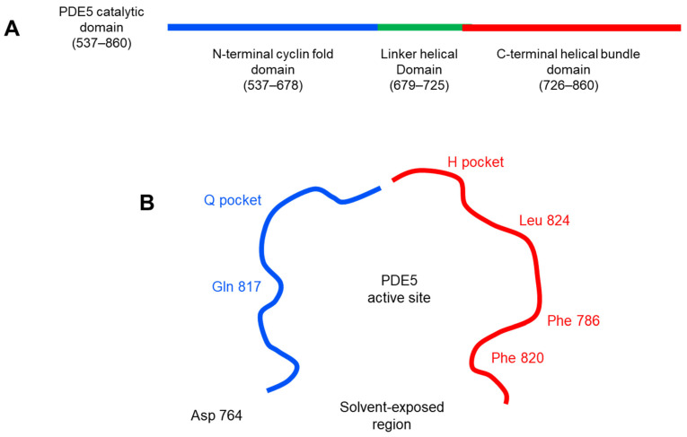
Figure 6.
2D diagram of PDE5 active site (PDB ID: 3HC8). (A) the catalytic domain of PDE5 enzyme can be divided into three subdomains: N-terminal cyclin-fold domain (residues 537–678 in blue), linker helical domain (residues 679–725 in green) and C-terminal helical bundle domain (residues 726–860 in red). (B) the active site of PDE5 can be described in 3 core regions: H pocket (hydrophobic pocket) in red color with key hydrophobic amino acid residues (Phe 786, Phe 820 and Leu 824), Q pocket (cGMP binding site) in blue color with Gln 817 as the key amino acid residue in that site and solvent-exposed region with Asp 764.

