Abstract
Genes encoding an ATP-binding cassette transporter system involved in hemin iron utilization from Corynebacterium ulcerans were cloned and characterized. The genes are homologous to a hemin transport system previously identified in Corynebacterium diphtheriae. Disruption of the hmuT gene, which encodes the putative hemin receptor, resulted in greatly reduced ability of C. ulcerans to use hemin or hemoglobin as an iron source. Inactivation of hmuT in C. diphtheriae by site-specific recombination had no effect on hemin utilization, which suggests that C. diphtheriae has an additional system for transporting hemin.
Corynebacterium diphtheriae is the cause of the severe respiratory disease diphtheria. The bacterium colonizes the upper respiratory tract of humans, where it synthesizes and secretes the potent exotoxin diphtheria toxin (18). Although C. diphtheriae is perhaps the best known pathogen of the genus Corynebacterium, numerous other species of Corynebacterium are known to cause disease in humans (3). Corynebacterium ulcerans, which is closely related to C. diphtheriae, is primarily associated with disease in animals; however, it occasionally causes a respiratory disease in humans that resembles diphtheria, and strains associated with this diphtheria-like disease produce diphtheria toxin (13).
The ability of bacterial pathogens to acquire iron during infection is essential for many organisms to cause disease (4, 30). A very limited amount of iron is available to an invading pathogen within the host, since most of the extracellular iron is sequestered by transferrin and lactoferrin, while much of the intracellular iron is associated with hemin (17). Mechanisms that are utilized by bacterial pathogens to acquire iron within the host include siderophores and a variety of systems for transporting and extracting iron from host compounds such as hemin, hemoglobin, transferrin, and lactoferrin (1, 11, 14).
Several genes involved in hemin utilization and transport have recently been characterized in the gram-positive pathogen C. diphtheriae C7 (−) (7). These include the hmuT, hmuU, and hmuV genes (hmuTUV), which encode an ATP-binding cassette (ABC) hemin transporter system (2), and the hmuO gene, which encodes a heme oxygenase that is involved in the degradation of hemin and the release of iron (24, 31). The hmuT gene encodes a lipoprotein that is proposed to function as a hemin receptor. The hmuU and hmuV genes are predicted to encode membrane proteins that function as a permease and an ATP-binding protein, respectively.
The C. diphtheriae hmuTUV genes were able to complement hemin utilization mutants in C. diphtheriae and C. ulcerans (2). The mutations in C. diphtheriae that resulted in an inability to use hemin as an iron source were shown by sequence analysis to reside only in the hmuO gene. The mechanism by which the hmuTUV transport genes were able to complement hmuO mutations has not been determined. All of the Corynebacterium mutants in these earlier studies were generated by chemical mutagenesis, and no mutations in the C. diphtheriae hmuTUV genes have ever been identified. While the hemin utilization mutants in C. ulcerans have not been genetically defined, the mutants can be separated into two distinct complementation groups. One group of mutants was complemented only by the C. diphtheriae hmuTUV genes, while the other group of mutants was complemented only by hmuO (2, 24). Surprisingly, DNA hybridization studies, which used the C. diphtheriae hmuTUV genes as probes, failed to detect any specific hybridization to C. ulcerans chromosomal DNA (2). This result suggests either that the C. ulcerans hemin transport genes have low homology with the C. diphtheriae hmuTUV genes or that C. ulcerans utilizes a different mechanism for transporting hemin.
Genetic analysis of C. diphtheriae has been frustrated by the lack of adequate genetic tools for this organism. No transposons, conjugation systems, or mechanisms for creating defined mutations have been developed for this species. Additionally, transformation into C. diphtheriae with DNA passaged through Escherichia coli or other species of Corynebacterium occurs at a very low frequency, presumably due to restriction barriers. In this study, a hemin transport system from C. ulcerans was identified, and a mechanism for creating defined mutations in both C. diphtheriae and C. ulcerans was developed and used to inactivate the hmuT genes in these organisms.
Cloning and characterization of the C. ulcerans hemin transport genes.
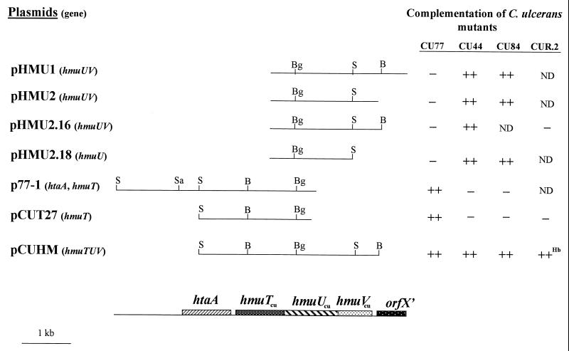
A chromosomal library of C. ulcerans Sau3AI DNA fragments was constructed in the E. coli-C. diphtheriae shuttle vector pKN2.6 (Knr) (25) to identify genes required for hemin transport (22). Three independent clones derived from the library, pHMU1, pHMU2, and p77-1, were able to complement the hemin utilization defect in various C. ulcerans mutants (Fig. 1). The C. ulcerans mutants harboring these clones were able to grow in low-iron agar medium that contained 2.5 μM hemin as the only iron source. The C. ulcerans mutants carrying only the vector, pKN2.6, were unable to grow on this medium (2, 24). The iron-depleted medium contained heart infusion agar with 0.2% Tween 80 (HIBTW) that was made low in iron by the addition of 200 μg of ethylenediamine di(o-hydroxyphenylacetic acid) (EDDA) per ml. Restriction maps of the three complementing clones, pHMU1, pHMU2, and p77-1, as well as various subclones, are shown in Fig. 1.
FIG. 1.
Restriction maps of the insert DNA in plasmids carrying the hmuTUVcu locus from wt C. ulcerans (CU712). Functional genes that are present on the various plasmids are indicated in parentheses. Restriction sites: B, BamHI; Bg, BglII; S, SalI; Sa, SacII. The genetic map of the hmuTUVcu locus is aligned with the restriction maps shown above it. Complementation of C. ulcerans hemin utilization mutants with plasmids carrying the hmuTUVcu locus is indicated to the right. −, inability to grow on low-iron agar medium in the presence of 2.5 μM hemin; ++, full complementation, i.e., growth is equivalent to that of the wt strain, CU712; ND, not determined. The superscript Hb indicates that CUR.2/pCUHM was also able to grow on low-iron agar medium with hemoglobin (0.5 μM) as the sole iron source.
The nucleotide sequence of the 5,262-bp region that extends from the SacII site in p77-1 to the right end of the insert in pHMU1 (Fig. 1) was determined by standard methods (23). The sequence of the insert of plasmid pHMU1 revealed two open reading frames (ORFs) that shared homology with the C. diphtheriae hmuU gene (80% identity at the amino acid level and 68% homology at the nucleotide level) and hmuV gene (69% identity at the amino acid level and 69% homology at the nucleotide level). A gene that is homologous to the C. diphtheriae hmuT gene (80% identity at the amino acid level and 70% homology at the nucleotide level) was identified from the partial nucleotide sequence of p77-1 (extending from the SacII site to the right end of the insert [Fig. 1]). The C. ulcerans genes are hereafter designated hmuTcu, hmuUcu, and hmuVcu, and they are predicted to encode proteins of 37.1, 37.1, and 30.0 kDa, respectively. HmuTcu is predicted to be a lipoprotein, since it contains a 20-amino-acid leader sequence that has a characteristic signal peptidase II processing site. The products encoded by the C. ulcerans hmuTUVcu genes have 25 to 40% identity to ABC hemin transport proteins from various gram-negative bacteria (8, 15, 16, 27, 32). Downstream from the hmuTUVcu genes is a partial ORF that is homologous to the orfX gene from C. diphtheriae (2), while upstream of the hmuTcu gene is an ORF designated htaA (for “hemin transport-associated gene A”) that is predicted to encode a protein of 423 amino acids (Fig. 1). HtaA has low homology to a putative membrane protein from Streptomyces coelicolor. The functions of OrfX and HtaA are not known.
Complementation experiments were done to determine if the cloned hmuTUVcu genes could correct the hemin utilization defect in the various C. ulcerans mutants. Approximately 104 cells from an overnight culture were plated on the surface of a low-iron HIBTW agar plate that contained 2.5 μM hemin. Plates were examined at 24 and 48 h for isolated colonies. C. ulcerans mutants carrying only the vector were unable to grow on this medium, while the C. ulcerans wild-type (wt) strain, CU712 (26), was able to grow on this medium carrying only vector sequences (reference 2 and data not shown). Complementation results revealed that plasmids pHMU1 and pHMU2 complemented the defect in the C. ulcerans hemin utilization mutants CU84 and CU44 but not CU77, while plasmid p77-1 was found to complement CU77 (Fig. 1). In an earlier study, strain CU77 was complemented by C. diphtheriae clones that carried only the hmuT gene (2), which suggests that CU77 is defective in HmuT activity. A subclone of plasmid p77-1, pCUT27, which carries only the hmuTcu gene, was also able to complement strain CU77 (Fig. 1). Plasmid pCUHM, which carries the entire hmuTUVcu locus, was able to fully complement all of the C. ulcerans mutants (Fig. 1). Plasmid pCUHM was constructed in vector pCM2.6 (Cmr) (25) by ligating the 2.26-kb SalI-BglII fragment from p77-1 to the 1.9-kb BglII-BamHI fragment from pHMU1. Plasmid pHMU2.18, which carries only the hmuUcu gene, was able to complement both CU44 and CU84, suggesting that these strains are defective in the HmuU permease.
In an earlier study, we found very weak promoter activity in the region 200 bp upstream of the C. diphtheriae hmuT gene, which includes the htaA-hmuT intergenic region (2). A search for promoter activity with cloned DNA from C. ulcerans using a promoter probe vector also detected only weak constitutive promoter activity upstream of both hmuT and htaA (data not shown). While most hemin transport genes in bacteria are repressed by high iron concentrations, the ABC hemin transporter in Yersinia pestis is transcribed at a low constitutive level (29), similar to what is observed here for C. ulcerans and what was observed previously in C. diphtheriae.
Immunoblot analysis of HmuT in strains of Corynebacterium.
In an earlier study, we reported the high-level expression and partial purification in E. coli of the C. diphtheriae C7 (−) HmuT protein (2). Isolation of large amounts of partially purified HmuT (>1 mg) for this study was accomplished as follows. The Triton X-100-soluble membrane fraction from E. coli containing HmuT was incubated with hemin agarose to allow binding of the HmuT protein. Nonadherent proteins were removed from the hemin agarose by washing with 50 mM Tris (pH 8.5) and 5 mM MgCl2 (TM). The HmuT protein was eluted from the hemin agarose with TM buffer containing 0.25 M NaCl and 1% octylglucoside. This partially purified HmuT protein was used to generate polyclonal serum in rabbits by standard methods (Cocalico Biologicals, Inc., Reamstown, Pa.). The polyclonal serum raised against the C. diphtheriae HmuT protein was used to detect HmuT in Corynebacterium. Whole-cell lysates of Corynebacterium strains were prepared from overnight cultures that were treated with 10 mg of lysozyme per ml and then resuspended in sodium dodecyl sulfate-polyacrylamide gel electrophoresis (SDS-PAGE) sample buffer and boiled (10). Proteins were separated by SDS-PAGE and then transferred to nitrocellulose as previously described (22). The polyclonal serum was diluted 1:40,000, and immunoreactive proteins were detected by incubating the blots with anti-rabbit horseradish peroxidase-conjugated antibodies followed by development of the blots as described previously (5). As shown in Fig. 2, the polyclonal serum detected the HmuT protein in whole-cell lysates from both C. diphtheriae C7 (−) (lane 2) and C. ulcerans 712 (lane 3). In C. ulcerans, the antiserum reacted with a doublet that had a faster migration than the C. diphtheriae HmuT protein. The doublet likely represents the mature and unprocessed forms of HmuTcu, which have predicted sizes of 35.1 and 37.1 kDa, respectively. The mature and unprocessed forms of C. diphtheriae HmuT are also 35.1 and 37.1 kDa, respectively. It is not known why the HmuT proteins from C. ulcerans and C. diphtheriae migrate differently on SDS-PAGE gels. A similar banding pattern was detected in the C. ulcerans hemin utilization mutants CU84 and CU44 (hmuU) (lanes 4 and 5, respectively). No band was detected in whole-cell lysates from CU77 (hmuT) (lanes 6 and 7). Detection of HmuT was restored when CU77 was transformed with plasmids carrying the cloned hmuT gene from either C. ulcerans (lane 8, pCUT27; lane 9, pCUHM) or C. diphtheriae (lane 10, p1.9Pst) (2).
FIG. 2.
Immunoblot analysis of HmuT production by various Corynebacterium species. Whole-cell lysates were subjected to SDS-PAGE, transferred to nitrocellulose, and probed with polyclonal antibodies raised against the C. diphtheriae C7 (−) HmuT protein. Lanes 4 through 14 are C. ulcerans hemin utilization mutants. Lane 1, purified C. diphtheriae HmuT; lane 2, C. diphtheriae C7 (−) (wt); lane 3, C. ulcerans 712 (wt); lane 4, CU84 (hmuU); lane 5, CU44 (hmuU); lane 6, CU77 (hmuT); lane 7, CU77/pCM2.6 (vector); lane 8, CU77/pCUT27 (hmuT+cu); lane 9, CU77/pCUHM (hmuTUV+cu); lane 10, CU77/p1.9Pst (hmuT+); lane 11, CUR.2/pCM2.6; lane 12, CUR.2/pCUT27 (hmuT+cu); lane 13, CUR.2/pCUHM (hmuTUV+cu); lane 14, CUR.2/p1.9Pst (hmuT+).
Construction and analysis of defined mutations in the hmuT gene in C. diphtheriae and C. ulcerans.
A mechanism for creating defined mutations in the chromosomal copy of the hmuT gene in both C. diphtheriae and C. ulcerans is described below. This method employs the integration of vector sequences and was initially utilized to create gene duplications or gene disruptions in Corynebacterium glutamicum, a nonpathogenic species of Corynebacterium (21). The system for the creation of defined mutations in C. diphtheriae and C. ulcerans involves a single crossover event between an internal portion of the cloned hmuT gene and the chromosomal copy of hmuT. The crossover results in the stable integration of the entire vector into the chromosome and disruption of the chromosomal copy of hmuT. (The vector was stably integrated into the chromosome for over 20 generations in the absence of antibiotic selection [M. Schmitt and E. S. Drazek, unpublished observation]).
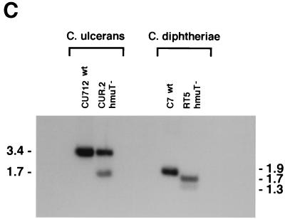
Since ColE1-type plasmids are unable to replicate in the genus Corynebacterium and can be transformed at high efficiency into C. ulcerans from DNA prepared in E. coli (24), we used these plasmids as suicide vectors to disrupt the chromosomal copy of hmuTcu. A derivative of the E. coli vector pBluescript KS (Stratagene, La Jolla, Calif.), pKS2.677, was used to disrupt the hmuTcu gene. Plasmid pKS2.677 was constructed as follows. A portion of the ampicillin resistance gene of pBluescript KS was excised and replaced with the kanamycin resistance marker from the vector pKN2.6 to create pKS2.6. A 552-bp fragment that contained an internal portion of the 5′ region of the hmuTcu gene (nucleotides 1645 to 2197) was amplified by PCR and ligated into the vector pCR-Blunt II-TOPO (Invitrogen, Carlsbad, Calif.) to create plasmid pCRTCU. A 480-bp fragment from pCRTCU was excised with BamHI and ligated into the BamHI site in pKS2.6 to create pKS2.677 (Fig. 3A). A stop codon was placed in the downstream primer to prevent the formation of fusion proteins (Fig. 3A). To disrupt the chromosomal hmuTcu gene, plasmid pKS2.677 was transformed by electroporation (6) into CU712 (wt), and kanamycin-resistant colonies were recovered. One isolate, CUR.2, was analyzed further. Chromosomal DNA from CUR.2 was digested with SalI and probed with a 3.4-kb SalI fragment from pCUHM (Fig. 1 and 3A) (9). The detection of 3.4- and 1.7-kb bands in the CUR.2 chromosomal digest confirmed that the entire vector had integrated into the chromosome within the hmuTcu gene (Fig. 3A and C).
FIG. 3.
(A) Schematic representation of the integration of plasmid pKS2.677 into the hmuTcu gene on the chromosome of C. ulcerans 712 (CU712). Relevant chromosomal regions of the hmuTUV locus of C. ulcerans 712 and the hmuTcu mutant CUR.2 are shown. Kn, kanamycin resistance gene; ColE1, E. coli origin of plasmid replication; hmuT′, truncated fragment of the C. ulcerans hmuTcu gene; ATG, start codon for hmuTcu; , stop codon. Restriction sites: B, BamHI; S, SalI. The distances between the SalI sites that were used for mapping in the DNA hybridization studies (panel C) are also shown. (B) Schematic representation of the integration of plasmid pBC842.Nco into the hmuT gene on the chromosome of C. diphtheriae C7 (−). Relevant chromosomal regions of the hmu locus of C. diphtheriae C7 (−) and the hmuT mutant strain, RT5, are shown. Kn, kanamycin resistance gene; hmuT′, truncated fragment of the C. diphtheriae hmuT gene; ATG, start codon for hmuT; ORI, origin of plasmid replication for the E. coli-C. diphtheriae shuttle vector pKN2.6; ΔORI, truncated origin of replication; , stop codon. Restriction sites: N, NcoI; P, PstI. Maps shown in panels A and B are not to scale. (C) DNA hybridization analysis of chromosomal DNA isolated from wt and recombinant hmuT mutant strains of C. diphtheriae and C. ulcerans. C. ulcerans chromosomal DNA was digested with SalI and probed with a 32P-labeled 3.4-kb SalI DNA fragment from plasmid pCUHM. C. diphtheriae DNA was digested with PstI and probed with a 32P-labeled 1.9-kb PstI fragment from plasmid p1.9Pst. Fragment sizes, in kilobases, are indicated.
Immunoblots using the HmuT antiserum did not detect immunoreactive proteins from whole-cell lysates of CUR.2 (Fig. 2, lane 11). When CUR.2 was transformed with clones carrying the hmuT gene from either C. ulcerans (pCUT27 and pCUHM) or C. diphtheriae (p1.9Pst), a protein that reacted with the HmuT antiserum was detected (Fig. 2, lanes 12 to 14). Hemin utilization studies revealed that CUR.2 had reduced ability to use hemin and hemoglobin as iron sources, similar to that seen with CU77 (hmuT) (Fig. 1 and Table 1). The data in Table 1 show that growth differences between mutant strains carrying only the vector and mutants harboring complementing clones are seen at hemin and hemoglobin concentrations of 0.1 μM. Growth of all strains was stimulated at 1 μM hemin or hemoglobin. Complementation studies indicated that the hmuT, hmuU, and hmuV genes on pCUHM were needed to complement CUR.2. The hmuT gene alone on pCUT27 or the hmuU and hmuV genes on pHMU1.26 (Cmr) failed to complement CUR.2 (Fig. 1 and Table 1). This result suggests that the disruption of the hmuT gene in CUR.2 has a strong polar effect on the expression of the downstream hmuU and hmuV genes. The hmuU gene on pHMU1.26 is functional, since this plasmid was able to complement CU44 (hmuU) (Fig. 1 and Table 1). These studies provide additional evidence that the HmuTcu protein in C. ulcerans has an essential role in the utilization of hemin and hemoglobin as iron sources.
TABLE 1.
Growth of C. ulcerans strains in the presence of hemin or hemoglobina
| Strain/plasmid (gene) |
A600 in the presence
of indicated iron source
|
||||
|---|---|---|---|---|---|
| None | Hemin
(μM)
|
Hemoglobin (μM)
|
|||
| 0.1 | 1.0 | 0.1 | 1.0 | ||
| CU712 (wt)/pCM2.6 (vector) | 0.1 | 1.8 | 6.4 | 4.4 | 6.1 |
| CU77/pCM2.6 | 0.2 | 0.2 | 4.8 | 0.6 | 4.9 |
| CU77/pCUT27 (hmuT) | 0.2 | 1.2 | 4.9 | 3.8 | 5.1 |
| CU77/pCUHM (hmuTUV) | 0.2 | 1.8 | 4.4 | 4.6 | 5.1 |
| CU77/pHMU1.26 (hmuUV) | 0.3 | 0.5 | 4.4 | 0.8 | 4.9 |
| CUR.2/pCM2.6 | 0.2 | 0.2 | 5.4 | 0.6 | 5.5 |
| CUR.2/pCUT27 (hmuT) | 0.2 | 0.2 | 5.3 | 0.8 | 5.4 |
| CUR.2/pCUHM (hmuTUV) | 0.3 | 1.3 | 5.4 | 3.3 | 5.9 |
| CUR.2/pHMU1.26 (hmuUV) | 0.2 | 0.2 | 5.1 | 0.4 | 5.2 |
| CU44/pCM2.6 | 0.1 | 0.1 | 2.8 | 0.2 | 3.0 |
| CU44/pCUHM (hmuTUV) | 0.1 | 1.6 | 4.0 | 2.7 | 3.9 |
| CU44/pHMU1.26 (hmuUV) | 0.1 | 1.4 | 3.6 | 2.2 | 4.2 |
Strains were initially grown overnight in HIBTW and then passaged 1/100 into HIBTW containing 200 μg of EDDA per ml and grown for 18 h. These cultures were then passaged 1/100 into HIBTW-EDDA medium containing hemin or hemoglobin or no iron source. Absorbance (A600) measurements were done on 18-h cultures, and the values shown are the averages of three independent experiments. Standard deviations did not vary by more than 20% from the mean.
ColE1 vectors, such as pKS2.6, cannot be used to recombine into the C. diphtheriae chromosome to disrupt the hmuT gene. Although ColE1 plasmids fail to replicate in Corynebacterium species, C. diphtheriae is transformed very poorly by these vectors because plasmid DNA that is prepared from E. coli (or C. ulcerans) appears to be restricted in C. diphtheriae. However, if the plasmid DNA is first passaged through C. diphtheriae, it can then be transformed back into C. diphtheriae at high efficiency (M. Schmitt, unpublished observation). Plasmids that have been passaged through C. diphtheriae can then be used for recombination if the vector is subsequently rendered replication defective. High-efficiency transformation and an inability to replicate are essential qualities for a plasmid to be used as a suicide vector for gene disruption in C. diphtheriae (and in C. ulcerans). A method for disrupting the C. diphtheriae hmuT gene is described below.
Inactivation of the C. diphtheriae C7 (−) hmuT gene was accomplished by integration of the replication-deficient plasmid pBC842.Nco into the chromosomal copy of hmuT (Fig. 3B). Plasmid pBC842.Nco was constructed as follows. A 563-bp fragment that contains an internal portion of hmuT near the 5′ end of the gene (nucleotides 243 to 806 [2]) was amplified by PCR and ligated into the pCR-Blunt II-TOPO vector to create plasmid pCRTd. The insert in pCRTd was excised with BglII and PuvII (sites incorporated into the PCR primers) and ligated into the BamHI-PvuII sites of the shuttle vector pKN2.6 to create plasmid pBC842 (Fig. 3B). Plasmid pBC842 was transformed into C. diphtheriae C7 (−). Plasmid pBC842 DNA was then prepared from C7 (−) and digested with NcoI, and a 2.6-kb fragment was purified and then ligated at low concentration (1 μg/ml) to generate circular molecules, pBC842.Nco (Fig. 3B). The NcoI digest of pBC842 removes a 1.2-kb portion of the origin of replication, which renders the vector unable to replicate in C. diphtheriae. To obtain recombinants, the ligated plasmid DNA (pBC842.Nco, Fig. 3B) was electroporated into C. diphtheriae C7 (−), from which kanamycin-resistant colonies were isolated, and one clone, RT5, was characterized further. RT5 chromosomal DNA was digested with PstI and then hybridized with an hmuT-specific probe (1.9-kb PstI fragment, Fig. 3B), and bands of 1.7 and 1.3 kb were detected, which confirmed that pBC842.Nco had integrated into the chromosomal copy of hmuT (Fig. 3B and C).
Western analysis of whole-cell lysates of RT5 did not detect any immunoreactive proteins (Fig. 4, lane 3) and thus confirmed the absence of the HmuT protein in this mutant. Production of the C. diphtheriae HmuT protein was restored by transformation of RT5 with plasmid p1.9Pst (hmuT+) (Fig. 4, lane 4).
FIG. 4.
Immunoblot analysis of HmuT production by various Corynebacterium species. Whole-cell lysates were subjected to SDS-PAGE, transferred to nitrocellulose, and probed with polyclonal antibodies raised against the C. diphtheriae HmuT protein. Lanes 2 through 8 are C. diphtheriae isolates. Lane 1, purified HmuT from C. diphtheriae; lane 2, C7 (−)/pKN2.6 (wt); lane 3, RT5; lane 4, RT5/p1.9Pst (hmuT+); lane 5, C7 (−)/pKN2.6; lane 6, 1716 (biotype Gravis; ribotype G1); lane 7, 1751 (biotype Mitis; ribotype M1v); lane 8, 1897 (biotype Gravis; ribotype G4); lane 9, C. ulcerans 712; lane 10, C. pseudotuberculosis; lane 11, C. renale; lane 12, C. glutamicum; lane 13, C. jeikeium.
Surprisingly, however, RT5 was unaffected in its ability to use hemin or hemoglobin as an iron source. Studies that compared the abilities of RT5 and the parent strain, C7 (−), to utilize hemin and hemoglobin over a wide range of concentrations failed to detect any significant differences in the growth of these strains (data not shown). This finding differs significantly from the observation for C. ulcerans, which requires HmuTcu in order to use hemin as an iron source (Fig. 1). The fact that a mutation in the C. diphtheriae hmuT gene has no effect on the ability to use hemin as an iron source suggests that C. diphtheriae has an additional mechanism for transporting hemin. Multiple hemin transport systems have been identified in other bacteria, including pathogenic strains of Neisseria (12, 28) and Haemophilus (19, 20).
Three C. diphtheriae clinical strains obtained from the Russian diphtheria epidemic all expressed the HmuT protein (Fig. 4, lanes 6 to 8). The migration of HmuT from the clinical strains was slightly faster than that observed for C. diphtheriae C7 (−). These Russian strains differ in their biotypes and ribotypes and were isolated from distinct geographical locations. These findings suggest that HmuT is conserved among virulent strains from diverse backgrounds. Four other Corynebacterium species were also examined for the presence of HmuT. Faint bands of various sizes were detected for Corynebacterium pseudotuberculosis (a pathogen of humans and animals) and Corynebacterium renale (an animal pathogen) (Fig. 4, lanes 10 and 11). Longer development of the blot allowed the detection of faint bands in C. glutamicum (commensal) and Corynebacterium jeikeium (human pathogen) (Fig. 4, lanes 12 and 13; longer exposure not shown).
This study describes, for the first time, the creation of defined mutations in the chromosome of the bacterial pathogens C. diphtheriae and C. ulcerans. Inactivation of hmuT in these organisms gave surprisingly different results: HmuT in C. ulcerans was essential for normal hemin utilization, while HmuT in C. diphtheriae was not required for the use of hemin as an iron source. Furthermore, these results provide an explanation of why earlier studies were unable to identify C. diphtheriae hemin utilization mutants that carried mutations in the hmuTUV locus. Since the C. diphtheriae hmuT mutant, RT5, is able to use hemin as an iron source, these findings suggest that C. diphtheriae has an additional mechanism for transporting hemin.
Nucleotide sequence accession number.
The nucleotide sequence of the 5,262-bp region of the hmuTUVcu locus has been assigned GenBank accession number AF304009.
Acknowledgments
We thank Tanja Popovic for providing C. diphtheriae clinical strains and Craig Hammack, Sr., for technical assistance. We are also grateful to Clare Schmitt, Tod Merkel, and Scott Stibitz for critical reading of the manuscript.
REFERENCES
- 1.Braun V, Hantke K, Koster W. Bacterial iron transport: mechanisms, genetics, and regulation. Met Ions Biol Syst. 1998;35:67–145. [PubMed] [Google Scholar]
- 2.Drazek E S, Hammack C A, Schmitt M P. Corynebacterium diphtheriaegenes required for acquisition of iron from hemin and hemoglobin are homologous to ABC hemin transporters. Mol Microbiol. 2000;36:68–84. doi: 10.1046/j.1365-2958.2000.01818.x. [DOI] [PubMed] [Google Scholar]
- 3.Funke G, Von Graevenitz A, Clarridge III J E, Bernard K A. Clinical microbiology of coryneform bacteria. Clin Microbiol Rev. 1997;10:125–159. doi: 10.1128/cmr.10.1.125. [DOI] [PMC free article] [PubMed] [Google Scholar]
- 4.Griffiths E. Iron and bacterial virulence—a brief overview. Biol Met. 1991;4:7–13. doi: 10.1007/BF01135551. [DOI] [PubMed] [Google Scholar]
- 5.Harlow E, Lane D. Antibodies: a laboratory manual. Cold Spring Harbor, N.Y: Cold Spring Harbor Laboratory Press; 1998. [Google Scholar]
- 6.Haynes J A, Britz M L. Electrotransformation of Brevibacterium lactofermentum and Corynebacterium glutamicum: growth in Tween 80 increases transformation frequencies. FEMS Microbiol Lett. 1989;61:329–334. [Google Scholar]
- 7.Holmes R K, Barksdale L. Genetic analysis of tox+ and tox− bacteriophages of Corynebacterium diphtheriae. J Virol. 1969;3:586–598. doi: 10.1128/jvi.3.6.586-598.1969. [DOI] [PMC free article] [PubMed] [Google Scholar]
- 8.Hornung J M, Jones H A, Perry R D. The hmu locus of Yersinia pestisis essential for utilization of free haemin and haem-protein complexes as iron sources. Mol Microbiol. 1996;20:725–739. doi: 10.1111/j.1365-2958.1996.tb02512.x. [DOI] [PubMed] [Google Scholar]
- 9.Kidd F J. In-situhybridization to agarose gels. Focus. 1983;6:3–4. [Google Scholar]
- 10.Laemmli U K. Cleavage of structural proteins during the assembly of the head of bacteriophage T4. Nature. 1970;227:680–685. doi: 10.1038/227680a0. [DOI] [PubMed] [Google Scholar]
- 11.Lee C B. Quelling the red menace: hemin capture by bacteria. Mol Microbiol. 1995;18:383–390. doi: 10.1111/j.1365-2958.1995.mmi_18030383.x. [DOI] [PubMed] [Google Scholar]
- 12.Lewis L A, Dyer D W. Identification of an iron-regulated outer membrane protein of Neisseria meningitidisinvolved in the utilization of hemoglobin complexed to haptoglobin. J Bacteriol. 1995;177:1299–1306. doi: 10.1128/jb.177.5.1299-1306.1995. [DOI] [PMC free article] [PubMed] [Google Scholar]
- 13.McDonald S, Cox D, Haute T, Alen R, Staggs W, Bixler D, Steele G. Respiratory diphtheria caused by Corynebacterium ulcerans—Terre Haute, Indiana, 1996. JAMA. 1997;277:1665–1666. [PubMed] [Google Scholar]
- 14.Mietzner T A, Morse S A. The role of iron-binding proteins in the survival of pathogenic bacteria. Annu Rev Nutr. 1994;14:471–493. doi: 10.1146/annurev.nu.14.070194.002351. [DOI] [PubMed] [Google Scholar]
- 15.Mills M, Payne S M. Genetics and regulation of heme iron transport in Shigella dysenteriae and detection of an analogous system in Escherichia coliO157:H7. J Bacteriol. 1995;177:3004–3009. doi: 10.1128/jb.177.11.3004-3009.1995. [DOI] [PMC free article] [PubMed] [Google Scholar]
- 16.Occhino D A, Wycoff E E, Henderson D P, Wrona T J, Payne S M. Vibrio cholerae iron transport: haem transport genes are linked to one of two sets of tonB, exbB, exbDgenes. Mol Microbiol. 1998;29:1493–1507. doi: 10.1046/j.1365-2958.1998.01034.x. [DOI] [PubMed] [Google Scholar]
- 17.Otto B R, Verweij-van Vught A M, MacLaren D M. Transferrins and heme compounds as iron sources for pathogenic bacteria. Crit Rev Microbiol. 1992;18:217–233. doi: 10.3109/10408419209114559. [DOI] [PubMed] [Google Scholar]
- 18.Pappenheimer A M., Jr Diphtheria toxin. Annu Rev Biochem. 1977;46:69–94. doi: 10.1146/annurev.bi.46.070177.000441. [DOI] [PubMed] [Google Scholar]
- 19.Reidl J, Mekalanos J J. Lipoprotein e(p4) is essential for hemin uptake by Haemophilus influenzae. J Exp Med. 1996;183:621–629. doi: 10.1084/jem.183.2.621. [DOI] [PMC free article] [PubMed] [Google Scholar]
- 20.Ren Z, Hongfan J, Morton D J, Stull T L. hgpB, a gene encoding a second Haemophilus influenzaehemoglobin- and hemoglobin-haptoglobin-binding protein. Infect Immun. 1998;66:4733–4741. doi: 10.1128/iai.66.10.4733-4741.1998. [DOI] [PMC free article] [PubMed] [Google Scholar]
- 21.Reyes O, Guyonvarch A, Bonamy C, Salti V, David F, Leblon G. “Integron”-bearing vectors: a method suitable for stable chromosomal integration in highly restrictive Corynebacteria. Gene. 1991;107:61–68. doi: 10.1016/0378-1119(91)90297-o. [DOI] [PubMed] [Google Scholar]
- 22.Sambrook J, Fritsch E F, Maniatis T. Molecular cloning: a laboratory manual. Cold Spring Harbor, N.Y: Cold Spring Harbor Laboratory; 1989. [Google Scholar]
- 23.Sanger F, Nicklen S, Coulson A R. DNA sequencing with chain-terminating inhibitors. Proc Natl Acad Sci USA. 1977;74:5463–5467. doi: 10.1073/pnas.74.12.5463. [DOI] [PMC free article] [PubMed] [Google Scholar]
- 24.Schmitt M P. Utilization of host iron sources by Corynebacterium diphtheriae: identification of a gene whose product is homologous to eukaryotic heme oxygenases and is required for acquisition of iron from heme and hemoglobin. J Bacteriol. 1997;179:838–845. doi: 10.1128/jb.179.3.838-845.1997. [DOI] [PMC free article] [PubMed] [Google Scholar]
- 25.Schmitt M P, Holmes R K. Iron-dependent regulation of diphtheria toxin and siderophore expression by the cloned Corynebacterium diphtheriae repressor gene dtxR in C. diphtheriaeC7 strains. Infect Immun. 1991;59:1899–1904. doi: 10.1128/iai.59.6.1899-1904.1991. [DOI] [PMC free article] [PubMed] [Google Scholar]
- 26.Serwold-Davis T M, Groman N B, Rabin M. Transformation of Corynebacterium diphtheriae, Corynebacterium ulcerans, Corynebacterium glutamicum, and Eschericia coli with the C. diphtheriaeplasmid pNG2. Proc Natl Acad Sci USA. 1987;84:4964–4968. doi: 10.1073/pnas.84.14.4964. [DOI] [PMC free article] [PubMed] [Google Scholar]
- 27.Stojiljkovic I, Hantke K. Transport of hemin across the cytoplasmic membrane through a hemin-specific periplasmic binding-protein-dependent transport system in Yersinia enterocolitica. Mol Microbiol. 1994;13:719–732. doi: 10.1111/j.1365-2958.1994.tb00465.x. [DOI] [PubMed] [Google Scholar]
- 28.Stojiljkovic I, Hwa V, Martin L S, O'Gaora P, Nassif X, Hefron F, So M. The Neisseria meningitidishemoglobin receptor: its role in iron utilization and virulence. Mol Microbiol. 1995;15:531–541. doi: 10.1111/j.1365-2958.1995.tb02266.x. [DOI] [PubMed] [Google Scholar]
- 29.Thompson J M, Jones H A, Perry R D. Molecular characterization of the hemin uptake locus (hmu) from Yersinia pestis and analysis of hmumutants for hemin and hemoprotein utilization. Infect Immun. 1999;67:3879–3892. doi: 10.1128/iai.67.8.3879-3892.1999. [DOI] [PMC free article] [PubMed] [Google Scholar]
- 30.Weinberg E D. Iron and infection. Microbiol Rev. 1978;42:45–66. doi: 10.1128/mr.42.1.45-66.1978. [DOI] [PMC free article] [PubMed] [Google Scholar]
- 31.Wilks A, Schmitt M P. Expression and characterization of a heme oxygenase (HmuO) from Corynebacterium diphtheriae. J Biol Chem. 1998;273:837–841. doi: 10.1074/jbc.273.2.837. [DOI] [PubMed] [Google Scholar]
- 32.Wyckoff E E, Duncan D, Torres A G, Mills M, Maase K, Payne S M. Structure of the Shigella dysenteriaehaem transport locus and its phylogenetic distribution in enteric bacteria. Mol Microbiol. 1998;28:1139–1152. doi: 10.1046/j.1365-2958.1998.00873.x. [DOI] [PubMed] [Google Scholar]


