Abstract
PARP inhibitors (PARPi) have increased treatment options in ovarian cancer, particularly in patients with BRCA1/2 mutations, although there are still marked differences in the duration of patients’ response to this targeted therapy. BRCA testing is routinely performed in tumor tissue of ovarian cancer patients. The resulting molecular pathological findings include the genetic nomenclature of the mutation, the frequency of the mutated allele (variant allele frequency, VAF), and the tumor cell content. VAF measures the percentage of mutated alleles from the total alleles in the cells of the examined tissue. The aim of this study was to investigate the significance of VAF on the therapeutic response to PARPis in ovarian cancer patients. Epithelial ovarian cancer patients harboring BRCA1/2 tumor mutations, who underwent germline testing and received PARPi therapy at the Medical University of Vienna (n = 41) were included in the study. Corrected VAF (cVAF) was calculated based on VAF, tumor cell content, and germline mutation. Patients were divided into two groups based on their cVAF. Median PFS under PARPi in patients with low cVAF was 13.0 months (IQR [10.3-not reached]) and was not reached in the high cVAF group. High cVAF was significantly associated with longer PFS in the multivariate analysis (HR = 0.07; 95% CI [0.01–0.63]; p = 0.017). In conclusion, high cVAF was associated with a significantly better response to PARPi in this study population.
Keywords: BRCA, PARP-inhibitor, ovarian cancer, allele frequency
1. Introduction
Ovarian cancer is the second most lethal gynecological malignancy, following breast cancer, in western countries [1]. One of the predisposing factors for epithelial ovarian cancer (EOC) is germline mutation in tumor suppressor genes, such as BRCA1 and BRCA2 (BRCA1/2). These mutations can be inherited as a germline mutation or arise as somatic mutation in the tumor [2,3]. Germline BRCA1/2 mutations are found in approximately 18% of high grade serous ovarian cancer (HGSOC) patients [4,5], while exclusive somatic mutations in BRCA1/2 occur in about 3–8% [6,7].
During diagnostic workup, BRCA testing is performed on tumor tissue routinely in patients with histologically confirmed epithelial ovarian cancer. Patients with a tumor BRCA mutation are subsequently referred to genetic counseling and are advised to additionally perform germline BRCA testing. Germline testing is of clinical importance, as patients with germline BRCA mutation have an increased risk for breast cancer. Furthermore, relatives of patients with germline BRCA mutation could potentially carry the mutation themselves, increasing the risk for breast cancer, ovarian cancer in female and prostate cancer in male relatives.
BRCA testing is a standardized procedure performed routinely. The molecular diagnostic workup includes specific information about the respective gene mutation, allelic frequency of the detected mutation and percentage of tumor tissue (tumor cellularity or purity) in the examined tissue. Variant allele frequency (VAF) describes the “relative frequency of an allele at a genetic locus” which has to be corrected for the tumor cell fraction of the analyzed tissue (tumor cellularity) [8].
Poly (ADP-ribose) polymerase (PARP)-inhibitors (PARPi) as maintenance therapy following chemotherapy have increased treatment options for patients with BRCA1/2 mutation in recent years. Although PARPis have been shown to significantly improve the prognosis of this patient cohort, PARPi response rates depend on BRCA mutation status, therapy line and platinum sensitivity of recurrent disease and type of PARPi. Response rates for first-line maintenance PARPi treatment were found to be approximately 60–81% [9,10]. In recurrent disease, response rates of BRCA1/2 mutated patients differ between 69% for platinum sensitive tumors and 23% for platinum-refractory tumors [11]. The SOLO-3 trial found objective response rates of 72% for BRCA1/2 mutated patients with at least 2 prior lines of chemotherapy and 59% for patients with more prior therapy lines [12]. An integrated analysis of data from Study 10 and ARIEL2 revealed a response rate of 68% in patients with a BRCA1/2 mutation for third-line rucaparib treatment and 45% for fourth-line or later rucaparib treatment [13]. The possibility of PARPi resistance has led to extensive research to elucidate the underlying resistance mechanism [14,15]. To reduce potential severe side effects in patients with PARPi resistance and to optimize cost-effectiveness [16,17], it would be of interest to identify those patients, who would profit most of PARPi treatment. Although PARPis are usually well tolerated with common side effects including nausea (74–76%), fatigue (59–69%), vomiting (34–37%), and anemia (37–50%), mostly grade 1–2, higher grade and serious adverse events including hematological toxicities (anemia, neutropenia, thrombocytopenia), gastrointestinal toxicities (nausea, vomiting, abdominal pain, intestinal obstruction), fatigue, and rare secondary malignancies (myelodysplastic syndrome, acute myeloid leukemia) (0.83%) have been reported [18,19,20].
The significance of variant allele frequencies of somatic BRCA 1/2 mutations in epithelial ovarian cancer has not yet been entirely evaluated or associated with response to PARPi. Therefore, the aim of this study was to evaluate the effect of BRCA1/2 tumor allele frequency on PARPi response in a unicentric cohort of ovarian cancer patients.
2. Materials and Methods
2.1. Study Population
A total of 41 patients with EOC treated between 2014 and 2020 at the Medical University of Vienna (Comprehensive Cancer Center), Austria, were included in the study. The study was conducted according to the guidelines of the Declaration of Helsinki and approved by the Institutional Review Board of the Medical University of Vienna (2067/2019). All patients with tumor and germline BRCA1/2 mutation status, who received PARPi in primary or recurrent setting, were included in this study. Tumor samples of all included patients were routinely obtained during diagnostic or debulking surgery. BRCA tumor testing was performed at the Department of Pathology, Medical University of Vienna. Patients were further referred to genetic counselling and germline BRCA mutation assessment, at the Department of Obstetrics and Gynecology, Medical University of Vienna. All patients signed an informed consent for BRCA tumor testing prior to surgery.
Clinical data were retrieved from medical records, histological subtype and grading were determined by gynecologic pathologists. All patients were treated according to national guidelines and standard institutional procedures, consisting of either primary debulking surgery with adjuvant platinum-based chemotherapy +/− bevacizumab or neoadjuvant chemotherapy followed by intervention debulking surgery. Surgical staging was performed according to FIGO guidelines. PARPi therapy was initiated according to national approval and the respective tumorboard recommendation.
2.2. BRCA Germline Testing
Patients, who fulfilled the clinical criteria for germline testing, were referred to genetic counseling and gave written informed consent for BRCA germline testing/molecular analyses of a blood sample. BRCA germline testing was performed as previously described [21].
Genetic testing was performed at the University Hospital of Vienna from 2007 to 2015 with Sanger Sequencing in conjunction with multiplex ligation-dependent probe amplification (MLPA) and thereafter the Illumina TruSight Cancer panel on the MiSeq instrument was used for multigene panel testing according to the manufacturer’s instructions (Illumina, San Diego, CA, USA). Data were analyzed with the Sophia DDM® software (Sophia Genetics, Boston, MA, USA)
2.3. BRCA Tumor Testing
BRCA tumor testing was performed as previously described [21]. Briefly, DNA was purified from FFPE tissue blocks with EZ1 DNA Tissue Kit (Qiagen, Hilden, Germany) on an EZ1 Advanced XL instrument (Qiagen). Next-generation sequencing libraries were generated with the Oncomine BRCA Research Assay (Thermo Fisher Scientific, Waltham, MA, USA) and sequenced with an Ion S5 system (Thermo Fisher Scientific). A threshold of 5% allele frequency was set for sequence variant calling. Variants were classified into pathogenic, likely pathogenic, variant of unknown significance (VUS), and likely benign/benign based on DNA sequence reference databases BRCA Exchange, ClinVar, COSMIC and dbSNP. The clinical report included only pathogenic/likely pathogenic sequence variants and VUS. To optimize data quality, only BRCA mutation reports with tumor cellularity greater than 10% were included in final analyses [6].
2.4. Calculation of Corrected Allele Frequency
Only the allele frequency of the tumor was of interest in this study; therefore, the specific corrected allele frequency of the tumor (VAFt = cVAF) had to be calculated from the overall allele frequency given in the molecular pathologic result (VAF). The given allele frequency is composed from the allele frequencies of the included tumor (VAFt = cVAF) and healthy (VAFg) tissues, according to the tumor cellularity (Pt) respective germline cellularity (Pg = 1 − Pt, as cellularity is given from 0 to 1).
To correct the allele frequency for tumor purity and germline mutation status, the following formula, adapted from Kanchi et al. [22], was used:
Germline mutations were only given as negative or positive results; therefore, VAFg was set as 0 in case of missing germline mutation or 0.5 (50% allele frequency) in case of detected BRCA germline mutation. cVAF was used for further analyses of treatment response.
2.5. Statistical Analysis
The corrected allele frequency (cVAF) was used for univariate and multiple Cox regression analyses. Time to progression under PARPi treatment (PFS) was defined as the time between start of PARPi treatment until tumor recurrence. Recurrence was confirmed radiologically according to RECIST 1.1 criteria. Patients who did not progress during follow up were censored respectively at the time point of last clinical contact. Median follow-up time of the cohort (only censored patients) was 30.0 months (interquartile range, IQR [15.9–37.3]). All statistical analyses were performed in GNU R 4.1.2 with following R-packages: survival 3.2-13, survMisc 0.5.5, and MASS 7.3-55. The optimal cut-off for a dichotomized cVAF predictor was determined by the cutp function from the R-package MASS and optically verified by a non-linear Cox regression modeling of the association between the cVAF and the hazard ratio for progression using smoothing splines, a penalized spline basis. Presented multiple Cox regression models were built by starting with the complete model (including all relevant clinicopathologic parameters) and selecting the most informative but smallest model by a backward elimination procedure minimizing the Akaike information criterion (AIC). To plot estimated survival curves for the optimally dichotomized cVAF from the final Cox-regression models, all correcting factors were averaged. For the predicted single patient survival curves, concrete values for the correcting factors were used. Continuous nominal values were compared between two groups with students’ t-test. Correlations of nominal variables with nominal or ordinal variables were performed using Spearman Correlations. Statistical significance was defined as p-value ≤ 0.05.
3. Results
3.1. Patient Characteristics
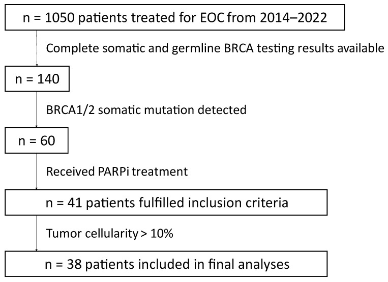
A total of 41 patients with EOC fulfilled the inclusion criteria. Thereof, three patients were excluded from all further analyses due to ≤10% tumor cellularity, leaving 38 remaining patients for final analyses [6] (Figure 1).
Figure 1.
Scheme of patient selection. In total, 41 patients were included in the study and 38 patients were included in the final analyses.
Of these 38 patients, 35 patients had high-grade serous histology, three patients had high-grade endometrioid ovarian cancer. The vast majority of patients suffered from advanced stage FIGO III or IV ovarian cancer (n = 37, 97.4%), one patient had a FIGO stage II disease at initial diagnosis. Further patient characteristics are shown in Table 1.
Table 1.
Patient characteristics at primary diagnosis of 38 patients with tumor cellularity >10%.
| Parameter | Variable | n (%) |
|---|---|---|
| Histology | Serous | 35 (92.1%) |
| Endometrioid | 3 (7.9%) | |
| Grade | high-grade | 38 (100.0%) |
| FIGO stage at initial diagnosis |
II | 1 (2.6%) |
| III | 27 (71.1%) | |
| IV | 10 (26.3%) | |
| Primary treatment | NACT + IDS | 13 (34.2%) |
| PDS + adj. CHT | 25 (65.8%) | |
| PARPi | Olaparib | 32 (84.2%) |
| Niraparib | 4 (10.5%) | |
| Rucaparib | 2 (5.3%) | |
| PARPi Treatment line | 1 | 12 (31.6%) |
| 2 | 20 (52.6%) | |
| 3 | 1 (2.6%) | |
| 4 | 3 (7.9%) | |
| 5 | 2 (5.3%) |
Thirteen patients (34.2%) received neoadjuvant chemotherapy and intervention debulking surgery, 25 patients (65.8%) were treated by primary debulking surgery and adjuvant chemotherapy.
Median follow-up was 30.0 months (IQR [15.9–37.3]).
3.2. BRCA Testing
All patients underwent somatic and germline BRCA testing. With respect to tumor testing, 25 (66%) patients had a BRCA1 mutation and 13 (34%) patients had a BRCA2 mutation (34%) (Table 2). With respect to germline BRCA testing, 6 patients (15.8%) had no germline BRCA1/2 mutation, 22 (57.9%) a BRCA1, and 10 (26.3%) a BRCA2 mutation. There were no cases with multiple BRCA mutations. In 30 patients, tumor testing was performed on primary tissue and in eight patients, samples from recurrent tumors were tested. Of the nine patients receiving neoadjuvant chemotherapy with BRCA testing in primary tumor, four BRCA tests were performed on tissue obtained prior to chemotherapy, and five tests were performed on tissue obtained at intervention debulking after neoadjuvant chemotherapy.
Table 2.
BRCA mutation results.
| Germline Mutation | Germline Wild-Type | |||
|---|---|---|---|---|
| BRCA1 | BRCA2 | |||
| Somatic mutation | BRCA1 (n = 25) | 22 | 0 | 3 |
| BRCA2 (n = 13) | 0 | 10 | 3 | |
| 22 | 10 | 6 | ||
Tumor cellularity ranged from 5% to 90%. Patients with ≤10% tumor cellularity were excluded from further analyses (n = 3) [6], resulting in 38 samples with 20% to 90% tumor cellularity.
Allele frequency evaluated in tumor tissue ranged from 6.4% to 89.8%, with 6.4% to 89.8% for germline BRCA1/2 wild-type patients compared to 49.9% to 88.8% for germline BRCA1/2 mutated patients. Corrected allele frequency (cVAF) ranged from 0.30 to 1.58, with a median of 0.60 [0.3;1.58] for germline wild-type and a median of 0.82 [0.56;1.27] for germline mutated patients. There was no significant difference in cVAF in patients with germline vs. wild-type BRCA mutation (p = 0.307) or in recurrent tumor samples compared to results obtained from primary tumor tissue (p = 0.155). (Figure S1). No significant correlations of cVAF with patients’ age or therapy-line were observed. (Figure S2).
3.3. PARPi Treatment
All patients received PARPi treatment, ranging from first-line to fifth-line therapy (Table 1). The vast majority (n = 11, 91.7%) of patients with first-line PARPi received bevacizumab prior to PARPi. In total, 32 (84.2%) patients received olaparib, four (10.5%) received niraparib, and two (5.3%) received rucaparib. Median duration of PARPi treatment was 12.94 months (IQR [9.6; 22.0]). Dose reduction was necessary in 12 (31.6%) patients and one (2.6%) patient discontinued PARPi treatment due to side effects. Of note, this patient developed acute myeloid leukemia (AML) and later died of that cause, while in complete remission of ovarian cancer. The majority of patients discontinued PARPi treatment due to progression (n = 19; 50.0%) and two (5.3%) due to other malignancy. A total of 16 (42.1%) patients were under PARPi treatment at time of censoring.
3.4. Progression-Free Survival
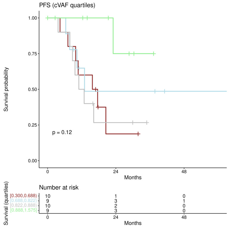
PFS under PARPi ranged from 2.6 to 82.1 months, with 19 (50.0%) patients reaching the study endpoint (progression). For first univariate and multiple Cox regression analyses patients were stratified according to the quartiles of cVAF. Patients in the three lower quartile groups (0–25%, 25–50%, and 50–75%) showed median PFS of 16.7 months, 13.0 months, and 11.9 months, respectively, while the median PFS was not reached in the group with the highest (75–100%) cVAF (Figure 2).
Figure 2.
PFS for cVAF quartiles: Kaplan–Meier estimates for PFS categorizing cVAF along quartiles (Borders for cVAF are shown bottom left with “[” indicating including, and “)” indicating excluding). The lowest quartile is shown in brown, the second in blue, the third in grey, and the highest quartile in green.
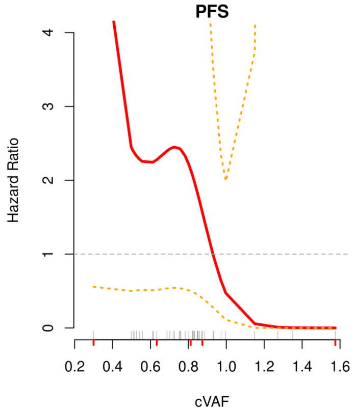
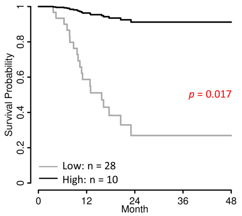
To determine the optimal cut-off of cVAF for PFS, the cutp function from R-package MASS was used, yielding 0.874 as the optimal cut-off with the lowest hazard ratio (HR) between the “high” (≥0.874; n = 10) and “low” (<0.874; n = 28) cVAF groups. To confirm this cut-off a non-linear modelling of the association of the cVAF value and the PFS was performed using smoothing splines, a penalized spline basis, shown in Figure 3. Here, the HR shows a steep decrease between 0.8 and 1.0, crossing the relative HR value of 1 by approx. 0.9 cVAF, confirming the optimal cut-off at 0.874.
Figure 3.
Non-linear modeling of the impact of cVAF on PFS using smoothing splines. Shown in red is the association of the hazard ratio with the cVAF with 95% confidence intervals (orange). Grey lines above the x-axis represent the single cVAF values, red lines below the x-axis the five-number summary (min, 25th percentile, median, 75th perc., max).
Further analyses were performed using this cut-off for the separation in a cVAF low and cVAF high group.
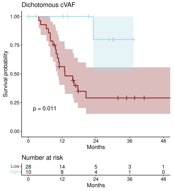
The median PFS in the cVAF low group was 13.0 months (IQR [10.3-not reached]), while it was not reached in the cVAF high group.
Univariate Cox regression analysis revealed a significantly association of the optimally dichotomized cVAF with PFS (HR 0.11; 95% CI [0.02–0.86]; p = 0.035) (Kaplan–Meier estimate, Figure 4). Using multiple Cox regression analysis starting with all relevant co-factors (age at start of PARPi treatment, FIGO stage at initial diagnosis, histology, BRCA mutation (BRCA 1 vs. 2), and the number of the PARPi treatment line) and selecting the minimal Cox-regression model by stepwise down-selection minimizing the AIC yielded a model comprising the two factors dichotomized cVAF (HR 0.07; 95% CI [0.01–0.63]; p = 0.017) and age. In Figure 5, survival curves of this model are shown and in Figure S3, a similar model (corrected for age) with the stratified cVAF quartiles is shown.
Figure 4.
Kaplan–Meier estimates for PFS categorizing cVAF optimally in a low and high cVAF group. Shading areas are 95% confidence intervals.
Figure 5.
Progression free survival curves for low and high cVAFs according to the multiple Cox regression model (Table 3), including age as a correcting factor. As this survival curve is generated from a Cox regression model, no censored patients are indicated.
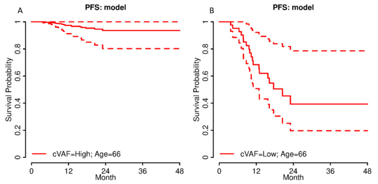
Using the multiple Cox regression model, individual survival curves for every putative patient can be estimated. In Figure 6, exemplary survival curves with 95% confidence intervals for patients with age 66 and either high or low cVAF are shown.
Figure 6.
Survival curves with 95% confidence intervals according to the multiple Cox regression model from Table 3 for single patients with either a (A) high or (B) low cVAF (Table 3) and an age of 66 years.
A subgroup analysis of only HGSOC patients (n = 35) also revealed a significant impact of cVAF on PFS, univariately (HR 0.12; 95% CI [0.02–0.88]; p = 0.037) as well as corrected for age (HR 0.08; 95% CI [0.01–0.69]; p = 0.022).
There was no significant difference in PFS in patients with germline vs. somatic BRCA mutations (HR 0.82; 95% CI [0.23–2.86]; p = 0.754).
PARPi dose reduction was necessary in 1/10 patients in the cVAF high group and 11/28 in the cVAF low group. The one patient, who discontinued PARPi treatment and developed lethal AML, in complete remission of ovarian cancer, was in the cVAF high group.
4. Discussion
In this study, we showed for the first time that response to PARPi treatment was improved in EOC patients harboring a higher cVAF. The optimal cut-off point for the highest discrimination between both groups was by the 74% percentile, yielding 26% of patients with high cVAF values and a significantly better PFS after PARPi treatment in our study population (HR 0.07).
To date, data on targeted treatment response dependent on VAF are still scarce. The impact of VAF on patient survival was for example studied in lung cancer [23,24], oral squamous carcinoma [25], cervical cancer [26], and melanoma [27]. Friedlaender et al. recently showed a PFS benefit (but no overall survival benefit) for epithelial growth factor receptor (EGFR) mutated non-small cell lung cancer (NSCLC) patients treated with EGFR tyrosine kinase inhibitors (TKIs) with EGFR mutation allele frequency higher than 30%. Although their patient population was rather small (n = 31) and VAF was assessed from tumor biopsies, but not corrected for tumor cellularity, this is a hint for the importance of allele frequency in targeted treatment response. Comparable to our results, they found EGFR VAF and age >65 to be associated with PFS in a bivariable model [28].
Zheng et al. investigated the effect of the allele frequency of the T790M TKI resistance mutation in 54 NSCLC patients treated with osimertinib (the targeted treatment against T790M mutation). They did not identify an association of maximum somatic allele frequency (MSAF) with objective response rate (ORR) or PFS, but found a significant association of ORR and PFS with relative mutation purity (RPM), defined as the ratio of T790M VAF to MSAF. In this study, VAF was defined as the percentage of mutant DNA allele reads relative to total DNA allele reads (mutant and wild type). EGFR mutations and allele frequency were assessed with NGS of blood samples. Results were validated with an independent NSCLC cohort (n = 34) [29].
In melanoma, BRAF VAF was identified as a potential prognostic and predictive biomarker for treatment response to BRAF inhibitors. DNA of primary and recurrent tumor samples from 327 melanoma patients was used for identification of BRAF mutations. In total, 156 BRAF mutated patients were treated with BRAF inhibitor +/− anti-MEK inhibitor and patients with high VAF showed prolonged PFS and overall survival in multivariate analyses. VAF was defined as the “percentage of the mutated peak in the BRAF sequence” [30].
Stagni et al. discovered a worse PFS for melanoma patients with low BRAF AF treated with MAPK-inhibitors [27].
These studies, while performed in lung cancer and melanoma, are in line with our findings in ovarian cancer of improved benefit for targeted treatment in patients with higher VAFs/cVAFs of targeted mutations.
Previous studies assessed ORR or PFS under PARPi for patients with germline vs. somatic BRCA mutations. A meta-analysis by Mohyuddin et al. reported no significant difference in PARPi treatment response for patients with germline vs. somatic BRCA mutations in ovarian, breast, prostate, and pancreatic cancer. [31] In comparison, our study focusses solely on the amount of BRCA mutated alleles in the tumor cells, creating groups with high and low cVAF, both containing patients with germline and purely somatic BRCA mutations.
Dose reductions were necessary in 12 of 38 (31.6%) patients in our study, which is comparable to previously published data [18,32]. A higher rate of dose reductions was observed in the low cVAF group, potentially influencing treatment response rates.
This study has several potential limitations. A rather small cohort of 38 patients in a single academic center was included, limiting the generalizability of the reported results. Of the 26 patients treated with second to fifth line PARPi, 8 samples for BRCA testing were obtained from recurrent tumors, while 18 BRCA results stem from primary tumor samples, limiting the comparability. Furthermore, five somatic BRCA testings were performed on tissue obtained after neoadjuvant chemotherapy. It has been shown, that patients with BRCA1/2 mutated tumors undergoing platinum based chemotherapy can acquire secondary BRCA1/2 mutations, restoring BRCA function and, therefore, contributing to platinum and PARPi resistance [33,34,35]. These secondary BRCA mutations have not been assessed in our study and could influence the treatment response.
The possibility of newly arising BRCA1/2 mutations at recurrent disease can be excluded in our study cohort, as all patients where recurrent tumor was tested had germline mutations.
PARPi therapy line ranged from first to fifth line therapy with a majority of patients receiving second line-PARPi. Nevertheless, therapy line was included in our cox regression model and, therefore, results were corrected for PARPi therapy line. A substantially larger patient cohort would allow for a more uniformed-approach (e.g., for further studies) to only include certain/a single therapy line(s).
Only one sample per patient was used for somatic BRCA mutation testing; therefore, the possibility of the influence of intratumor heterogeneity on VAF and PARPi response remains.
Despite some limitations, this study represents a rather big and uniform cohort of patients all tested and treated at one single institution. Considering that PARPi treatment is a relatively new therapy approved for EOC patients, the patient number is appropriate. Patients underwent regular follow-up visits, granting good data quality and comprehensive follow-up.
For further investigation of the influence of cVAF on treatment response to PARPi, larger (multi-center) studies with bigger patient cohorts and more uniformed criteria (such as only first or second line PARPi) would be needed. A comparison of low cVAF patients to non-BRCA1/2mut patients receiving PARPi treatment would be of interest.
5. Conclusions
We showed, for the first time, a tremendously improved progression free survival time for 26% of PARPi treated epithelial ovarian cancer patients (with one to four preceding chemotherapy lines) with high corrected variant allele frequencies (cVAF) of either a BRCA1 or a BRCA2 mutation. Age was the only confounding factor, significantly improving the progression free survival (PFS) Cox regression model.
Table 3.
Univariate and multiple Cox-Regression for progression-free survival.
| Parameters (n = 38, 19 Events) |
Univariate HR [95% CI] | p-Value | Multiple HR [95% CI] | p-Value |
|---|---|---|---|---|
| cVAF | 0.11 [0.02–0.86] | 0.035 | 0.07 [0.01–0.63] | 0.017 |
| Age 1 | 0.99 [0.96–1.03] | 0.733 | 0.96 [0.91–1.01] | 0.145 |
| Therapy-line | 1.21 [0.83–1.78] | 0.327 | - 4 | - |
| FIGO at initial diagnosis 2 | 1.02 [0.37–2.86] | 0.964 | - 4 | - |
| Histology 3 | 0.43 [0.05–3.49] | 0.428 | - 4 | - |
1 Age at start of PARPi treatment. 2 FIGO: stage IV (metastatic) vs. stage II/III at initial diagnosis. 3 Histology: serous vs. endometrioid. 4 removed from the model by step-wise down-selection, minimizing the Akaike Information Criterion (AIC).
Supplementary Materials
The following supporting information can be downloaded at: https://www.mdpi.com/article/10.3390/jpm12091467/s1, Figure S1. Boxplot of cVAF in primary and recurrent tumor tissue; Figure S2: Correlation coefficient matrix for cVAF, age and therapy line; Figure S3: Progression free survival curves for cVAFs quartiles according to the multiple Cox regression model.
Author Contributions
Conceptualization, C.T.G., C.B., S.A. and L.M.; data curation, C.T.G. and C.B.; formal analysis, C.T.G., D.P., T.R. and C.B.; methodology, L.M.; project administration, C.T.G., C.B. and S.A.; resources, C.G., S.P., L.M. and A.R.; supervision, S.A., C.G., S.P. and A.R.; visualization, C.T.G. and D.P.; writing—original draft, C.T.G., D.P. and C.B.; writing—review & editing, C.T.G., S.A., D.P., C.B., T.R., C.G. and L.M. All authors have read and agreed to the published version of the manuscript.
Institutional Review Board Statement
The study was conducted according to the guidelines of the Declaration of Helsinki and approved by the Institutional Review Board of the Medical University of Vienna (2067/2019).
Informed Consent Statement
Patient consent was waived due to the retrospective study design.
Data Availability Statement
The data presented in this study are available upon reasonable request from the corresponding author. The data are not publicly available due to privacy and ethical reasons.
Conflicts of Interest
The authors declare no conflict of interest. The funders had no role in the design of the study; in the collection, analyses, or interpretation of data; in the writing of the manuscript; or in the decision to publish the result.
Funding Statement
The current study was supported by an unrestricted research grant of Astra Zeneca, Vienna, Austria (Project number C22-AZA-AL-QF-027).
Footnotes
Publisher’s Note: MDPI stays neutral with regard to jurisdictional claims in published maps and institutional affiliations.
References
- 1.Siegel R.L., Miller K.D., Jemal A. Cancer Statistics. CA Cancer J. Clin. 2020;70:7–30. doi: 10.3322/caac.21590. [DOI] [PubMed] [Google Scholar]
- 2.Antoniou A., Pharoah P.D., Narod S., Risch H.A., Eyfjord J.E., Hopper J.L., Loman N., Olsson H., Johannsson O., Borg Å., et al. Average risks of breast and ovarian cancer associated with BRCA1 or BRCA2 mutations detected in case Series unselected for family history: A combined analysis of 22 studies. Am. J. Hum. Genet. 2003;72:1117–1130. doi: 10.1086/375033. [DOI] [PMC free article] [PubMed] [Google Scholar]
- 3.Sowter H.M., Ashworth A. BRCA1 and BRCA2 as ovarian cancer susceptibility genes. Carcinogenesis. 2005;26:1651–1656. doi: 10.1093/carcin/bgi136. [DOI] [PubMed] [Google Scholar]
- 4.Norquist B.M., Harrell M.I., Brady M.F., Walsh T., Lee M.K., Gulsuner S., Bernards S.S., Casadei S., Yi Q., Burger R.A., et al. Inherited Mutations in Women with Ovarian Carcinoma. JAMA Oncol. 2016;2:482–490. doi: 10.1001/jamaoncol.2015.5495. [DOI] [PMC free article] [PubMed] [Google Scholar]
- 5.Zhang S., Royer R., Li S., McLaughlin J.R., Rosen B., Risch H.A., Fan I., Bradley L., Shaw P.A., Narod S.A. Frequencies of BRCA1 and BRCA2 mutations among 1342 unselected patients with invasive ovarian cancer. Gynecol. Oncol. 2011;121:353–357. doi: 10.1016/j.ygyno.2011.01.020. [DOI] [PubMed] [Google Scholar]
- 6.Turashvili G., Lazaro C., Ying S., Charames G., Wong A., Hamilton K., Yee D., Agro E., Chang M., Pollett A., et al. Tumor BRCA Testing in High Grade Serous Carcinoma: Mutation Rates and Optimal Tissue Requirements. Cancers. 2020;12:3468. doi: 10.3390/cancers12113468. [DOI] [PMC free article] [PubMed] [Google Scholar]
- 7.Weren R.D., Mensenkamp A.R., Simons M., Eijkelenboom A., Sie A.S., Ouchene H., van Asseldonk M., Gomez-Garcia E.B., Blok M.J., de Hullu J.A., et al. Novel BRCA1 and BRCA2 Tumor Test as Basis for Treatment Decisions and Referral for Genetic Counselling of Patients with Ovarian Carcinomas. Hum. Mutat. 2016;38:226–235. doi: 10.1002/humu.23137. [DOI] [PMC free article] [PubMed] [Google Scholar]
- 8.Rezaei N., Hedayat M. Allele Frequency. In: Maloy S., Hughes K., editors. Brenner’s Encyclopedia of Genetics. 2nd ed. Academic Press; Cambridge, MA, USA: 2013. pp. 77–78. [Google Scholar]
- 9.Moore K., Colombo N., Scambia G., Kim B.-G., Oaknin A., Friedlander M., Lisyanskaya A., Floquet A., Leary A., Sonke G.S., et al. Maintenance Olaparib in Patients with Newly Diagnosed Advanced Ovarian Cancer. N. Engl. J. Med. 2018;379:2495–2505. doi: 10.1056/NEJMoa1810858. [DOI] [PubMed] [Google Scholar]
- 10.González-Martín A., Pothuri B., Vergote I., DePont Christensen R., Graybill W., Mirza M.R., McCormick C., Lorusso D., Hoskins P., Freyer G., et al. Niraparib in Patients with Newly Diagnosed Advanced Ovarian Cancer. N. Engl. J. Med. 2019;381:2391–2402. doi: 10.1056/NEJMoa1910962. [DOI] [PubMed] [Google Scholar]
- 11.Fong P.C., Yap T.A., Boss D.S., Carden C.P., Mergui-Roelvink M., Gourley C., De Greve J., Lubinski J., Shanley S., Messiou C., et al. Poly(ADP)-ribose polymerase inhibition: Frequent durable responses in BRCA carrier ovarian cancer correlating with platinum-free interval. J. Clin. Oncol. 2010;28:2512–2519. doi: 10.1200/JCO.2009.26.9589. [DOI] [PubMed] [Google Scholar]
- 12.Penson R.T., Valencia R.V., Cibula D., Colombo N., Leath C.A., 3rd, Bidzinski M., Kim J.W., Nam J.H., Madry R., Hernandez C., et al. Olaparib Versus Nonplatinum Chemotherapy in Patients with Platinum-Sensitive Relapsed Ovarian Cancer and a Germline BRCA1/2 Mutation (SOLO3): A Randomized Phase III Trial. J. Clin. Oncol. 2020;38:1164–1174. doi: 10.1200/JCO.19.02745. [DOI] [PMC free article] [PubMed] [Google Scholar]
- 13.Oza A.M., Tinker A.V., Oaknin A., Shapira-Frommer R., McNeish I.A., Swisher E.M., Ray-Coquard I., Bell-McGuinn K., Coleman R.L., O’Malley D.M., et al. Antitumor activity and safety of the PARP inhibitor rucaparib in patients with high-grade ovarian carcinoma and a germline or somatic BRCA1 or BRCA2 mutation: Integrated analysis of data from Study 10 and ARIEL2. Gynecol. Oncol. 2017;147:267–275. doi: 10.1016/j.ygyno.2017.08.022. [DOI] [PubMed] [Google Scholar]
- 14.Li H., Liu Z.-Y., Wu N., Chen Y.-C., Cheng Q., Wang J. PARP inhibitor resistance: The underlying mechanisms and clinical implications. Mol. Cancer. 2020;19:107. doi: 10.1186/s12943-020-01227-0. [DOI] [PMC free article] [PubMed] [Google Scholar]
- 15.Janysek D.C., Kim J., Duijf P.H., Dray E. Clinical use and mechanisms of resistance for PARP inhibitors in homologous recombination-deficient cancers. Transl. Oncol. 2021;14:101012. doi: 10.1016/j.tranon.2021.101012. [DOI] [PMC free article] [PubMed] [Google Scholar]
- 16.Wolford J.E., Bai J., Moore K.N., Kristeleit R., Monk B.J., Tewari K.S. Cost-effectiveness of niraparib, rucaparib, and olaparib for treatment of platinum-resistant, recurrent ovarian carcinoma. Gynecol. Oncol. 2020;157:500–507. doi: 10.1016/j.ygyno.2020.02.030. [DOI] [PMC free article] [PubMed] [Google Scholar]
- 17.Muston D., Hettle R., Monberg M., McLaurin K.K., Gao W., Swallow E., Zhang S., Kalemaj I., Signorovitch J., Moore K. Cost-effectiveness of olaparib as a maintenance treatment for women with newly diagnosed advanced ovarian cancer and BRCA1/2 mutations in the United States. Gynecol. Oncol. 2020;159:491–497. doi: 10.1016/j.ygyno.2020.08.013. [DOI] [PubMed] [Google Scholar]
- 18.Bitler B.G., Watson Z.L., Wheeler L.J., Behbakht K. PARP inhibitors: Clinical utility and possibilities of overcoming resistance. Gynecol. Oncol. 2017;147:695–704. doi: 10.1016/j.ygyno.2017.10.003. [DOI] [PMC free article] [PubMed] [Google Scholar]
- 19.LaFargue C.J., Molin G.Z.D., Sood A.K., Coleman R.L. Exploring and comparing adverse events between PARP inhibitors. Lancet Oncol. 2019;20:e15–e28. doi: 10.1016/S1470-2045(18)30786-1. [DOI] [PMC free article] [PubMed] [Google Scholar]
- 20.Morice P.-M., Leary A., Dolladille C., Chrétien B., Poulain L., González-Martín A., Moore K., O’Reilly E.M., Ray-Coquard I., Alexandre J. Myelodysplastic syndrome and acute myeloid leukaemia in patients treated with PARP inhibitors: A safety meta-analysis of randomised controlled trials and a retrospective study of the WHO pharmacovigilance database. Lancet Haematol. 2021;8:e122–e134. doi: 10.1016/S2352-3026(20)30360-4. [DOI] [PubMed] [Google Scholar]
- 21.Bekos C., Grimm C., Kranawetter M., Polterauer S., Oberndorfer F., Tan Y., Müllauer L., Singer C. Reliability of Tumor Testing Compared to Germline Testing for Detecting BRCA1 and BRCA2 Mutations in Patients with Epithelial Ovarian Cancer. J. Pers. Med. 2021;11:593. doi: 10.3390/jpm11070593. [DOI] [PMC free article] [PubMed] [Google Scholar]
- 22.Kanchi K.L., Johnson K.J., Lu C., McLellan M.D., Leiserson M.D.M., Wendl M.C., Zhang Q., Koboldt D.C., Xie M., Kandoth C., et al. Integrated analysis of germline and somatic variants in ovarian cancer. Nat. Commun. 2014;5:3156. doi: 10.1038/ncomms4156. [DOI] [PMC free article] [PubMed] [Google Scholar]
- 23.Li X.-M., Yang J.-J., Wu Y.-L. Association of allele frequency of EGFR mutation with efficacy of EGFR TKIs in advanced non-small cell lung cancer. J. Clin. Oncol. 2019;37:e20678. doi: 10.1200/JCO.2019.37.15_suppl.e20678. [DOI] [Google Scholar]
- 24.Al-Dherasi A., Liao Y., Al-Mosaib S., Hua R., Wang Y., Yu Y., Zhang Y., Zhang X., Jalayta R., Mousa H., et al. Allele frequency deviation (AFD) as a new prognostic model to predict overall survival in lung adenocarcinoma (LUAD) Cancer Cell Int. 2021;21:451. doi: 10.1186/s12935-021-02127-z. [DOI] [PMC free article] [PubMed] [Google Scholar]
- 25.Ma J., Fu Y., Tu Y.-Y., Liu Y., Tan Y.-R., Ju W.-T., Pickering C.R., Myers J.N., Zhang Z.-Y., Zhong L.-P. Mutation allele frequency threshold does not affect prognostic analysis using next-generation sequencing in oral squamous cell carcinoma. BMC Cancer. 2018;18:758. doi: 10.1186/s12885-018-4481-8. [DOI] [PMC free article] [PubMed] [Google Scholar]
- 26.Tian J., Geng Y., Lv D., Li P., Cordova-Delgado M., Liao Y., Tian X., Zhang X., Zhang Q., Zou K., et al. Using plasma cell-free DNA to monitor the chemoradiotherapy course of cervical cancer. Int. J. Cancer. 2019;145:2547–2557. doi: 10.1002/ijc.32295. [DOI] [PubMed] [Google Scholar]
- 27.Stagni C., Zamuner C., Elefanti L., Zanin T., Del Bianco P., Sommariva A., Fabozzi A., Pigozzo J., Mocellin S., Montesco M.C., et al. BRAF Gene Copy Number and Mutant Allele Frequency Correlate with Time to Progression in Metastatic Melanoma Patients Treated with MAPK Inhibitors. Mol. Cancer Ther. 2018;17:1332–1340. doi: 10.1158/1535-7163.MCT-17-1124. [DOI] [PubMed] [Google Scholar]
- 28.Friedlaender A., Tsantoulis P., Chevallier M., De Vito C., Addeo A. The Impact of Variant Allele Frequency in EGFR Mutated NSCLC Patients on Targeted Therapy. Front. Oncol. 2021;11:644472. doi: 10.3389/fonc.2021.644472. [DOI] [PMC free article] [PubMed] [Google Scholar]
- 29.Zheng Q., Hong S., Huang Y., Zhao H., Yang Y., Hou X., Zhao Y., Ma Y., Zhou T., Zhang Y., et al. EGFR T790M relative mutation purity predicts osimertinib treatment efficacy in non-small cell lung cancer patients. Clin. Transl. Med. 2020;9:17. doi: 10.1186/s40169-020-0269-y. [DOI] [PMC free article] [PubMed] [Google Scholar]
- 30.Berrino E., Balsamo A., Pisacane A., Gallo S., Becco P., Miglio U., Caravelli D., Poletto S., Paruzzo L., Debernardi C., et al. High BRAF variant allele frequencies are associated with distinct pathological features and responsiveness to target therapy in melanoma patients. ESMO Open. 2021;6:100133. doi: 10.1016/j.esmoop.2021.100133. [DOI] [PMC free article] [PubMed] [Google Scholar]
- 31.Mohyuddin G.R., Aziz M., Britt A., Wade L., Sun W., Baranda J., Al-Rajabi R., Saeed A., Kasi A. Similar response rates and survival with PARP inhibitors for patients with solid tumors harboring somatic versus Germline BRCA mutations: A Meta-analysis and systematic review. BMC Cancer. 2020;20:507. doi: 10.1186/s12885-020-06948-5. [DOI] [PMC free article] [PubMed] [Google Scholar]
- 32.Francis K., Kim S., Friedlander M., Gebski V., Ray-Coquard I., Clamp A., Penson R., Oza A., Perri T., Huzarski T., et al. The impact of olaparib dose reduction and treatment interruption on treatment outcome in the SOLO2/ENGOT-ov21 platinum-sensitive recurrent ovarian cancer. Ann. Oncol. 2022;33:593–601. doi: 10.1016/j.annonc.2022.02.222. [DOI] [PubMed] [Google Scholar]
- 33.Norquist B., Wurz K.A., Pennil C.C., Garcia R., Gross J., Sakai W., Karlan B.Y., Taniguchi T., Swisher E.M. Secondary somatic mutations restoring BRCA1/2 predict chemotherapy resistance in hereditary ovarian carcinomas. J. Clin. Oncol. 2011;29:3008–3015. doi: 10.1200/JCO.2010.34.2980. [DOI] [PMC free article] [PubMed] [Google Scholar]
- 34.Swisher E.M., Sakai W., Karlan B.Y., Wurz K., Urban N., Taniguchi T. Secondary BRCA1 mutations in BRCA1-mutated ovarian carcinomas with platinum resistance. Cancer Res. 2008;68:2581–2586. doi: 10.1158/0008-5472.CAN-08-0088. [DOI] [PMC free article] [PubMed] [Google Scholar]
- 35.Sakai W., Swisher E.M., Karlan B.Y., Agarwal M.K., Higgins J., Friedman C., Villegas E., Jacquemont C., Farrugia D.J., Couch F.J., et al. Secondary mutations as a mechanism of cisplatin resistance in BRCA2-mutated cancers. Nature. 2008;451:1116–1120. doi: 10.1038/nature06633. [DOI] [PMC free article] [PubMed] [Google Scholar]
Associated Data
This section collects any data citations, data availability statements, or supplementary materials included in this article.
Supplementary Materials
Data Availability Statement
The data presented in this study are available upon reasonable request from the corresponding author. The data are not publicly available due to privacy and ethical reasons.


