Abstract
A 29-year-old preeclamptic postpartum patient with no symptoms of hypertension in her medical history before pregnancy was referred to the ophthalmology outpatient clinic with the complaint of sudden bilateral vision loss. Slit-lamp fundus examination and B-scan ultrasonography showed serous retinal detachment (SRD) in both eyes. She was diagnosed with bullous SRD due to preeclampsia (PE). The patient’s fundoscopy findings regressed spontaneously, and visual acuities improved within one month. SRD should be considered in case of vision loss before or after birth in patients with PE, and such patients should undergo retinal examination.
Keywords: preeclampsia, serous retinal detachment, sudden vision loss, pregnancy
Introduction
Hypertension is a complication of 5–10% of all gravidities. Pregnancy-induced hypertension (PIH) is a challenging disorder in obstetrics that is one of the significant reasons for maternal and perinatal mortality. PIH may exist after the 20th week of gravidity when there are no other causes of high blood pressure (BP) (eg, measured higher 140/90 mmHg twice at 6-hour intervals).1 Gestational hypertension, preeclampsia, severe preeclampsia, and eclampsia are the subgroups of PIH. BP that reaches 140/90 mmHg or higher for the first time after mid-pregnancy is considered as gestational hypertension. Gestational hypertension together with remarkable proteinuria (higher than 300 mg per 24 hours) is described as preeclampsia (PE).1
Preeclamptic patients may have retinal and choroidal circulatory abnormalities. Therefore, different fundoscopic signs and following visual impairment may exist. Retinal hemorrhage, subretinal serous fluid accumulation, papilledema, and Elschnig spots may develop in these patients, which are signs of severe hypertensive retinopathy.2 In less than 1% of PE patients, serous retinal detachment (SRD) is an extraordinary reason for vision loss.3,4
In this case report from Somalia, we present spontaneous resolution of bilateral bullous SRD in a postpartum patient with PE. Written informed consent for publication of their details was obtained from the patient and institutional approval was not required to publish the case details.
Case Report
A 29-year-old preeclamptic postpartum patient was referred to the ophthalmology outpatient clinic with the complaint of sudden vision loss in both eyes. She applied to the obstetrics and gynecology clinic with headache and cloudy vision the day before. In her first examination, systemic BP was 165/105 mmHg, and there was bilateral (++) pretibial edema. It was unclear when her BP began to rise, as the patient had no regular pregnancy follow-up. She was not at high-risk for either PE or SRD and did not have clinical high-risk predictors such as pre-existing chronic hypertension or chronic kidney disease. In her obstetric ultrasonography findings, there was anhydramnios, and the fetus was compatible with 39 weeks of gestation. While thrombocyte count was 232x103/mm3; hemoglobin was 10.2 g/dL, ALT was 142 IU/L, and AST was 142 IU/L. There was (+++) proteinuria with the dipstick. Renal function and coagulation tests were normal. She was diagnosed as PE, hospitalized and had a vaginal delivery. Antihypertensive (nifedipine) and anticonvulsant therapy for eclampsia prophylaxis (MgSO4) was started following hospitalization.
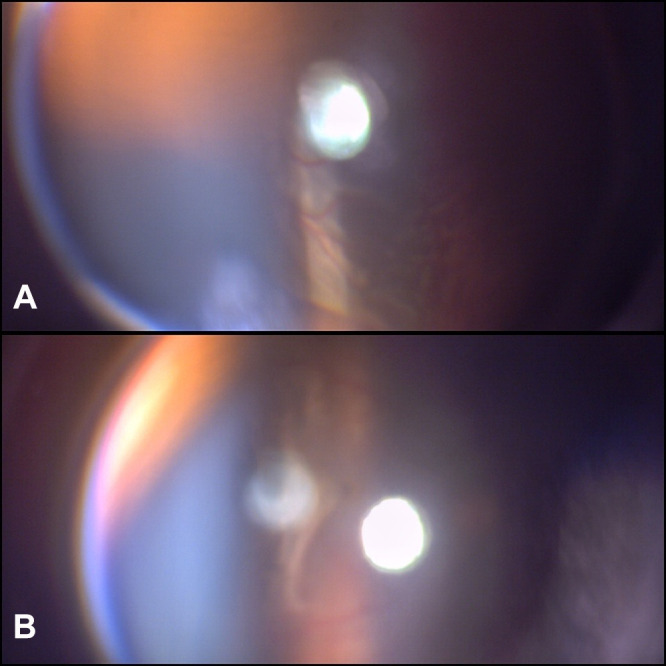
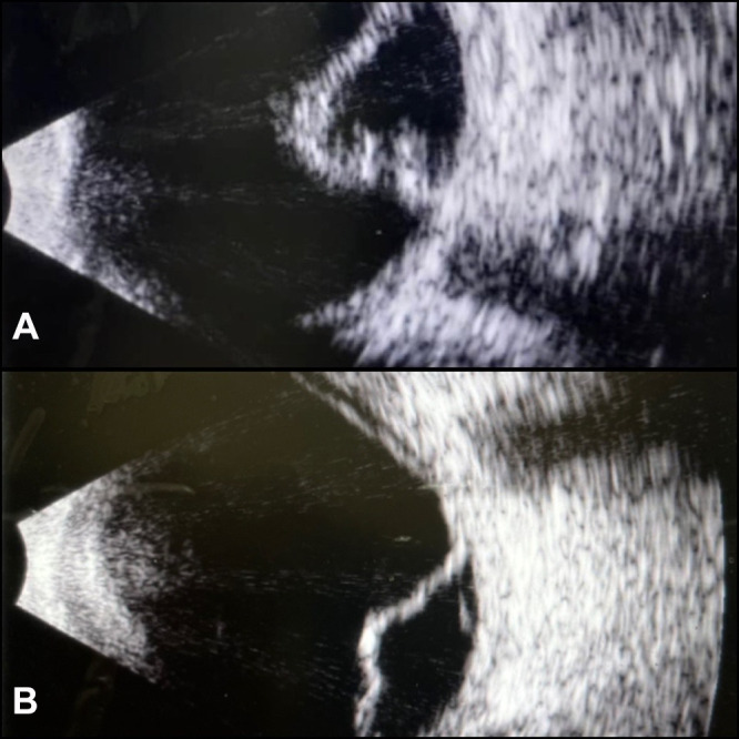
On the first postnatal day, the visual acuity of the patient was finger counting at one meter for the right eye, while it was two meters for the left eye. She did not have any abnormalities in her anterior segment examination for both eyes and had normal intraocular pressure. In the slit-lamp fundus examination, there was bullous SRD involving the retina’s superior aspect and extending to the optic disk in the right eye. In the examination of the left eye, there was a similar SRD including the temporal aspect of the retina and it was enlarging into the macula (Figure 1A and B). Unfortunately, we did not have the opportunity to do a retinography or optical coherence tomography (OCT) screening in our department, thus we confirmed the SRD in both eyes via B-scan ophthalmic ultrasonography in accordance with fundus examination (Figure 2A and B). Both before and after the resolution of the detachment, the patient was asked to undergo an OCT examination at another hospital. We informed the patient that this disorder had a self-resolving manner and had a good prognosis.
Figure 1.

Bullous serous retinal detachment with slit lamp funduscopy in the right eye (A) and in the left eye (B).
Figure 2.

B-scan ultrasonography confirms bullous serous retinal detachment in both eyes (A): right, (B): left.
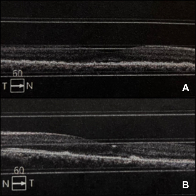
We were able to get the patients’s OCT imaging in the second week of follow-up, her visual acuity improved to 8/10 for the right eye, while it was 4/10 for the left eye. Except for a minimal subretinal fluid in both eyes and a localized retinal pigment epithelium disruption in the left eye, OCT was considered normal (Figure 3A and B). While the patient’s right eye vision had fully recovered, her left eye vision improved to 8/10, after 1-month follow-up. She did not provide us with a second OCT imaging after her vision completely recovered.
Figure 3.

Optical coherence tomography (OCT) indicates minimal subretinal fluid in the right eye (A), minimal subretinal fluid and localized retinal pigment epithelium disruption in the left eye (B).
Discussion
Diffuse arteriolar spasm, which is a result of segmental or generalized narrowing of retinal arterioles on the retina, is the most visible ocular sign of PE. Severe hypertensive patients rarely have SRD as a complication.5 Researchers such as Jayashree et al6 reported a significant positive correlation between blood pressure levels and disease severity. Whether unilateral or bilateral, SRD may occur before or after birth.7
The exact SRD mechanism in PE cases remains unknown. There are different theories explaining the development of SRD. Serious generalized vasospasm, which is considered to be secondary to increased sensitivity to circulating prostaglandins, is assumed as the underlying pathophysiology.8 Choroidal dysfunction is considered to exist after the arteriolar vasospasm. Retinal pigment epithelium damage and breakdown of the blood-retina barrier are caused by choroidal dysfunction, especially by choriocapillaris ischemia. The compromised fluid and ion transfer cause subretinal liquid accumulation and following serous detachment.9,10 Studies with fluorescence angiography and green indocyanine angiography reported that changes in the choroid vasculature, including occlusion of the choroid and choriocapillaris arterioles, caused most of the retinal harm.11 There is a recent publication suggesting that the initial injury in many women with de novo hypertensive disorders of pregnancy was passive edema of the retina due to hyperperfusion in pregnancy. If this continues, the authors propose edema is likely to cause BP elevation in PE and resolves with delivery of the placenta.12 In light of this, our case of SRD is likely to be an exaggerated form of retinal edema of PE.
OCT is a safe, non-invasive, and commonly used screening method in this period. However, we did not have the chance to perform OCT examination at the time of diagnosis and therefore confirmed the bullous SRD through B-scan ultrasonography. After two weeks of follow-up, we had the chance to get OCT images.
The management of the serous retinal detachment is the treatment of underlying reason. Further observations and systematic antihypertensive drugs therapy may be useful for such cases. The breakdown of the blood‑retinal barrier and failure of the retinal pigment epithelium pump function recover spontaneously at the postpartum period.2 In most of the patients, SRD shows complete resolution within 2–12 weeks after delivery.14 In our patient, the BP spontaneously decreased after birth, and the patient was discharged from the obstetrics and gynecology clinic with a prescription including methyldopa (Alfamet, Ibrahim Ethem, Turkey). In line with the literature, we observed that SRD gradually resolved, and visual acuity recovered completely.13,14
Conclusion
SRD should be considered in case of vision loss before or after birth in patients with PE, and such patients should undergo retinal examination. Even giant bullous SRDs regress spontaneously within a few weeks after BP is regulated following delivery.
Abbreviations
PIH, pregnancy-induced hypertension; BP, blood pressure; PE, preeclampsia; SRD, serous retinal detachment; OCT, optical coherence tomography.
Disclosure
The authors report no conflicts of interest in this work.
References
- 1.Gary FC, Leveno KJ, Bloom SL. Williams Obstetrics. 24th ed. United States of America: MC Graw-Hill Education; 2014. Vol. 1: 728–730. [Google Scholar]
- 2.Celik O, Hascalık Ş, Gokdeniz R. Bilateral serous retinal detachment in preeclampsia: report of two cases. Gynecol Obstet Reprod Med. 2002;8:61–62. [Google Scholar]
- 3.Roos NM, Wiegman MJ, Jansonius NM, Zeeman GG. Visual disturbances in (pre) eclampsia. Obstet Gynecol Surv. 2012;67(4):242–250. doi: 10.1097/OGX.0b013e318250a457 [DOI] [PubMed] [Google Scholar]
- 4.Hussain SA, O’Shea BJ, Thagard AS. Preeclamptic serous retinal detachment without hypertension: a case report. Case Rep Womens Health. 2019;21:e00098. doi: 10.1016/j.crwh.2019.e00098 [DOI] [PMC free article] [PubMed] [Google Scholar]
- 5.Hutchings K, Sangalli M, Halliwell T, Tuohy J. Bilateral retinal detachment in pregnancy. Aust NZ J Obstet Gynaecol. 2002;42:409‑11. doi: 10.1111/j.0004-8666.2002.409_3.x [DOI] [PubMed] [Google Scholar]
- 6.Jayashree MP, Niveditha RK, Kuntoji NG, et al. Ocular fundus changes in pregnancy induced hypertension – a case series study. J Clin Res Ophthalmol. 2018;5(2):37–41. [Google Scholar]
- 7.Gundlach E, Junker B, Gross N, Hansen LL, Pielen A. Bilateral serous retinal detachment. Br J Ophthalmol. 2013;97:939–940. doi: 10.1136/bjophthalmol-2012-302528 [DOI] [PubMed] [Google Scholar]
- 8.Ober RR. Pregnancy-induced hypertension (preeclampsia-eclampsia). In: Ryan SJ, editor. Retina. 2nd ed. St Louis, MO: Mosby; 1994:1405–1411. [Google Scholar]
- 9.Spaide RF, Goldbaum M, Wong DWK, Tang KC, Lida T. Serous detachment of the retina. Retina. 2003;23:820–846. doi: 10.1097/00006982-200312000-00013 [DOI] [PubMed] [Google Scholar]
- 10.Saito Y, Tano Y. Retinal pigment epithelial lesions associated with choroidal ischemia in preeclampsia. Retina. 1998;18:103–108. doi: 10.1097/00006982-199818020-00002 [DOI] [PubMed] [Google Scholar]
- 11.Srećković SB, Janićijević-Petrović MA, Stefanović IB, Petrović NT, Šarenac TS, Paunović SS. Bilateral retinal detachment in a case of preeclampsia. Bosn J Basic Med Sci. 2011;11(2):129–131. doi: 10.17305/bjbms.2011.2598 [DOI] [PMC free article] [PubMed] [Google Scholar]
- 12.Herman RJ, Ambasta A, Williams RG, et al. Sequential measurement of the neurosensory retina in hypertensive disorders of pregnancy: a model of microvascular injury in hypertensive emergency. J Hum Hypertens. 2021. doi: 10.1038/s41371-021-00617-1 [DOI] [PMC free article] [PubMed] [Google Scholar]
- 13.Çelik G, Eser A, Günay M, Yenerel NM. Bilateral vision loss after delivery in two cases: severe preeclampsia and HELLP syndrome. Turk J Ophthalmol. 2015;45:271–273. doi: 10.4274/tjo.45722 [DOI] [PMC free article] [PubMed] [Google Scholar]
- 14.Limon U. Spontaneous resolution of serous retinal detachment in preeclampsia. Saudi J Ophthalmol. 2020;34:313–315. doi: 10.4103/1319-4534.322603 [DOI] [PMC free article] [PubMed] [Google Scholar]


