Abstract
Titanium dioxide nanoparticles (TiO2 NPs) were prepared by Caricaceae (Papaya) Shell extracts. The Nanoparticles were analyzed by UV–Vis spectrums, X-ray diffractions, and energy-dispersive X-rays spectroscopy analyses with a scanning electron microscope. An antifungal study was carried out for TiO2 NP in contradiction of S. sclerotiorums, R. necatrixs and Fusarium classes that verified a sophisticated inhibitions ratio for S. sclerotiorums (60.5%). Germs of pea were individually preserved with numerous concentrations of TiO2 NPs. An experience of TiO2 NPs (20%, 40%, 80% and 100%), as well as mechanisms that instigated momentous alterations in seed germinations, roots interval, shoot lengths, and antioxidant enzymes, were investigated. Associated with controls, the supreme seeds germinations, roots and plant growth were perceived with the treatments of TiO2 NPs. Super-oxide dis-mutase and catalase activities increased because of TiO2 NPs treatments. This advocates that TiO2 Nanoparticles may considerably change antioxidant metabolisms in seed germinations.
Subject terms: Biophysics, Plant sciences, Engineering, Materials science, Physics
Introduction
Nanotechnologies are utilized in the areas of medicines, chemistry, environments, energies, agronomy, communications as well as consumer possessions1. Metallic oxides with Nanostructures have getting significant interest in numerous fields of technologies2. Attentiveness in titanium dioxide (TiO2), a metallic oxide, has been growing in current times. Titanium dioxide (Titania) is the furthermost hopeful mineral oxide that is broadly being utilized for fabrications of instruments and other applications3,4. TiO2 is encouraging for application in the light-emitting device (LCD and LED) that operates in the short wavelength array, from blue light to ultra-violet, as well as in photovoltaic solar cells detectors thin films5. Additionally, it is broadly utilized for colourant explained fabrications of transistors as well as field-effect transistors, hybrids and QDSCs (quantum dots solar cells), and Nano generators6–8. TiO2 Nano Structure of numerous surface morphologies, comprising Nanorods; Nano ropes; Nano threads; Nano ranks; Nano girdles; Nano pointers; Nano prisms; Nano pipes; Nano buds; quantum dot; Nanoparticles; Nanofilms, Nanosheets and Nano plates; Nano microspheres; Nano pyramids; and Nano tetra-pods have applied in varies investigations9–23.
Many investigators have conveyed the influences of Titania Nanoparticles on plant germinations as well as development. Titanium as a valuable element rises and helps growth24, increases plants productivity by 10–20%25 and bio-mass as well as the growth of different plants class26 and productions of free radical in propagated seed27. The unpredictable outcomes attained from the application of titanium dioxide nanoparticles can designate the positive as well as negative influences of this matter28 Other reports stated that titanium dioxide nanoparticles reserved chlorophyll as well as carotenoid at optimum temperature29.
Therefore, fabrications of Titanium dioxide Nanostructures are greatly interested all over the biosphere. Titanium dioxide Nanoparticles have received significant consideration because of their exceptional antibacterial, antifungal, UV-filtering characteristics, extraordinary catalytic as well as photochemical activities30,31. Fabrications of titanium dioxide nanoparticles are often luxurious, and methods used in the procedure need high energies32. Additionally, poisonous diluters and poisonous chemicals are used in these approaches. The substitute technique to prepare these Nanoparticles is biological synthesis. The green approach of nanoparticles through plant’s extract is presently drawing pronounced deals of attention due to their eco-friendly and financial dispensation, scalable, pure surfaces in chemical and greatest considerably their utilization in biology as well as medicines. Several intra-cellular and extra-cellular biological extracts (bacterial, yeasts, fungal, algae and plant) were investigated for the biological synthesis of Nanoparticles and stated their properties such as size shapes compositions chemically towards stability in a medium33–40. Biological technologies are used in Bio-synthesis, like usages of plant extracts; it could be a favourite to other techniques. Amongst the biologically objects stated above, plant or their extract appear to be the paramount proxies due to their simply available, appropriate for masses productions of Nanoparticles and wastes product is environmentally friendly dissimilar some micro-organismal extract41,42. Phyto constituents in plant extract can concurrently purpose as dropping agents because of the kindly and multipurpose function43,44. TiO2 Nanoparticles enter the eco-system with incorrect disposals of manufacturing wastes and prevent seeds germinations, seedling development and plant growth.
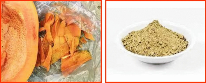
Several techniques can be useful to avoid these victims. Though, these techniques also have various restrictions on the environment as well as humankind fitness. The utility of Nanoparticles in pathogens controls is recognized as an eco-friendly as well as cost-effective substitute45. Nanoparticles are extremely significant in the treatments of plants46–49. Carica papaya fits into the family of Caricaceae and is usually utilized in treatment as well as control worldwide, particularly in a humid and sub-tropical part of the biosphere. Diverse portions of Carica papaya, like leaves, bark, root, latexes, fruits, flowers, as well as seeds, were used in societies medicals to pleasure diversities of infections50–53. Comprising different significant ingredients like vitamins, vitamins (A, E and C) that are a gorgeous basis of antioxidants as well as a mineral-like magnesium (Mg) and potassium (K), vitamin B pantothenic acid as well as foliate and fibres54. In the present research, bio-synthesis and analyses of TiO2 Nanoparticles through Shell extracts of Carica papaya L. and its antifungal activities and seeds germinations were investigated. Since ten years ago, research in the biological synthesis of metallic Nanoparticles through plant extract has released novel views in the area of Nanomedicines55. Carica papayas are widespread through the biosphere as well as yield fruits obtainable in every period (Fig. 1). In the present research, Titanium dioxide Nanoparticle was prepared through leafs extracts of Carica papayas in cleans as well as bio-Synthesization technique.
Figure 1.
Carica shell preparation.
Materials and methods
Plants preparations
Titanium Iso-propoxide was bought from Merck Chemicals Ltd, Ethiopia. Carica papaya Shell was peeled and washed. They were scratched into small bits as well as dehydrated at 50 °C. Twenty grams of dry Carica papaya Shells were heated in sanitized water for 30 min. The extracts gained were cleaned via What-man paper Number one and kept in a fridge for more utilization. The preparation of Carica papaya Shell extracts is as Fig. 1. The plant we have used in this report was cultivated in the local area of Dambi Dollo Town, Oromia, Ethiopia. This study complies with relevant international, national, institutional and legislative guidelines.
Biosynthesis of TiO2 nanoparticles
A 65 mL 0.2 M titanium Iso-propoxide (99.98%) was equipped in triple distilled water. 15 mL of Carica papaya Shell extracts were gradually mixed dropwise to the solutions at 85 °C with a magnetic stirrer for 5 h, attuned to pH value 11. The occasioning mixtures were centrifuged at 15,000 rpm for 15 min. Pills were splashed as well as centrifuged at 4000 rpm for 15 min. The cleaned pills attained after centrifugations were dehydrated at 55 °C for 5 h and calcite in a soft oven at 455 °C to prepare TiO2 Nanoparticles56.
Physical characterization of TiO2 nanoparticles
The bio-synthesized Titanium dioxide Nanoparticles were analyzed through the next procedures. Extreme absorbances of the sample were examined through the usage of UV–Visible Spectrophotometry. The physical characterizations of the optical characteristics of titanium oxide nano particle were carried out through ultraviolets and visible absorptions spectroscopy (spectro-photo-meter, Cary-E500 in the ranges of 250 nm–800 nm. X-Ray Diffractions (XRDs) analyses of powders of TiO2 nanoparticles were conducted PANalytical X-ray diffractometer functioned at 40 k-V with a current of 30 m-A under Cu-Ka radiations of 2 range between 10–80. Dynamics light scatterings (DLSs) were accomplished with Dyna-Pro Plate Readers (Wyatt-Technology). The prepared output was analyzed through transmission electron microscopy [(TEM) Tecnai G2-200 kV with microanalysis]. Scanning-Electron-Microscope (SEM) micrograph was verified through JEOL-JSM-6390 systems as well as elemental plotting was using a similar instrument.
Preparations of fungal
Platters Potatoes Dextrose Agars (PDAs) Petri plates were subculture for Sclerotinias sclerotiorums, Rossellini’s necatrixs and Fusariums spp. Separately, the fungal mycelium tads were provided by the Molecular Plants Microbes Interaction Laboratories. A fungal mycelia tad, scratch through a steriles dagger blades were inoculated at centres of each coagulated steriled PDAs Petri plates that were protected at 20 °C until the fungi matured over an entire surface. The platters were then kept in the fridge at 4 °C for supplementary experimentation uses after being wrapped by Para films.
Preparations of fungal deferments
Liquefied Culture of three (3) fungal strains-Sclerotinia sclerotium, Rosellinias necatrixs and Fusarium spp. were equipped through potatoes dextroses broths (PDBs) Mediums, for that fungal mycelium tads were scratch from master platters by using sterilized penknife blades. Each PDBs test tube was protected with 15 mL sterilized PDBs and fungus mycelium bits. This was protected at 20 °C in incubator shakers at 170 rpm for 4–6 days until an adequate development of the fungus mycelium. The test tube was then kept in the fridge at 4 °C. For Spacemen preparations, concentrations of 1 mg/mL of TiO2 Nanoparticles were added with 1 mL ultrapure water, in a sterilized eppen-dorf, by energetic shaky for approximately 35 min; after that, the eppen-dorf was centrifuged at 5500 rpm for 15 min. The pills were then wasted, and the supernatants were used for experimentations.
Determinations of antifungal potentials
The antifungal potentials of syringes filters pasteurized spacemen of TiO2 Nanoparticles on Sclerotinias sclerotiorums, Rosellinia necatrixs and Fusariums spp. were measured in a nearby context57. Sclerotinias sclerotiorums, Rosellinias necatrixs as well as Fusariums spp. were equipped in 15 mL sanitized PDBs, separately. 2 mL of these suspensions were mixed to each pasteurized yarn persevered test tubes comprising 15 mL of the pasteurized soup mediums to gives finishing volumes of 12 mL. 55 L of TiO2 Nanoparticles (needle filters pasteurized) were mixed to define sets of this test-tube for the fortitude of the anti-fungus potentials. A certain identical set of test tubes without a specimen were utilized as a control for the experiments. The test tubes were protected at 20 °C in an incubator shaker at 120 rpm until an adequate development of the fungus mycelium. After 4–6 days, the fungal deferments of wholly test tubes were cleaned usage of What man Filters Papers and weights verified consequently58,59.
Seeds germinations
Seeds feasibility tests were conducted by the floatation techniques. The pea (Cicerarietinum) seeds attained from local markets were laid in beakers of water as was allowable to stand for 6–9 min. Seeds that descended were deliberated variables. Approximately 55 seeds of peas were superficially pasteurized with 0.1% Mercury chloride (HgCl2) and cleaned exhaustively with double deionized water several ways60. Then seeds were saturated in changed TiO2 Nanoparticles suspensions (20%, 40%, 80% and 100%) as well as controlled (water treatments) for an hour at incubators (155 rpm) in 55 mL of solutions. After 1 h, the seed was covered in Petridish comprising moistened filter papers. The Petridish were then positioned in development chambers at 37 °C under a 4: 2 h light: dark photo-period for 12 days. Each petri-dish 10seeds was protected. After the incubations of 12 days, the plantlets germination percentages, roots sizes and shoots sizes were determined for all spacemen61.
Enzymes extractions and analyzes
Shoot sample and root of 550 mg Cicerarietinum were standardized with 2 mL (0.2)M sodium phosphates buffers comprising 0.1% polyvinylpyrrolidones and 20 L 0.05 mM phenylmethanes sulfonyl fluorides. These extracts were centrifuged at 15,000 rpm for 10 min at 4 °C as well as supernatants were utilized to analyze the enzymes.
Catalases analyze (CAT)
In the present research, catalases analyzes were calculated using the approach of Cakmak and Horst62. The reaction mixtures contained 55 L of H2O2 (0.3%) with 0.1 mL of enzyme extracts, and the final volumes were completed up to 3 mL by mixing 50 mM phosphates buffers (pH value = 7). The decreases in absorbances were taken for 0–2 min at 240 nm. The CATs activities were communicated as nmol min−1 g−1 of proteins.
Result and discussions
The prepared TiO2 Nanoparticles through green synthesis show colour change, as shown in Fig. 2.
Figure 2.
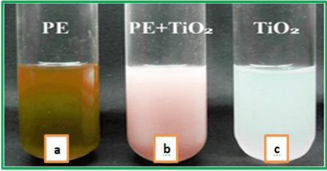
Graphic observations of TiO2 nanoparticle preparation (a) Titanium Iso-propoxide solution, (b) caricca papaya Shell extracts, (c) changed color.
Analysis of TiO2 nanoparticles
The captivation spectrums of bio-synthesized TiO2 nanoparticles by Carica papaya Shell extracts displayed a maximum optical absorptions band at 350 nm (Fig. 3). This absorptions peaks attained were the same as earlier reports. According to the absorption, edges frequently shifted to inferior wavelength or higher energies with declining sizes of nanoparticle63.
Figure 3.
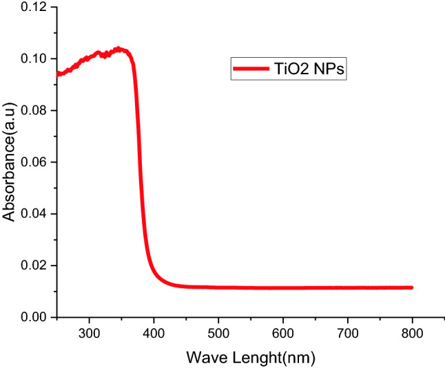
UV-spectra of TiO2 nanoparticle from Carica papaya shell extracts.
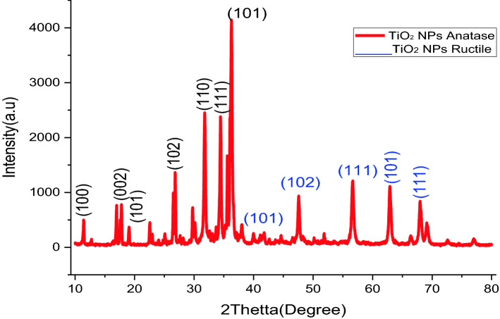
The phenolics groups prohibited agglomerations so that they can be forms metallic Nanoparticles to steady the environment. This advocates that biological molecule is bi-functional in the formations as well as steadying of TiO2 nanoparticles in an aqueous intermediate64. The X-ray diffraction patterns of biosynthesized TiO2 nanoparticles from Shell extracts of C. papaya are presented in (Fig. 4). The separate diffractions peak at 2 = 12.76, 18.2, 20.01, 28.34, 32.91, 35.32, 36.57, 40.21, 49.74, 58.34, 64.56 and 70.5 were corresponded to (100), (002), (101), (102), (101), (102),(110),(111), (102), (111), (101), and (111) crystal planes separately. Wholly the deflection peaks were finely indexed to hexagonal phases of both anatase and ructile of TiO2. The deflection patterns corresponded to the standards jointly committees on powders diffractions standard (JCPDS) No. 80 to 0075. The X-ray diffraction peaks with great strength disguised that Nanoparticles were greatly crystallized.
Figure 4.
X-ray diffraction patterns of TiO2 Nanoparticles from Carica papaya shell extracts.
The average crystal sizes of biosynthesized spacemen were deliberate by Debye Scherer’s formulas, that is
| 1 |
D is crystal size, λ stands for the length of wave (0.154 nm), β is FWHM (full width at half maximums), as well as θ (Theta) stands for Bragg diffraction angles. The average crystal size of TiO2 nanoparticles exists to be 15 nm65. X-ray diffraction patterns gained by present research are identical to the X-R-D pattern attained for previously stated TiO2 nanoparticles preparations.
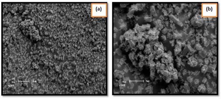
The morphologies of biosynthesized nanoparticles were assessed through scanning electron microscopy (SEM). Figure 5a,b display the superficial morphologies of TiO2 Nanoparticles under varied magnification. The scanning electron microscopy (SEM) image shows the agglomeration of separate TiO2 nanoparticles. The accumulated images display that convinced particles are semispherical (Fig. 5a) and some monoclinic spherical (Fig. 5b). The formations of floret-like morphologies of TiO2 nanoparticles with petals like Nano-sheets can be perceived66,67.
Figure 5.
Scanning electron microscope (SEM) images of TiO2 nanoparticles from Carica papaya shell extracts.
An energy dispersive x-ray diffractive (E-D-X) analysis was conducted for biosynthesized TiO2 nanoparticles to identify elementals compositions. A dispersive energy spectrum of sample attained from the scanning electron microscope (SEM) to energy dispersive X-ray diffractive (EDX) analysis displays that samples synthesized by the route have pure TiO2 anatase and ructile phases68–70. An energy dispersive x-ray diffractive (EDX) approves the existence of Ti and oxygen indications of Titanium dioxide Nanoparticles as displayed in Fig. 6, and its analyses displayed peaks that correspond to optical absorptions of prepared Nanoparticle. The basis of these elements deceits in the bio-components, habitually algae towards TiO2 Nanoparticles. Elementals analysis of nanoparticle produced 63.9% of Ti and 36.1% of oxygen (O2), which shows the synthesized Nano particle is in its maximum decontaminated forms. The energy dispersive X-ray diffractive (E-D-X) analyses in this research show identical outcomes to previous reports, elemental analyzes of the Nanoparticle produced 36.1% of Titanium and of oxygen 63.9%, respectively71–73.
Figure 6.
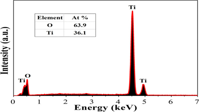
EDX analyzes TiO2 nanoparticle biosynthesized from Carica papaya shell extracts.
Transmissions electron microscopes (TEMs) depend on the imaging of high energies electron that is passed via a very thin sample. The images acted by the interaction of an electron with a prepared sample are inflamed and absorbed on sensors such as fluorescences screens and photographic film layers cameras. Bisynthesized TiO2 nanoparticles were strong-minded in a JEOL 1220 JEM brands transmissions electron microscopes74. Transmissions electron microscopes (TEMs) have been utilized for additional studies on the particle sizes, crystal and morphologies of the sample. Transmissions electron microscopes (TEMs) black spherical images of TiO2 nanoparticle micro-powders in rutile and anatase phase are given in Fig. 775–77.
Figure 7.

TEM analysis of TiO2 nanoparticles biosynthesized from Carica papaya shell extracts.
Antifungal activities
Phyto pathogens ground an excessive reduction in crop yields. Fungicide might be the solution for these, but over time, problems of resistance occur78–81. Nanoparticles have just the focus of attention with their special antimicrobial effects.
The sets of experimentations for the antifungals potentials purposes of TiO2 nanoparticle on Sclerotinias sclerotium, Rosellinia necatrixs, as well as Fusariums spp. Revealed fungal mycelial development inhibitions to certain extents in the test tube that was protected with TiO2 nanoparticles, as associated with the controls test tube82. The outcome was verified after comparisons of the dehydrated weights of the fungal that were on the test-tubes, with as well as without TiO2 nanoparticles, consequently, which advocated that TiO2 nanoparticles show antifungals activities on the three fungal strains, such that weights (in grams) of parched fungal were bigger for the controls in each case, while associated to test-tube. The comparatives outcomes described graphically (Figs. 8, 9) evidently show the fungal myceliums developments inhibition to some amount in the tests tubes that were immunized with extracts as associated with the controls tests tube.
Figure 8.
PDBs test-tube for anti-fungus potentials of TiO2 nanoparticles (a) Sclerotinias sclerotiorums, (b) Rosellinia necatrixs, (c) Fusariums strains.
Figure 9.
(a) Sclerotinias sclerotiorums, (b) Rosellinia necatrixs, (c) Fusariums strains.
Nanoparticles could be utilized as a potential anti-fungus agent and help overcome hurdles in fungal disease management modelled by the growth of resistances to conservative fungicide, but various from other where there were little sizes influence, like for PcO6s, Caenorhabditis-elegans, and soils bacterial community83. Due to the furthermost of the Nanoparticles accumulated in liquefied broths, it is pretty possible that supplementary modifications of the metal oxide nanoparticles in mediums may have happened after additions of agars to coagulate the mediums.
Seeds germinations
Seed germinations are speedily increasing procedure and broadly utilized for phytotoxicity analyses, and also have the advantage of sensitivities, simplicities, cost-effective as well as appropriateness for verified chemicals specimens84. In the present research, the influences of TiO2 nanoparticles on germinations of peas were studied. Figure 10 displays the influence of TiO2 nanoparticles biosynthesized from Caricas papaya shell extracts on pea germinations over 12 days. All concentrations of TiO2 nanoparticles increased shoots as well as roost elongated. In these experiments, the lengths of the roots after 12 days increased, owing to bigger absorptions. Figure 10 showed that a concentration of TiO2 NP 100% observed has a higher performance of roots lengths. The growths in roots and shoot lengths have gotten supreme with TiO2 nanoparticles (100%). Nevertheless, for the seeds preserved with 20%, 40% and 80% of the TiO2 nanoparticles, the standard errors were unimportantly inferior for roots and shoot lengths. In identical research85 Titanium oxide nanoparticle was prepared from liquefied extracts of Elaeagnus-Angustifolia.
Figure 10.
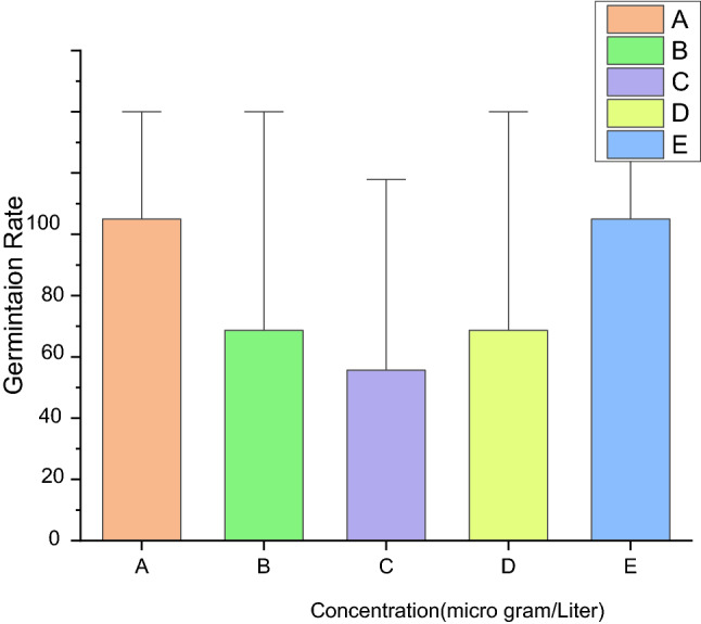
Germinations of antifungals potential (A) Controls (B) TiO2 20%, (C) TiO2 40%, (D) TiO2 80% (E)TiO2 100%.
Conclusion
In the present study, Titanium dioxide nanoparticles (TiO2NPs) were biosynthesized by using Carica papaya Shell extract, as well as nanoparticles prepared as well as analyzed. Crystalline average sizes measured 15 nm. In addition, it was observed that TiO2 NPs were semispherical as well as mono-clinic not spherical. In antifungal studies was perceived that biosynthesized TiO2 nanoparticles displayed antifungal influence in contradiction of Sclerotinias sclerotiorums Fusariums spp, as well as Rosellinia necatrixs. The effects of TiO2 NPs on seeds germinations, 100% of TiO2 NPs are furthermost appropriate for cultivating the roots as well as shoot lengths. TiO2 NPs were biosynthesized through Carica-papaya shell extracts in cheaper, eco-friendly techniques with biological preparation techniques. These biosynthesized TiO2 NPs could be utilized to control the reproductions of pathogenic fungus that damages plant growths. The outcomes of this study will give novel comprehensions into the effectiveness of biological methods. Additionally, this research will tile the ways for an optimistic step in the direction of a green strategy for the preparations of metallic oxides nanoparticles and the utilization of their biopotentials in agricultural areas. However, the special properties of various influences like doses, toxicities, real ecological circumstances etc., on germinations and plantlet growths of floras need to be investigated auxiliary in relation to biological methods.
Author contributions
Conceptualization, A.S., Y.S., LT.J., B.B., N.N., S.R., L.PD., V.V. and K.R.; Data curation, A,S., Y.S., LT.J., B.B., N.N., S.R., L.PD., V.V. and K.R.; Analysis and Validation, A,S., Y.S., LT.J., B.B., N.N., S.R., L.PD., V.V. and K.R.; Formal analysis, A.S., Y.S., LT.J., B.B., N.N., S.R., L.PD., V.V and K.R.; Investigation, A.S., Y.S., LT.J., B.B., N.N., S.R., L.PD., V. V and K .R.; Methodology, A.S., Y.S., LT.J., B.B., N.N., S.R., L.PD., V.V. and K.R.; Project administration, LT.J., and K.R. Resources, A.S., Y.S, LT.J., B.B., N.N., S.R., L.PD., V.V. and K.R.; Software, A.S., Y.S., LT.J., B.B., N.N., S.R., L.PD.,V.V. and K.R., Supervision, K.R. and L.T.J.; Validation, A.S., Y.S., LT.J., B.B., N.N., S.R., L.PD., V.V and K.R.; Visualization, A.S., Y.S., LT.J., B.B., N.N., S.R., L.PD., V.V. and K.R.; Writing—original draft, A.S., Y.S., LT.J., B.B., N.N., S.R., L.PD., V.V. and K.R., Data Visualization, Editing and Rewriting, A.S., Y.S., LT.J., B.B., N.N., S.R., L.PD., V.V. and K.R.
Data availability
The datasets used and analyzed during the current study are available from the corresponding author on request.
Competing interests
The authors declare no competing interests.
Footnotes
Publisher's note
Springer Nature remains neutral with regard to jurisdictional claims in published maps and institutional affiliations.
References
- 1.Vani P, Manikandan N, Vinitha G. A green strategy to synthesize environment friendly metal oxide nanoparticles for potential applications: A review. Asian J. Pharm. Clin. Res. 2017;10:337. [Google Scholar]
- 2.Dwivedi, M.K., Pandey, S.K. & Singh, P.K. Larvicidal activity of green synthesized zinc oxide nanoparticles from Carica papaya leaf extract. Inorg. Nano Metal Chem. 1–11. 10.1080/24701556.2022.2072340 (2022).
- 3.Alam MW, Al Qahtani HS, Aamir M, Abuzir A, Khan MS, Albuhulayqah M, Mushtaq S, Zaidi N, Ramya A. Phyto synthesis of manganese-doped zinc nanoparticles using carica papaya leaves: Structural properties and its evaluation for catalytic. Antibact. Antioxid. Act. Polym. 2022;14(9):1827. doi: 10.3390/polym14091827. [DOI] [PMC free article] [PubMed] [Google Scholar]
- 4.Mogazy AM, Hanafy RS. Foliar spray of biosynthesized zinc oxide nanoparticles alleviate salinity stress effect on vicia faba plants. J. Soil Sci. Plant Nutr. 2022;22:1–16. [Google Scholar]
- 5.Jeevanandam J, Kiew SF, Boakye-Ansah S, Lau SY, Barhoum A, Danquah MK, Rodrigues J. Green approaches for the synthesis of metal and metal oxide nanoparticles using microbial and plant extracts. Nanoscale. 2022;14(7):2534–2571. doi: 10.1039/d1nr08144f. [DOI] [PubMed] [Google Scholar]
- 6.Ukidave, V.V. & Ingale, L.T. Green synthesis of zinc oxide nanoparticles from coriandrum sativum and their use as fertilizer on Bengal gram, Turkish gram, and green gram plant growth. Int. J.Agro.10.1155/2022/8310038 (2022).
- 7.Alturki AM. Facile synthesis route for chitosan nanoparticles doped with various concentrations of the biosynthesized copper oxide nanoparticles: Electrical conductivity and antibacterial properties. J. Mol. Struc. 2022;1263:133108. [Google Scholar]
- 8.Mandal S, Alankar T, Hughes R, Marpu SB, Omary MA, Shi SQ. Removal of hazardous dyes and waterborne pathogens using a nanoengineered bioadsorbent from hemp–Fabrication, characterization and performance investigation. Surf. Interfaces. 2022;29:101797. [Google Scholar]
- 9.Sunny NE, Mathew SS, Chandel N, Saravanan P, Rajeshkannan R, Rajasimman M, Vasseghian Y, Vasseghian Y, Rajamohan N, Kumar SV. Green synthesis of titanium dioxide nanoparticles using plant biomass and their applications—A review. Chemosphere. 2022;300:134612. doi: 10.1016/j.chemosphere.2022.134612. [DOI] [PubMed] [Google Scholar]
- 10.Srivastava, S. & Bhargava, A. Biological Synthesis of Nanoparticles: Dicotyledons. In Green Nanoparticles: The Future of Nanobiotechnology. 10.1007/978-981-16-7106-7_12 (2022).
- 11.Shahbaz F, Akhter N, Shahid M, Riaz M, Anjum F, Hussain F. Ultrasound assisted extraction and characterization of bioactives from v erbascum thapsus roots to evaluate their antioxidant and medicinal potential. Dose Response. 2022;20(2):15593258221097665. doi: 10.1177/15593258221097665. [DOI] [PMC free article] [PubMed] [Google Scholar]
- 12.Subbiah R, Muthukumaran S, Raja V. Phyto-assisted synthesis of Mn and Mg Co-doped ZnO nanostructures using Carica papaya leaf extract for photocatalytic applications. BioNanoScience. 2021;11(4):1127–1141. [Google Scholar]
- 13.Thakare Y, Kore S, Sharma I, Shah M. A comprehensive review on sustainable greener nanoparticles for efficient dye degradation. Environ. Sci. Pollut. Res. 2022;29:1–22. doi: 10.1007/s11356-022-20127-y. [DOI] [PubMed] [Google Scholar]
- 14.Suvaitha SP, Sridhar P, Divya T, Palani P, Venkatachalam K. Bio-waste eggshell membrane assisted synthesis of NiO/ZnO nanocomposite and its characterization: Evaluation of antibacterial and antifungal activity. Inorg. Chimica. Acta. 2022;536:120892. [Google Scholar]
- 15.Farshori NN, Al-Oqail MM, Al-Sheddi ES, Al-Massarani SM, Saquib Q, Siddiqui MA, Wahab R, Al-Khedhairy AA. Green synthesis of silver nanoparticles using Phoenix dactylifera seed extract and its anticancer effect against human lung adenocarcinoma cells. J. Drug Deliv. Sci. Technol. 2022;70:103260. [Google Scholar]
- 16.Jayapriya, J. & Rajeshkumar, S. Eco-friendly synthesis of metal nanoparticles for smart nanodevices in the treatment of diseases. In Smart Nanodevices for Point-of-Care Applications.10.1201/9781003157823 (2022)
- 17.Navada KM, Nagaraja GK, D'Souza JN, Kouser S, Nithyashree BR, Manasa DJ. Bio-fabrication of multifunctional nano-ceria mediated from Pouteria campechiana for biomedical and sensing applications. J. Photochem. Photobio. A Chem. 2022;424:113631. [Google Scholar]
- 18.Olga M, Jana M, Anna M, Irena K, Jan M, Alena C. Antimicrobial properties and applications of metal nanoparticles biosynthesized by green methods. Biotechnol. Adv. 2022;58:107905. doi: 10.1016/j.biotechadv.2022.107905. [DOI] [PubMed] [Google Scholar]
- 19.Gole, A. et al. Role of phytonanotechnology in the removal of water contamination. J. Nanomater.10.1155/2022/7957007 (2022).
- 20.Abel, S. et al. Investigating the influence of bath temperature on the chemical bath deposition of nanosynthesized lead selenide thin films for photovoltaic application. J. Nanomater. 10.1155/2022/3108506 (2022).
- 21.Sharma RR, Deep A, Abdullah ST. Herbal products as shellcare therapeutic agents against ultraviolet radiation-induced shell disorders. J. Ayur. Int. Med. 2021;13:100500. doi: 10.1016/j.jaim.2021.07.016. [DOI] [PMC free article] [PubMed] [Google Scholar]
- 22.Deepika, S., Roopan, S.M. & Selvaraj, C.I. Bionanocomposite assembly with larvicidal activity against Aedes aegypti. In Applications for Nanobiotechnology Neglected Tropical Diseases10.1016/B978-0-12-821100-7.00001-7 (2021).
- 23.Dhivya B, Sujatha K, Sudha AP. Facile synthesis of calcium oxide nanoparticles from the carica papaya leaf extract with the significantly enhanced antibacterial activity. Nanoscale Rep. 2020;3(1):1–9. [Google Scholar]
- 24.Nor SM, Ding P. Trends and advances in edible biopolymer coating for tropical fruit: A review. Food Res. Int. 2020;134:109208. doi: 10.1016/j.foodres.2020.109208. [DOI] [PubMed] [Google Scholar]
- 25.Rajam RP, Kannan S, Kajendran D. Cosmeceuticals an emerging technology—A review. World J. Pharma. Res. 2019;8(12):664–685. [Google Scholar]
- 26.Verma V, Al-Dossari M, Singh J, Rawat M, Kordy MG, Shaban M. A review on green synthesis of TiO2 NPs: Photocatalysis and antimicrobial applications. Polymers. 2022;14(7):1444. doi: 10.3390/polym14071444. [DOI] [PMC free article] [PubMed] [Google Scholar]
- 27.Tharani, M. & Rajeshkumar, S. Antimicrobial applications of nanodevices prepared from metallic nanoparticles and their role in controlling infectious diseases. In Smart Nanodevices for Point of Care Applications. 10.1201/9781003157823 (2022).
- 28.Rehman K, Zaman U, Tahir K, Khan D, Khattak NS, Khan SU, Khan WU, Nazir S, Bibi R, Gul R. A Coronopus didymus based eco-benign synthesis of Titanium dioxide nanoparticles (TiO2 NPs) with enhanced photocatalytic and biomedical applications. Inorg. Chem. Commun. 2022;137:109179. [Google Scholar]
- 29.Abel S, Tesfaye JL, Nagaprasad N, Shanmugam R, Dwarampudi LP, Deepak T, Zhang H, Krishnaraj R, Stalin B. Examining impacts of acidic bath temperature on nano-synthesized lead selenide thin films for the application of solar cells. Bioinorg. Chem. Appl. 2022;2022:1003803. doi: 10.1155/2022/1003803. [DOI] [PMC free article] [PubMed] [Google Scholar] [Retracted]
- 30.Vieira IRS, de Carvalho APAD, Conte‐Junior CA. Recent advances in biobased and biodegradable polymer nanocomposites, nanoparticles, and natural antioxidants for antibacterial and antioxidant food packaging applications. Comprehensive Rev. Food Sci. Food Saf. 2022;21(4):3673–3716. doi: 10.1111/1541-4337.12990. [DOI] [PubMed] [Google Scholar]
- 31.Suhag, R. et al. Fruit peel bioactives, valorization into nanoparticles and potential applications: A review. Crit. Rev. Food Sci. Nutr. 10.1080/10408398.2022.2043237 (2022). [DOI] [PubMed]
- 32.Zhang S, Lin L, Huang X, Lu YG, Zheng DL, Feng Y. Antimicrobial properties of metal nanoparticles and their oxide materials and their applications in oral biology. J. Nanomat. 2022;2022(4):1–18. [Google Scholar]
- 33.Shyamalagowri S, Charles P, Manjunathan J, Kamaraj M, Anitha R, Pugazhendhi A. In vitro anticancer activity of silver nanoparticles phyto-fabricated by Hylocereus undatus peel extracts on human liver carcinoma (HepG2) cell lines. Process Biochem. 2022;116:17–25. [Google Scholar]
- 34.Dzulkharnien NSF, Rohani R. A review on current designation of metallic nanocomposite hydrogel in biomedical applications. Nanomaterials. 2022;12:1629. doi: 10.3390/nano12101629. [DOI] [PMC free article] [PubMed] [Google Scholar]
- 35.Bhavyasree PG, Xavier TS. Green synthesized copper and copper oxide-based nanomaterials using plant extracts and their application in antimicrobial activity. Cur. Res. Green. Sustain. Chem. 2022;5:100249. [Google Scholar]
- 36.Silva, L.P. et al. Sustainable exploitation of agricultural, forestry, and food residues for green nanotechnology applications. In Biogenic Nanomaterials10.1201/9781003277149.
- 37.Basumatary, I.B., Kalita, S., Katiyar, V., Mukherjee, A. & Kumar, S. Edible Films and Coatings. Biopolymer‐Based Food Packaging: Innova. Tech. App. 445–475 (2022).
- 38.Zare, M. et al. Emerging trends for Zno nanoparticles and their applications in food packaging. ACS Food Sci. Technol. 10.1021/acsfoodscitech.2c00043 (2022).
- 39.Islam, S.U. & Sun, G. Biological chemicals as sustainable materials to synthesize metal and metal oxide nanoparticles for textile surface functionalization. Available at SSRN 4074544.
- 40.Dwivedi, M.K., Pandey, S.K. & Singh, P.K. Larvicidal activity of green synthesized zinc oxide nanoparticles from Carica papaya leaf extract. Inorg. Nano Metal Chem. 10.1080/24701556.2022.2072340 (2022).
- 41.Saka A, Jule LT, Soressa S, Gudata L, Nagaprasad N, Seenivasan V, Ramaswamy K. Biological approach synthesis and characterization of iron sulfide (FeS) thin films from banana peel extract for contamination of environmental remediation. Sci. Rep. 2022;12(1):1–8. doi: 10.1038/s41598-022-14828-0. [DOI] [PMC free article] [PubMed] [Google Scholar]
- 42.Dadkhah M, Tulliani JM. Green synthesis of metal oxides semiconductors for gas sensing applications. Sensors. 2022;22(13):4669. doi: 10.3390/s22134669. [DOI] [PMC free article] [PubMed] [Google Scholar]
- 43.Felicia WXL, Rovina K, Nur’Aqilah MN, Vonnie JM, Erna KH, Misson M, Halid NFA. Recent advancements of polysaccharides to enhance quality and delay ripening of fresh produce: A review. Polymers. 2022;14(7):1341. doi: 10.3390/polym14071341. [DOI] [PMC free article] [PubMed] [Google Scholar]
- 44.Ballesteros, L.F. et al. Active packaging systems based on metal and metal oxide nanoparticles. Nanotechnol. Enhanc. Food Pack. 10.1002/9783527827718.ch7 (2022).
- 45.Dzulkharnien NSF, Rohani R. A review on current designation of metallic nanocomposite hydrogel in biomedical applications. Nanomaterials. 2022;12(10):1629. doi: 10.3390/nano12101629. [DOI] [PMC free article] [PubMed] [Google Scholar]
- 46.Saka, A., Jule, L.T., Gudata, L., Seeivasan, V., Nagaprasad, N. & Ramaswamy, K. Preparation of Environmentally Friendly Method of zinc oxide (ZnO) nanoparticles from osmium gratissimum (Damaakasee) plant leaf extracts and its antibacterial Activities (2022).
- 47.Ramesh R, Parasaran M, Mubashira GF, Flora C, Khan FLA, Almaary KS, Elbadawi YB, Chen TW, Kanimozhi K, Bashir AKH, Hussein AA. Biogenic synthesis of ZnO and NiO nanoparticles mediated by fermented Cocos nucifera (L) deoiled cake extract for antimicrobial applications towards gram positive and gram negative pathogens. J. King Saud Univ. Sci. 2022;34(1):101696. [Google Scholar]
- 48.Donga S, Chanda S. Caesalpinia crista seeds mediated green synthesis of zinc oxide nanoparticles for antibacterial, antioxidant, and anticancer activities. BioNanoScience. 2022;12(2):451–462. [Google Scholar]
- 49.Kumar A, Choudhary A, Kaur H, Guha S, Mehta S, Husen A. Potential applications of engineered nanoparticles in plant disease management: A critical update. Chemosphere. 2022;295:133798. doi: 10.1016/j.chemosphere.2022.133798. [DOI] [PubMed] [Google Scholar]
- 50.Chaerun SK, Prabowo BA, Winarko R. Bionanotechnology: the formation of copper nanoparticles assisted by biological agents and their applications as antimicrobial and antiviral agents. Environ. Nanotechnol. Monit.Manag. 2022;18:100703. [Google Scholar]
- 51.Kaur, N., Sharma, R. & Sharma, V. Advances in the synthesis and antimicrobial applications of metal oxide nanostructures. In Advanced Ceramics for Versatile Interdisciplinary Applications.10.1016/B978-0-323-89952-9.00015-4 (2022).
- 52.Parmar M, Sanyal M. Extensive study on plant mediated green synthesis of metal nanoparticles and their application for degradation of cationic and anionic dyes. Environ. Nanotechnol. Monit. Manag. 2022;17:100624. [Google Scholar]
- 53.Mustapha T, Misni N, Ithnin NR, Daskum AM, Unyah NZ. A review on plants and microorganisms mediated synthesis of silver nanoparticles, role of plants metabolites and applications. Int. J. Environ. Res. Public Heal. 2022;19(2):674. doi: 10.3390/ijerph19020674. [DOI] [PMC free article] [PubMed] [Google Scholar]
- 54.Mogazy AM, Mohamed HI, El-Mahdy OM. Calcium and iron nanoparticles: A positive modulator of innate immune responses in strawberry against Botrytis cinerea. Process Biochem. 2022;115:128–145. [Google Scholar]
- 55.Cao Y, Dhahad HA, El-Shorbagy MA, Alijani HQ, Zakeri M, Heydari A, Bahonar E, Slouf M, Khatami M, Naderifar M, Iravani S. Green synthesis of bimetallic ZnO–CuO nanoparticles and their cytotoxicity properties. Sci. Rep. 2021;11(1):1–8. doi: 10.1038/s41598-021-02937-1. [DOI] [PMC free article] [PubMed] [Google Scholar]
- 56.Deshmukh, R.K. & Gaikwad, K.K. Natural antimicrobial and antioxidant compounds for active food packaging applications. Biomass. Conv. Bioref. 10.1007/s13399-022-02623-w (2022).
- 57.Luzala MM, Muanga CK, Kyana J, Safari JB, Zola EN, Mbusa GV, Nuapia YB, Liesse JMI, Nkanga CI, Krause RW, Balčiūnaitienė A. A critical review of the antimicrobial and antibiofilm activities of green-synthesized plant-based metallic nanoparticles. Nanomaterials. 2022;12(11):1841. doi: 10.3390/nano12111841. [DOI] [PMC free article] [PubMed] [Google Scholar]
- 58.Saka A, Tesfaye JL, Gudata L, Shanmugam R, Dwarampudi LP, Nagaprasad N, Krishnaraj R, Rajeshkumar S. Synthesis, characterization, and antibacterial activity of ZnO nanoparticles from fresh leaf extracts of Apocynaceae, Carissa spinarum L. (Hagamsa) J. Nanomater. 2022;2022:1–6. [Google Scholar]
- 59.Suhag, R. et al. Fruit peel bioactives, valorization into nanoparticles and potential applications: A review. Crit. Rev. Food Sci. Nutr. 10.1080/10408398.2022.2043237 (2022). [DOI] [PubMed]
- 60.Sathiyavimal S, Vasantharaj S, Bharathi D, Saravanan M, Manikandan E, Kumar SS, Pugazhendhi A. Biogenesis of copper oxide nanoparticles (CuONPs) using Sida acuta and their incorporation over cotton fabrics to prevent the pathogenicity of Gram negative and Gram positive bacteria. J. Photochem. Photobiol. B. 2018;188:126–134. doi: 10.1016/j.jphotobiol.2018.09.014. [DOI] [PubMed] [Google Scholar]
- 61.Murugadoss G. Bio-Mediated Synthesis of Nanomaterials for Dye-Sensitized Solar Cells. Mater. Res. Found. 2022;121:175–210. [Google Scholar]
- 62.Srivastava, S.K., Srivastava, S. & Chauhan, N. Nanotechnology as an effective tool for antimicrobial applications: Current research and challenges.
- 63.Rajkumar S, Venkatraman MR, Suguna K, Karuppasamy P, Pandian MS, Ramasamy P. Synthesis of Ag-incorporated TiO2 nanoparticles by simple green approach as working electrode for dye-sensitized solar cells. J. Mater. Sci. Mater. Elect. 2022;33(8):4965–4973. [Google Scholar]
- 64.Dulta K, Koşarsoy Ağçeli G, Chauhan P, Jasrotia R, Chauhan PK. Ecofriendly synthesis of zinc oxide nanoparticles by carica papaya leaf extract and their applications. J. Clus. Sci. 2022;33(2):603–617. [Google Scholar]
- 65.Oyeshola, H. Evaluation of photovoltaic properties of green synthesized zinc oxide nanoparticles from extract of carica papaya for anode buffer film layer of polymer solar cells. (2022).
- 66.Priyadharsini N, Bhuvaneswari N, Joshy J. Plant mediated synthesis of Zno and Mn doped Zno nanoparticles using carica papaya leaf extract for antibacterial applications. Asian J. Appl. Sci. Technol. (AJAST) 2021;5(4):69–81. [Google Scholar]
- 67.Sabouri Z, Sabouri M, Amiri MS, Khatami M, Darroudi M. Plant-based synthesis of cerium oxide nanoparticles using Rheum turkestanicum extract and evaluation of their cytotoxicity and photocatalytic properties. Mater. Technol. 2022;37(8):555–568. [Google Scholar]
- 68.Hamrayev H, Shameli K, Korpayev S. Green synthesis of zinc oxide nanoparticles and its biomedical applications: A review. J. Res. Nanosci. Nanotechnol. 2021;1(1):62–74. [Google Scholar]
- 69.Barkat, M.A. Therapeutic Importance of Phyto-assisted Green Synthesis of Zinc Oxide Nanoparticles in Burn Wound Management. Nanotechnology Driven Herbal Medicine for Burns: From Concept to Application10.2174/9789815039597121010009 (2021).
- 70.Yasmin H, Mazher J, Azmat A, Nosheen A, Naz R, Hassan MN, Noureldeen A, Ahmad P. Combined application of zinc oxide nanoparticles and biofertilizer to induce salt resistance in safflower by regulating ion homeostasis and antioxidant defence responses. Ecotoxicol. Environ. Saf. 2021;218:112262. doi: 10.1016/j.ecoenv.2021.112262. [DOI] [PubMed] [Google Scholar]
- 71.Abdullah FH, Bakar NA, Bakar MA. Comparative study of chemically synthesized and low temperature bio-inspired Musa acuminata peel extract mediated zinc oxide nanoparticles for enhanced visible-photocatalytic degradation of organic contaminants in wastewater treatment. J. Haz. Mater. 2021;406:124779. doi: 10.1016/j.jhazmat.2020.124779. [DOI] [PubMed] [Google Scholar]
- 72.Nabi G, Majid A, Riaz A, Alharbi T, Kamran MA, Al-Habardi M. Green synthesis of spherical TiO2 nanoparticles using Citrus Limetta extract: Excellent photocatalytic water decontamination agent for RhB dye. Inorg. Chem. Commun. 2021;129:108618. [Google Scholar]
- 73.Aslam M, Abdullah AZ, Rafatullah M. Recent development in the green synthesis of titanium dioxide nanoparticles using plant-based biomolecules for environmental and antimicrobial applications. J. Ind. Eng. Chem. 2021;98:1–16. [Google Scholar]
- 74.Silva-Osuna ER, Vilchis-Nestor AR, Villarreal-Sanchez RC, Castro-Beltran A, Luque PA. Study of the optical properties of TiO2 semiconductor nanoparticles synthesized using Salvia rosmarinus and its effect on photocatalytic activity. Optic. Mater. 2022;124:112039. [Google Scholar]
- 75.Irshad MA, Nawaz R, UrRehman MZ, Adrees M, Rizwan M, Ali S, Ahmad S, Tasleem S. Synthesis, characterization and advanced sustainable applications of titanium dioxide nanoparticles: A review. Ecotoxicol. Environ. Saf. 2021;212:111978. doi: 10.1016/j.ecoenv.2021.111978. [DOI] [PubMed] [Google Scholar]
- 76.Akbarizadeh MR, Sarani M, Darijani S. Study of antibacterial performance of biosynthesized pure and Ag-doped ZnO nanoparticles. Rendiconti Lincei. Scienze Fisiche e Naturali. 2022;33:1–9. [Google Scholar]
- 77.Siddaramappa M, Latha HKE, Lalithamba HS, Udayakumar A. The effect of tin concentration on microstructural, optical and electrical properties of ITO nanoparticles synthesized using green method. Iran. J. Mater. Sci. Eng. 2021;18(4):1–12. [Google Scholar]
- 78.Zadeh FA, Bokov DO, Salahdin OD, Abdelbasset WK, Jawad MA, Kadhim MM, Qasim MT, Kzar HH, Al-Gazally ME, Mustafa YF, Khatami M. Cytotoxicity evaluation of environmentally friendly synthesis Copper/Zinc bimetallic nanoparticles on MCF-7 cancer cells. Rendiconti Lincei. Scienze Fisiche e Naturali. 2022;33:1–7. doi: 10.1007/s12210-022-01064-x. [DOI] [PMC free article] [PubMed] [Google Scholar]
- 79.Haghighat M, Alijani HQ, Ghasemi M, Khosravi S, Borhani F, Sharifi F, Iravani S, Najafi K, Khatami M. Cytotoxicity properties of plant-mediated synthesized K-doped ZnO nanostructures. Bioprocess Biosyst. Eng. 2022;45(1):97–105. doi: 10.1007/s00449-021-02643-2. [DOI] [PubMed] [Google Scholar]
- 80.Alijani HQ, Iravani S, Pourseyedi S, Torkzadeh-Mahani M, Barani M, Khatami M. Biosynthesis of spinel nickel ferrite nanowhiskers and their biomedical applications. Sci. Rep. 2021;11(1):1–7. doi: 10.1038/s41598-021-96918-z. [DOI] [PMC free article] [PubMed] [Google Scholar]
- 81.Hassan AMS, Mahmoud AS, Ramadan MF, Eissa MA. Microwave-assisted green synthesis of silver nanoparticles using Annona squamosa peels extract: Characterization, antioxidant, and amylase inhibition activities. Rendiconti Lincei. Scienze Fisiche e Naturali. 2022;33(1):83–91. [Google Scholar]
- 82.Mandhata, C.P., Sahoo, C.R. & Padhy, R.N., Biomedical applications of biosynthesized gold nanoparticles from cyanobacteria: An overview. Biol. Trace Elem Res. 10.1007/s12011-021-03078-2 (2022). [DOI] [PubMed]
- 83.Vijayakumar S, Chen J, Divya M, Durán-Lara EF, Prasannakumar M, Vaseeharan B. A review on biogenic synthesis of selenium nanoparticles and its biological applications. J. Inorg. Organomet. Polym. Mater. 2022;32:1–16. [Google Scholar]
- 84.Jasim SA, Patra I, Opulencia MJC, Hachem K, Parra RMR, Ansari MJ, Jalil AT, Al-Gazally ME, Naderifar M, Khatami M, Akhavan-Sigari R. Green synthesis of spinel copper ferrite (CuFe2O4) nanoparticles and their toxicity. Nanotechnol. Rev. 2022;11(1):2483–2492. [Google Scholar]
- 85.Behera, A., Pradhan, S.P., Ahmed, F.K. & Abd-Elsalam, K.A. Enzymatic synthesis of silver nanoparticles: Mechanisms and applications. In Green Synthesis of Silver Nanomaterials (pp. 699–756). Elsevier 10.1016/B978-0-12-824508-8.00030-7 (2022).
Associated Data
This section collects any data citations, data availability statements, or supplementary materials included in this article.
Data Availability Statement
The datasets used and analyzed during the current study are available from the corresponding author on request.


