Abstract
BACKGROUND:
Pregnant women experience falls, particularly in the third trimester. In this population, physiological changes, as well as ligament laxity, might influence joint proprioception and do not return to normal during the postpartum period. The prevalence of falls during pregnancy and postpartum periods imposes a need to study proprioception in pregnant women and the postpartum period.
MATERIALS AND METHOD:
An observational longitudinal study was conducted in June 2018 in outpatient clinic Chinchwad Pune. A total of 36 primiparous women were included in the study by using purposive sampling. The mean and standard deviation of the age was 25.92 (2.59). Proprioception was assessed for the knee joint and the ankle joint during the third trimester of pregnancy and 6th and 12th week postpartum. Outcomes included were the Joint Reposition Test for both knee and ankle joints using UTHSCSA Image Tool Software 3.0. Repeated-measure ANOVA was performed for the normally distributed data, and nonparametric test Friedman's test was performed for data that were not distributed normally. The data was statistically analyzed using the SPSS software version 26. The level of significance was set at 0.05, confidence intervals of 95% were used.
RESULT:
The result revealed significant (P < 0.05) improvement in both knee and ankle proprioception during the third trimester of pregnancy and postpartum period 6th and 12th week.
CONCLUSION:
Knee proprioception and ankle proprioception were found to improve significantly during the postpartum period 6th and 12th weeks compared to the third trimester of pregnancy but do not return to the prepregnancy state.
Keywords: Ankle proprioception, knee proprioception, postpartum period, pregnancy, reposition error
Introduction
Proprioception is the whole neural input to the central nervous system from specific nerve terminals termed mechanoreceptors found in ligaments, muscles, joints, capsules, tendons, and skin.[1] Proprioception is an important component of the sensory-motor system because it gives information regarding joint position sensation, kinesthesia, and force perception.[2] For motor learning and movement anticipation, conscious knowledge of proprioception is required.[3] The afferent information that arises from the muscle spindles is most important for the mediation of proprioception, while the other sources of proprioceptive information, which includes the cutaneous receptors and the joint mechanoreceptors are also very important for determining the position of the distal body segments and also for determining the limits of the range of motion.[4]
At the brain stem level, afferent information is integrated with visual and vestibular inputs in order to control automatic and stereotypical movement patterns, balance, and posture.[5]
At the higher levels of the central nervous system, the cerebral cortex and cerebellum elicit the conscious awareness of proprioception, thus contributing to the voluntary movements.[6] The integration of proprioceptive input at the mentioned three levels of the central nervous system aims to coordinate body stability ahead of movement execution as well as to correct the velocity and timing errors during the execution.[7]
A woman's body undergoes several physiological changes throughout pregnancy. Placental development, belly extension, weight gain, glandular development, breast enlargement, and posture modifications are among the changes that occur to the female body in order to accommodate the growing fetus. One of the less investigated changes is in the foot of pregnant women. These changes could be due to the accumulation of fluid or fat well as changes in the ligaments caused by the extra weight gain that is carried during pregnancy or by hormonally induced changes of the connective tissue in the ligaments. Among the musculoskeletal changes, the effect of hormones on the ligament and joint laxity is well known.[8] The changing shape and the inertia of the lower trunk require compensatory postural adjustments like the elevation of the head, the hyperextension of the lumbar spine, and the extension of knee and ankle joints.[9]
Sway alterations in pregnant women have been investigated, and it has been discovered that pregnant women rely on visual signals rather than proprioceptive cues for postural stability.[10] The ankle plantar flexors function significantly to maintain postural stability in pregnant women, according to the inverted pendulum framework theory that describes the control of postural sway.[11,12] This could result in early exhaustion of the muscles surrounding the ankle joint. The association between impaired postural control and fatigue in the plantar flexors was studied by Roerdink et al.[13] and explained that tiredness in the plantar flexors could be one of the reasons for the reduced postural control seen during pregnancy.
As the changes due to the center of gravity affect the mechanical arrangement of the body, every change in one joint or part of the mechanical system affects the other parts as well. Thus, studying ankle and knee proprioception together is a need to better understand the influence of hormones and postural changes on the affection of proprioception in pregnant women and the risk factors associated with its affection. The increasing prevalence of falls among pregnant women is imposing an alarming threat to the overall health of the pregnant woman and her fetus.[14]
Due to a loss in balancing ability, a fall rate of 27% was found during pregnancy, notably during the third trimester.[15] The altered sensory input from the vestibular and proprioceptive systems is considered as one of the intrinsic factors leading to falls during pregnancy. Along with this joint laxity and increased interstitial fluid volume may lead to reduced coordination and kinaesthetic sensation of the weight-bearing joints.[16]
The study conducted by Solomon et al. on ankle proprioception demonstrated a significant difference in ankle proprioception between pregnant and nonpregnant women.[17] Despite the fact that balance difficulties and visual dependence have been documented in pregnant women, their loss of proprioception during pregnancy and the postpartum period has been less investigated. Hence, the aim of the study was to assess the ankle and knee proprioception of pregnant women during the third trimester and 6 and 12 week postpartum and to check whether both, the knee and ankle proprioception return to the prepregnancy state.
Material and Method
The study was approved by the Institutional Ethical Committee of the institute where the study was conducted. It was an observational longitudinal study.
Study design and setting
This was an observational longitudinal study conducted in an outpatient clinic of the CMFs College of Physiotherapy Chinchwad Pune. The study was conducted between June 2018 and March 2019. The study was approved by the Institutional Ethical Committee of the institute where the study was conducted.
Study participants and sampling
Purposive sampling was used for collecting the sample. Using GPower Software version 3, assuming an effect size of 0.25 with an alpha error of 0.05 with a power of 0.95, the sample obtained was 36. Pregnant women who are in the third trimester of pregnancy were included in the study.
Inclusion criteria were as follows
Primiparous women
Age group: 21–35 years
The women ready to expose up to 10 cm above the knee joint.
Exclusion criteria were as follows
Individuals having any recent history of fracture or surgery lower limb
Any fixed spinal deformities
Ankle deformities/any contractures in lower limb
Any neurological disorders related to the upper or lower limb.
Data collection tool and technique
A total of 36 participants were enrolled in the research. Prior to the start of the evaluation, written consent was obtained. Proprioception was assessed for the knee and ankle joint during the third trimester, 6th week, and 12th week postpartum. All the assessment was done by using the knee and ankle reposition test during pregnancy (third trimester), 6 week, and 12 week postpartum.
For knee proprioception assessment
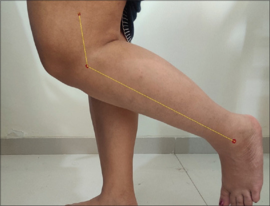
Assessment for knee proprioception on the dominant side was done using Knee Reposition Test. The subject was instructed to stand erect with the support. The patient was then asked to expose up to 10 cm above the knee joint for placing the marker and capturing the image. Three locations were marked, that is, the lateral epicondyle, greater trochanter to the lateral epicondyle 5 cm above the knee, and the lateral malleolus [Figure 1].
Figure 1.

Position of markers for the assessment of knee reposition error
The subject had explained the procedure, and the therapist will move the knee in available ranges of flexion and extension 10 times and position the knee at a particular angle; this angle was now addressed as the target angle. This angle was then photographed. The subject then had to feel this position for 15 s. Now, the subject was instructed to move the knee by herself 10 times in the available ranges of knee flexion and extension and reposition the knee joint as accurately to the target angle as possible. This angle was then referred to as the estimated angle. This angle was also photographed. Now, using these two pictures in the UTHSCSA Image Tool Software 3.0, the difference between these two angles was calculated which is the knee reposition error. It was done three times, and the average was taken.
For ankle proprioception assessment
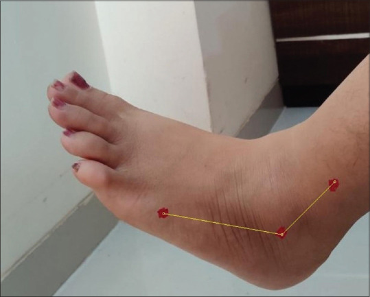
The assessment for ankle proprioception was done on the dominant side using the Ankle Reposition test. The subject was asked to sit at the edge of the plinth with their feet hanging. The markers were placed at three points, that is, at the tip of the lateral malleolus, at the lateral aspect of the base of the fifth metatarsal, and 5 cm above lateral malleolus on the shaft of the fibula as shown in the picture alongside [Figure 2].
Figure 2.

Position of markers for the assessment of ankle reposition error
The subject has then explained the procedure to the patient. The therapist will move the ankle in available ranges of plantar flexion and dorsiflexion 10 times and position the ankle at a particular angle; this angle was now addressed as the target angle. This angle was then photographed. The subject then had to feel this position for 15 s. Now, the subject was instructed to move the ankle by herself 10 times in the available ranges of ankle plantar flexion and dorsiflexion and reposition the ankle joint as accurately to the target angle as possible. This angle was then referred to as the estimated angle. This angle was also photographed. Now, using these two pictures in the UTHSCSA Image Tool Software 3.0, the difference between these two angles was calculated, which is the ankle reposition error. It was done three times, and the average was taken as the mean reposition error of the ankle joint. This data was recorded during the third trimester, 6 week postpartum, and 12 week postpartum. This data was analyzed using SPSS software version 26.
The outcome measure used was as follows
Joint reposition test: the test was done using UTHSCSA (University of Texas Health Science Centre at San Antonio's) Image Tool Software 3.0. The test's reliability for the ankle and knee joints is 0.81.[18] This test is used to find out the affection of proprioception or the joint position sense. A target angle is set for the individual which the person has to feel for 15 s and has to reproduce the same angle themselves, which is called the estimated angle. The difference between the estimate and the target angle is measured and is called joint reposition error, which is used to analyze the affection of proprioception in the individual.
Ethical consideration
The present study was approved by the Institutional Ethical Committee of Chaitanya Medical Foundations College of Physiotherapy Chinchwad Pune (ethics code: CMF/MPT/201819). The written consent was taken from all the women who participated in the study. Participants were assured that their details would be kept confidential, and all ethical principles were followed throughout the study. They had informed that they can withdraw from the study if they are not satisfied with the procedure.
Data analysis
The data were statistically analyzed using IBM Corporation's SPSS software version 26 and Microsoft Excel 2007. The normality test (Shapiro–Wilk test) was used to determine if the data for both knee and ankle mean reposition error were distributed normally. For the normally distributed data, a repeated measure ANOVA was used. Friedman's test was used for the data which is not distributed normally. The significance level was set at 0.05, and 95% confidence intervals were employed.
Results
Forty-three subjects were screened for eligibility criteria and included in the study. There were seven dropouts during the period between the third trimester and 12 week postpartum. Thus, 36 subjects were assessed during the third trimester, 6th week postpartum, and 12th week postpartum. The mean age of the subjects in the study was 25.92±2.59 [Table 1]. Table 2 and 3 shows mean knee and ankle reposition error during pregnancy and postpartum period respectively.
Table 1.
Mean age of subjects in the study
| Number of Subjects | Mean±Standard Deviation (in years) |
|---|---|
| 36 | 25.92±2.59 |
Table 2.
Knee reposition error during pregnancy and postpartum period
| Time of assessment | Mean±Standard Deviation |
|---|---|
| During the third trimester | 7.13±1.96 |
| During the 6th week postpartum | 4.64±1.47 |
| During the 12th week postpartum | 3.23±1.20 |
Table 3.
Ankle reposition error during pregnancy and the postpartum period
| Time of assessment | Mean |
|---|---|
| During third trimester | 6.41±2.23 |
| During 6th week postpartum | 5.01±1.42 |
| During 12th week postpartum | 3.65±1.67 |
Table 4 compares the mean difference of knee reposition error during pregnancy and postpartum period. There is a statistically significant difference in knee proprioception error during pregnancy and postpartum period (P < 0.05). This means that there is improvement in the knee proprioception from the third trimester to 6th week postpartum as well as between the 6th week and the 12th week postpartum.
Table 4.
Knee reposition error between the third trimester of pregnancy and the sixth week postpartum
| Time Period | Mean Difference | Std. Error | Significance (P) | 95% CI | |
|---|---|---|---|---|---|
|
| |||||
| Lower bound | Upper bound | ||||
| 3rd Trimester to 6th week postpartum | 2.49 | 0.24 | 0.0001 | 1.89 | 3.09 |
| 3rd Trimester to 12th week postpartum | 3.90 | 0.27 | 0.0001 | 3.23 | 4.57 |
| 6th Week to 12th week postpartum | 1.41 | 0.17 | 0.0001 | 0.98 | 1.83 |
Table 5 shows that there is a statistically significant difference in ankle proprioception during the different trimesters of pregnancy and the postpartum period (P < 0.05). This shows that there is improvement in the ankle proprioception from the third trimester to 6th week postpartum as well as between the 6th week and 12thweek postpartum.
Table 5.
Ankle reposition error between the third trimester of pregnancy and 6th and 12th week postpartum
| Time Period | Mean Difference | Std. Error | Significance (P) | 95% CI | |
|---|---|---|---|---|---|
|
| |||||
| Lower bound | Upper bound | ||||
| 3rd Trimester and 6th week postpartum | 1.40 | 0.25 | 0.0001 | 0.78 | 2.03 |
| 3rd Trimester and 12th week postpartum | 2.76 | 0.29 | 0.0001 | 2.02 | 3.50 |
| 6th Week and 12th week postpartum | 1.36 | 0.21 | 0.0001 | 0.83 | 1.88 |
Discussion
The present study was carried out to assess the proprioception during the third trimester of pregnancy and 6th week and 12th week postpartum.
The results of the present study show that there was a reduction of the repositioning error from the third trimester of pregnancy to the postpartum period. Proprioception in the knees and ankles declines during pregnancy, which includes hormonal changes, postural alterations, and the presence of edema. The coordinated function of the somatosensory system, visual system, and vestibular system is also required for proprioception.[10] Hormonal changes during pregnancy have been observed to produce changes in the homeostasis of labyrinthine fluids, which have a direct impact on neurotransmitter function. Vestibular disorders such as dizziness, vertigo, and instability are linked to hormones such as estrogen and progesterone. There was a link between hormonal alterations and vestibular system affection, according to a study undertaken by PM Schmidt.[19]
Variations in serum relaxin hormone may cause abnormalities in knee proprioception during pregnancy. Relaxin levels have been shown to be 10-fold higher, with a wide range of impacts on the body's connective tissues. It has been linked to collagen fiber remodeling, which triggers fibroblasts to manufacture new collagen fibers. Ligament laxity and the weakening of soft tissue structures are caused by changes in relaxin levels, which contribute to a loss of proprioception.[20]
Proprioception is necessary for smooth and coordinated movements, maintaining a proper body posture, regulating balance and postural control, and influencing the motor learning process.[1] Increased lumbar curvatures and pelvic inclinations, decreased dissociations between pelvic and trunk motions, knee valgus misalignments, and reduction in the height of the longitudinal arch of the foot are all examples of postural adaptations that occur during pregnancy. Postural instability, increased ankle plantarflexion, and a loss in lower limb proprioception have all been observed as a result of these lower limb postural abnormalities.[20]
PM Schmidt explained that the postural instability is a common symptom of the second and third trimesters of pregnancy. The likelihood of falling into this phase is also said to be higher during the third trimester.[19] This is confirmed by a study conducted by Cakmak B, who found that pregnant women in the third trimester have a larger anterior–posterior oscillation than those in the first trimester, explaining the loss of balance.[21]
Additionally, due to increased body mass and superior and anterior center of gravity displacements, the lower limb joints are overworked during pregnancy (COG). This additional loss of postural stability in pregnant women has been shown to impact balance and movement control, increasing the risk of falling during pregnancy. All of these factors could explain why the knee and ankle proprioception error is higher in the third trimester than in the postpartum period.[22]
A rise in estrogen levels alters the ACL ligament, causing it to lose its tensile characteristics, resulting in laxity. This has been observed to happen during the second part of pregnancy when the muscles of the knee joint become more affectionate. As the pregnancy progresses from the first to the third trimester, the laxity of the joint increases.[23]
Because of the constriction of joint space, joint discomfort and altered muscle activity, as well as insufficient ligamentous tension, have been demonstrated to contribute to the interruption of afferent signals for proprioception and neuromuscular control. T Banyai discovered that a weaker proprioceptive perception of the anterior–posterior orientation of the tibia at the knee joint was found, resulting in increased instability and weaker proprioceptive perception.[24]
In addition to the loss of capsule-ligamentous stability, proprioceptive deficiencies have been found to result in insufficient activation of mechanoreceptors, resulting in a delayed muscle reaction time. Damage to the mechanoreceptors within the capsulo-ligamentous structures of the joint, as well as interruptions to the afferent sensory pathways that play a critical part in producing these smooth and coordinated motions, results in a loss of neuromuscular control.[25]
Instability along with the shift of the center of gravity during pregnancy reduces neuromuscular coordination and balance, which in turn causes a decrease in proprioception. This postural sway has been found to increase over the three trimesters of pregnancy, thus increasing the women's reliance on the visual cues to maintain her balance which further suggests a decrease in proprioception.[11]
Apart from these factors, repositioning errors during the third trimester may be attributed to edema that occurs around the ankles and feet, which can obscure proprioceptor inputs. As a result, the loss of proprioception in the third trimester of pregnancy could be due to this. The present study shows that the proprioception improves in the postpartum period when assessed at 6th week and 12th week postpartum. The return of the hormonal levels to normal during a postnatal period can explain the improvement in knee and ankle proprioception during the postpartum period. The postural and structural changes take time to return back the prepregnancy state, thus explaining why the proprioception was still mildly affected even 12 week postpartum. In this present study, the proprioception was found to improve during the 6th week and 12th week postpartum, which is supported by the study conducted by P Ramchandra. In his study, he concluded that the knee proprioception improves during the postpartum period as compared to the third trimester, but the knee proprioception did not reach baseline after the 6th week postpartum.[26] The study was not without limitations:
The study's sample size is one of its limitations. Only knee and ankle joint proprioception was tested in this study. Other bodily afflictions, such as impaired balance and postural sway, which can contribute to falls, were discovered in pregnant women but were not assessed.
In order to better understand the pattern of proprioceptive deficit reported in pregnant women during pregnancy, studies must incorporate data from the women's whole 9-month gestational period up to 12 week postpartum.
Limitation and recommendation
The study's small sample size is one of its limitations. Only knee and ankle joint proprioception was tested in this study. Other bodily afflictions, such as impaired balance and postural sway, which can contribute to falls, were discovered in pregnant women but were not assessed. In order to better understand the pattern of proprioceptive deficit reported in pregnant women during pregnancy, studies must incorporate data from the women's whole 9-month gestational period up to 12 week postpartum.
Conclusions
From this study, it is concluded that the knee, as well as ankle proprioception, was found to improve significantly during postpartum 6th and 12th week compared to the third trimester. However, the ankle and knee proprioception did not reach back to prepregnant state even after the 12-week postpartum. This indicates that pregnant women should perform proprioceptive training to improve balance and to prevent fall risk during pregnancy and the postpartum period.
Financial support and sponsorship
Nil.
Conflicts of interest
There are no conflicts of interest.
Acknowledgment
The authors would like to thank all the study participants for their participation and for giving their valuable time to the study. The authors are grateful to the CMFs College of Physiotherapy for their support and assistance in performing this research.
References
- 1.Riemann BL, Lephart SM. The sensorimotor system, part I: The physiologic basis of functional joint stability. J Athl Train. 2002;37:71–9. [PMC free article] [PubMed] [Google Scholar]
- 2.Proske U. Kinesthesia: The role of muscle receptors. Muscle Nerve. 2006;34:545–58. doi: 10.1002/mus.20627. [DOI] [PubMed] [Google Scholar]
- 3.Ribeiro F, Oliveira J. Factors influencing proprioception: what do they reveal? In: Klika V, editor. Biomechanics in applications. Rijeka: Intec; 2011. [Google Scholar]
- 4.Goble DJ, Coxon JP, Wenderoth N, Van Impe A, Swinnen SP. Proprioceptive sensibility in the elderly: Degeneration, functional consequences and plastic-adaptive processes. Neurosci Biobehav Rev. 2009;33:271–8. doi: 10.1016/j.neubiorev.2008.08.012. [DOI] [PubMed] [Google Scholar]
- 5.Myers JB, Lephart SM. The role of the sensorimotor system in the athletic shoulder. J Athl Train. 2000;35:351–63. [PMC free article] [PubMed] [Google Scholar]
- 6.Riemann BL, Lephart SM. The sensorimotor system, Part II: The role of proprioception in motor control and functional joint stability. J Athl Train. 2002;37:80–4. [PMC free article] [PubMed] [Google Scholar]
- 7.Batson G. Update on proprioception: Considerations for dance education. J Dance Med Sci. 2009;13:35–41. [PubMed] [Google Scholar]
- 8.Marghmaleki FG, Dehnavi ZM, Beigi M. Investigating the relationship between cognitive emotion regulation and the health of pregnant women. J Educ Health Promot. 2019;8:175. doi: 10.4103/jehp.jehp_10_19. [DOI] [PMC free article] [PubMed] [Google Scholar]
- 9.Opala-Berdzik A, Błaszczyk JW, Bacik B, Cieślińska-Świder J, Świder D, Sobota G. Static postural stability in women during and after pregnancy: A prospective longitudinal study. PLoS One. 2015;10:e0124207. doi: 10.1371/journal.pone.0124207. [DOI] [PMC free article] [PubMed] [Google Scholar]
- 10.Butler EE, Colón I, Druzin ML, Rose J. Postural equilibrium during pregnancy: Decreased stability with an increased reliance on visual cues. Am J Obstet Gynecol. 2006;195:1104–8. doi: 10.1016/j.ajog.2006.06.015. [DOI] [PubMed] [Google Scholar]
- 11.Kawakami O, Sudoh H, Koike Y, Mori S, Sobue G, Watanabe S. Control of upright standing posture during low-frequency linear oscillation. Neurosci Res. 1998;30:333–42. doi: 10.1016/s0168-0102(98)00016-9. [DOI] [PubMed] [Google Scholar]
- 12.Kang H, Lipsitz L. Stiffness control of balance during quiet standing and dual task in older adults: The MOBILIZE Boston study. J Neurophysiol. 2010:3510–7. doi: 10.1152/jn.00820.2009. [DOI] [PMC free article] [PubMed] [Google Scholar]
- 13.Roerdink M, Hlavackova P, Vuillerme N. Effects of plantar-flexor muscle fatigue on the magnitude and regularity of center-of-pressure fluctuations. Exp Brain Res. 2011;212:471–6. doi: 10.1007/s00221-011-2753-5. [DOI] [PMC free article] [PubMed] [Google Scholar]
- 14.Okeke TC, Ugwu EO, Ikeako LC, Adiri CO, Ekwuazi KE, Okoro OS. Falls among pregnant women in Enugu, Southeast Nigeria. Niger J Clin Pract. 2014;17:292–5. doi: 10.4103/1119-3077.130228. [DOI] [PubMed] [Google Scholar]
- 15.Shultz SJ, Kirk SE, Johnson ML, Sander TC, Perrin DH. Relationship between sex hormones and anterior knee laxity across the menstrual cycle. Med Sci Sports Exerc. 2004;36:1165–74. doi: 10.1249/01.MSS.0000132270.43579.1A. [DOI] [PMC free article] [PubMed] [Google Scholar]
- 16.Wu X, Yeoh HT. Intrinsic factors associated with pregnancy falls. Workplace Health Saf. 2014;62:403–8. doi: 10.3928/21650799-20140902-04. [DOI] [PubMed] [Google Scholar]
- 17.Solomon J, Ramachandra P. Comparison of Ankle Proprioception Between Pregnant and Non Pregnant Women. Online J Health Allied Sci. 2011 Aug;30:10. [Google Scholar]
- 18.Pausić J, Pedisić Z, Dizdar D. Reliability of a photographic method for assessing standing posture of elementary school students. J Manipulative Physiol Ther. 2010;33:425–31. doi: 10.1016/j.jmpt.2010.06.002. [DOI] [PubMed] [Google Scholar]
- 19.Schmidt PM, Flores Fda T, Rossi AG, Silveira AF. Hearing and vestibular complaints during pregnancy. Braz J Otorhinolaryngol. 2010;76:29–33. doi: 10.1590/S1808-86942010000100006. [DOI] [PMC free article] [PubMed] [Google Scholar]
- 20.Ribeiro AP, João SM, Sacco IC. Static and dynamic biomechanical adaptations of the lower limbs and gait pattern changes during pregnancy. Womens Health (Lond) 2013;9:99–108. doi: 10.2217/whe.12.59. [DOI] [PubMed] [Google Scholar]
- 21.Cakmak B, Ribeiro AP, Inanir A. Postural balance and the risk of falling during pregnancy. J Matern Fetal Neonatal Med. 2016;29:1623–5. doi: 10.3109/14767058.2015.1057490. [DOI] [PubMed] [Google Scholar]
- 22.Rejali M, Soghra AS, Hassanzadeh A, Yazdani R, Ahmadi SN. The relationship between weight gain during pregnancy and urinary tract infections in pregnant women of Shahrekord, by using the “Nested case-control study”. J Educ Health Promot. 2015;4:84. doi: 10.4103/2277-9531.171797. [DOI] [PMC free article] [PubMed] [Google Scholar]
- 23.Danna-Dos-Santos A, Magalhães AT, Silva BA, Duarte BS, Barros GL, Silva MFC. Upright balance control strategies during pregnancy. Gait Posture. 2018;66:7–12. doi: 10.1016/j.gaitpost.2018.08.004. [DOI] [PubMed] [Google Scholar]
- 24.Bányai T, Haga A, Gera L, Molnár B, Tóth K, Nagy E. Knee joint stiffness and proprioception during pregnancy. J Orthop. 2009;1:29–32. [Google Scholar]
- 25.Abichandani D, Gupta V. Comparison of knee proprioception during the three trimesters of pregnancy. Int J Sci Res. 2016;4:223–7. [Google Scholar]
- 26.Ramachandra P, Maiya AG, Kumar P, Kamath A. Ankle proprioception pattern in women across various trimesters of pregnancy and postpartum. Online J Health Allied Sci. 2015;14:7. [Google Scholar]


