Abstract
Feet are often the most neglected part of the body, all the while being the highly dependent part of daily work and mobility. The lack of attention to them can lead to painful conditions such as calcaneal spurs and associated conditions. Calcaneal spurs are bony projections that form around the calcaneal bone, the strongest, most significant, and posterior-most bone in the feet. The classic symptom of the calcaneal spur is talalgia, commonly known as heel pain. There are many causes of heel pain, which are usually associated with calcaneal spurs. Hence it becomes imperative to diagnose and treat them effectively. The development of calcaneal spur is shrouded in mystery, and why a few individuals are more prone to developing the condition than others depends on their gender, age, occupation, and lifestyle. Calcaneal spurs are seen in association with many diseases. It is also regarded as the etiological factor in plantar fasciitis and increasing body weight and as a complication in arthropathies, Gout, pes cavus, and pes planus. This review article aims to highlight a relationship between those factors while also summarizing the treatment modalities present today. Hence, it promotes the usage of a model for administering treatment based on a tier-wise follow-up procedure, where the response to a particular treatment is recorded. If it does not resolve the spur, the treatment progresses to the next tier. This review article hopes to shed light on the understanding and treatment of calcaneal spurs.
Keywords: bony outgrowth, calcaneal tuberosity, gout, heel pain, plantar fasciitis, calcaneal spur
Introduction and background
Calcaneal spurs are fibro-cartilaginous triangular projections that vary in size present on the calcaneum. There are two types of calcaneal spur based on their location, dorsal surface of the calcaneum is the dorsal calcaneal spur, and on the plantar surface, plantar calcaneal spur [1]. Plantar calcaneal spurs arise from the calcaneal tuberosity either from medial or lateral tuberosity. The calcaneum is the posterior pillar of the arch of the foot apart from being the largest, strongest and longest tarsal. It is the first foot bone to ossify [2]. It is related to the plantar fascia, dense connective tissue rich in fibrocytes. Plantar fascia is connected medially to the calcaneal tuberosity and extends to the digits of the foot. It maintains the arch of the foot and absorbs the tension created by weight bearing [3].
Traction of plantar fascia
According to this theory, the chronic traction at the insertion of the plantar fascia into the calcaneum leads to inflammation and subsequent ossification leading to enthesitis [4]. Studies have shown the relationship between increasing plantar fascial tension and the lowering of the medial longitudinal arch. There is also a positive correlation between the presence of heel pain and flatfoot [5,6]. This model of causation is not supported due to factors such as firstly, the direction of trabeculae is vertical- indicative of vertical compression; secondly, excision of the spur is followed by reformation and thirdly, there are no signs of inflammation seen at the site after histological examination in surgical excision [7,8].
Vertical compression
This hypothesis puts forth the pathology of micro-fractures, also called calcaneal stress fractures in the tendon, caused due to the repetitive compression of the plantar fascia. The formation of the fibrocartilaginous growths is a protective mechanism [9]. This corresponds to the association found in patients with increased weight and professions requiring long standing hours [10].
The heel pad and fascial thickness
According to a study, there is a gradual increase in the thickness of the heel pad with age and weight, which is accompanied by a decrease in the elasticity of the heel fascia. Hence the sub-calcaneal spur is directly associated with heel pain, also called talalgia [10]. Plantar calcaneal spur and fascial thickening are both established to cause plantar heel pain. However, their association is increased in the presence of both together. Plantar heel pain is a compound, multiple causative condition with varied tissue involvement [1].
Age variation
A study showed an incidence of plantar calcaneal spur of 11.2% and 9.3% of a dorsal calcaneal spur from a sample of 1026 lateral ankle X-rays [11]. Newer studies with larger and more age-appropriate samples, with lateral ankle X-rays of 1335 patients with a mean age of 46.5 years, showed an increased occurrence of either spur of 38% and both together of 11% [8]. The change in the gait of the geriatric age group is also a contributing factor to the increased incidence of the development of calcaneal spur. It is due to the heel and mid-foot contact time, reduced stride, and hence increased relative step count [3]. These studies also showed an increase in incidence and size of both calcaneal spurs types with an increase in age.
Gender variation
One study reviewed 1228 calcanei lateral X-rays with the frequency of plantar calcaneal spur as 14.6%, including 17.7% in females and 13% in males [12]. Conversation on the gender association of calcaneal spur is ongoing, with a few studies showing no gender disparities in occurrence, and other studies with a younger population showed an increased occurrence of the plantar calcaneal spur in females as compared to males and an increased occurrence of the dorsal calcaneal spur in males [13]. A gender difference was detected in only patients above the age of 50 when observed in cohort studies. The disparity may be due to wearing high-heeled shoes [14].
Review
Associations of calcaneal spurs
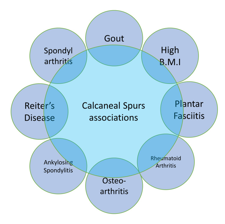
Calcaneal spurs occur in varied types of disorders, a few are shown in the Figure 1.
Figure 1. Associations of calcaneal spurs.
B.M.I.: Body mass index
The figure is created by the author.
Gout
Gout is a type of inflammation in arthritis caused due to the gradual and chronic deposition of uric acid crystals. These crystals, which get deposited in the joints, cause severe pain, leading to inflammatory conditions in the affected joint. Deposition of these crystals in the joints serves as the diagnosis of gout. Sometimes the mono sodium urate crystals may also get deposited in soft tissues such as Achilles tendon. These conditions lead to the formation of calcaneal spurs at the site of insertion. A study also shows a significant relationship in patients with gout developing calcaneal, and Achilles spur. The study had a sample size of 181 patients. 44.7% of patients in the study showed the presence of either Achilles or plantar spur, and 22.1% of patients had both Achilles and calcaneal spur. The patients with either of the two spurs showed a greater association with high mean serum uric acid, metabolic comorbidities, duration after diagnosis of gout till the appearance of the spur, disease duration and hypertension. They concluded that the presence of comorbidities increased the disposition to development of spurs pertaining due to the pathology of faulty microvasculature and hypoxia, resulting in calcification on the site of insertion of the ligament and tendon [15].
Increased body weight
Obesity is the direct causative factor for the development of calcaneal spurs, with studies showing a positive correlation between increased body weight and calcaneal spurs conducted in military recruits [16]. Forty-five percent of the population had obesity and calcaneal spurs, compared to 9% who weren't obese. The concept of this association is backed by the theory of vertical compression in the development of calcaneal spur, which is affected by the altered gait of the person in obesity. Excessive body mass may cause the process of the increase in the heel pad and the toughening of the plantar fascia and even cause a faster age-related degeneration process [17]. Obesity has also been mentioned in a few studies as the cause of loss of the foot's medial arch, creating more traction and supporting the tractional theory of spur formation [9]. No clear causality exists between increased BMI according to studies but a higher degree of association with heel pain. There may be heel pain due to the increased BMI as the vertical forces are higher due to gravity, or there may be an increase in BMI because of heel pain [6,18].
Plantar fasciitis
From the literature reviewed, only one article said there isn't enough evidence to establish a connection to say that calcaneal spurs cause plantar fasciitis [3]. Other articles highlighted that there is a direct link between calcaneal spurs as one of the causative factors of plantar fasciitis [18-20]. Calcaneal spurs can be classified based on size, shape, and position in patients with plantar fasciitis. Plantar fasciitis is said to be a benign and self-limiting condition due to a degenerative process instead of an inflammation condition [18]. A study including thirty patients with heel pain who also had calcaneal spurs were treated surgically by removing the spur and the fascia removed, called endoscopic plantar fasciotomy, and the relationship between them was studied to classify the types of spurs. They classified them into two types, type A and type B. Type A was superior to the fascia, and type B was located within the fascia. Type B showed more severity in magnetic resonance imaging of plantar fasciitis before the surgical intervention. They also found a higher granulocyte count in type B rather than type A spurs. The patients did not show spur recurrence [19].
Rheumatoid arthritis, osteoarthritis, ankylosis spondylitis, spondylarthritis, and reactive arthritis
Studies show a frequent association of calcaneal spurs in patients with rheumatoid arthritis, ankylosing spondylitis, and Reiter's disease (Reactive arthritis). One of the studies showed that 21.6% of patients with rheumatoid arthritis had developed calcaneal spurs and 16% in controls owing to erosive property of rheumatoid arthritis and an increase in the incidence of osteoarthritis with increasing age [13]. Frequencies of each rheumatoid arthritis, Reiter's disease, osteoarthritis, and ankylosis spondylitis were calculated, and the highest incidence was seen in patients with Reiter's disease of 31% out of 16 patients with a complete triad (nongonococcal urethritis, arthritis, and conjunctivitis) for talalgias of moderate to severe type. Calcaneal spurs are the characteristic feature of Reiter's disease. The type of pain in Reiter's disease was more severe as compared to the other conditions [21]. Another study showed the association between spondylarthritis {SpA} and Achille's tendon enthesitis and bone erosion being separate processes, and SpA is related to the normal mechanic only. Still, these individuals showed smaller spurs at the initial stages of SpA and then larger spurs if the disease is chronic. The regions which were expected to undergo erosion showed a low bone-to-marrow ratio [22].
Clinical symptoms
Calcaneal spurs usually present as moderate to high heel pain, also called talalgia. Hence, the clinical symptoms are majorly pertaining to plantar heel pain. Plantar heel pain can arise from three broad origins. One is from the skeletal origin, such as calcaneal stress fractures, and inflammatory conditions. The second is soft tissue pathology, such as previously discussed foot fat pad atrophy, plantar fascia injury, and plantar fasciitis. Finally, heel pain may be induced because of neural causes such as compression of nerves, specifically Baxter's Nerve or lateral plantar nerve and medial calcaneal nerve, and tarsal tunnel syndrome [23].
Pain
According to studies, the patients with calcaneal spurs and plantar fasciitis reported early morning pain for 88% out of the study group of 250 patients. When they take their first steps, the pain gets relieved after walking almost half of them. The patients with foot pad atrophy had intense, severe pain, tingling, burning sensation, and cold. The pain usually increases after long walks, especially at night or after work while resting [23].
Foot anatomy and position
Ankle dorsiflexion was found to be painful and difficult in patients, the cause behind it being the biomechanical problem of heel cord tightness. There was an increased association seen in patients with pes planus, also known as flat foot, than patients with pes cavus [23].
Treatment
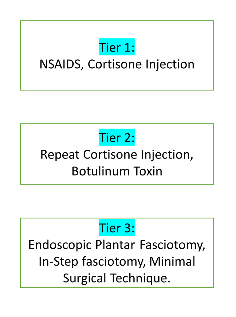
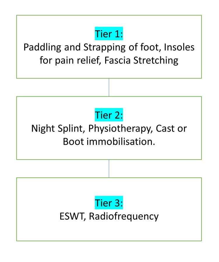
There do exist many treatment modalities in research for calcaneal enthesophytes. Still, patients are usually known to suffer from the symptoms due to the lack of actual treatment modalities used clinically. This research aims to compile data for all the research treatment modalities present, to apply. Conventional treatment includes radiofrequency, ultrasound, laser, non-steroidal anti-inflammatory drugs, steroids, local anesthesia, extracorporeal therapy, splints, modified shoes with silicon shoe implants, physiotherapy, stretching, and taping (Figures 2, 3) [24].
Figure 2. Medical management of calcaneal spur.
NSAIDs: Non-steroidal anti-inflammatory drugs
The figure is created by the author.
Figure 3. Conservative treatment of calcaneal spurs.
ESWT: Extracorporeal shockwave therapy
The figure is created by the author.
Gastrocnemius-soleus stretching over other stretching methods
A study found the increased effectiveness of gastrocnemius-soleus stretching in patients aged 30-70 suffering from painful heel spur syndrome. It established that gastrocnemius-soleus stretching is more effective than Tendo-Achille's stretching, as the group of patients who practiced gastrocnemius-soleus stretching showed a significant amount of reduction in pain score than what was the initial pain score before intervention. The stretching of gastrocnemius and soleus muscles causes an increased range of dorsiflexion movement. This stretching relieves rigidity or lack of flexibility due to excessive pronation and the overcompensation of the first metatarsal joint's plantar fascia. This inflexibility also caused increased tension at the calcaneum insertions. Stretching is a type of conservative treatment for heel spur. This type of therapy is cost-effective and patient-centric; hence may become a better form of therapy in the long run [25].
Extracorporeal shock wave therapy and laser treatment therapy
Studies show that extracorporeal shock wave therapy has been effectively used to treat patients with calcaneal enthesophytes. They do so by using shocks of 0.03mJ/mm2 energy applied, which is gradually increased to 0.4mJ/mm2 since the patient may experience pain at the start. These variations in symptoms were measured using the visual analogue scale {VAS} [26]. Other diagnostic tests used were X- rays for dimension monitoring throughout the treatment and grade of enthesis by sonography [27,28]. These studies concluded that extracorporeal shock wave therapy was a safe treatment modality because the results showed a significant decrease in the visual analog scale of about 68% along with morphological changes on X-rays, but there were no changes in grades observed on sonography [24]. Another study was based on the efficacy comparison of extracorporeal shock wave therapy and laser treatment therapy. Two groups were subjected to different treatment modalities, and the results were evaluated based on the visual analog scale. There was no significant difference in their score [29]. Hence establishing both are equally good treatment modalities for calcaneal enthesophytes.
Cryo-ultrasound therapy
A study of a sample size of 102 patients with chronic plantar fasciitis and heel spurs debated the effectiveness of using cryo-ultrasound therapy or using just cryotherapy. They used different types of treatment on each subject group and evaluated their visual analog score. Treatment was administered 10 times, each lasting for 20 minutes. The patients were evaluated at time intervals of three months, 12 months, and 18 months. There was a difference of 3.00, 4.35, and 4.81 points, respectively, in the score observed after each follow-up in the group who had undergone cryo-ultrasound. Hence after reviewing the effectiveness index of both, a better response was seen in patients who had undergone cryo-ultrasound with long-lasting effects in relieving plantar fasciitis [30].
Radiological treatment
Various studies are present on radiological treatment as it is one of the least invasive therapy and is emerging as one of the most effective therapy with the least side effects. The effectiveness of the therapy is measured in terms of the reduction of pre-radiation pain [31]. In cases of intralesional radiofrequency treatment is used when they are not responding to anti-inflammatory drugs and corticosteroid injections or other mechanical management such as shoe insoles [32]. A low dose of radiation is used, and after compiling the articles under study, the frequency used ranges from 3-12 Gy with less or almost no side effects [24,33].
Surgery
The failure of conservative therapy leaves only surgical intervention, which can only be done after 9/12 months. The operations to treat plantar fasciitis due to calcaneal spur involve endoscopic plantar fascial resection, the most common surgical process done. It may be done along with calcaneal spur resection or alone. Both processes have been known to be effective. Endoscopic and fluoroscopic calcaneal spur resection without plantar fascial release also had favourable outcomes. The older literature shows another approach to creating a workspace while resectioning the plantar fascia and calcaneal spur. This creates a more invasive approach than endoscopic surgery. The post-operative care in the endoscopic approach is much faster than in other surgeries [34,35].
Conclusions
Calcaneal spurs, also known as calcaneal enthesophytes, is a condition whose cause has long been debated. This article intends to provide a summary of the theories put forth previously to hone in on the exact etiology and mechanism of the formation of a spur. The majority of cases reported have been theorized under vertical compression theory, which corresponds to the high association of increased BMI. After reviewing the literature, we found that there was also an increase in incidence seen with progressing age, which was also accompanied by cases of gout and other arthropathies in older patients. The patients are known to experience heel pain, also known as talalgia, throughout their life without seeking effective treatment. The new treatment modalities have been researched to find their effectiveness and their application to become standardized. Currently, there is a surgical procedure that shows the lowest rate of reformation of the spur and safer treatment, but it can only be done after 9-12 months. This research highlights the fact that there is a need for a treatment modality other than conservative treatment for the period before 9-12 months to reduce and alleviate the patient's condition suffering in pain. Extracorporeal shock and laser treatment are one of the best currently in study to become standardized for clinical practice. Calcaneal spurs cause many lifestyle changes, associated as a cause of increased BMI, affect heavy lifting, create inconvenience to a pregnant mother, and adversely affect the performance of athletes. It may become debilitating in patients for whom even walking may become a challenge worsening the condition itself. It also affects the footwear the patients need to use. This article puts forth the need to create a treatment and management paradigm against the wait-and-see approach in the calcaneal enthesophytes background.
Acknowledgments
Namrata R. Velagala, Tanishq Kumar, Arihant Singh and Ashok M. Mehendale contributed equally to the article and should be considered second authors.
The content published in Cureus is the result of clinical experience and/or research by independent individuals or organizations. Cureus is not responsible for the scientific accuracy or reliability of data or conclusions published herein. All content published within Cureus is intended only for educational, research and reference purposes. Additionally, articles published within Cureus should not be deemed a suitable substitute for the advice of a qualified health care professional. Do not disregard or avoid professional medical advice due to content published within Cureus.
Footnotes
The authors have declared that no competing interests exist.
References
- 1.Coexistence of plantar calcaneal spurs and plantar fascial thickening in individuals with plantar heel pain. Menz HB, Thomas MJ, Marshall M, et al. Rheumatology (Oxford) 2019;58:237–245. doi: 10.1093/rheumatology/key266. [DOI] [PMC free article] [PubMed] [Google Scholar]
- 2.A study of calcaneal enthesophytes (spurs) in Indian population. Kullar JS, Randhawa GK, Kullar KK. Int J Appl Basic Med Res. 2014;4:0–6. doi: 10.4103/2229-516X.140709. [DOI] [PMC free article] [PubMed] [Google Scholar]
- 3.The plantar calcaneal spur: a review of anatomy, histology, etiology and key associations. Kirkpatrick J, Yassaie O, Mirjalili SA. J Anat. 2017;230:743–751. doi: 10.1111/joa.12607. [DOI] [PMC free article] [PubMed] [Google Scholar]
- 4.History and mechanical control of heel spur pain. Bergmann JN. https://europepmc.org/article/med/2189536. Clin Podiatr Med Surg. 1990;7:243–259. [PubMed] [Google Scholar]
- 5.Biomechanics of longitudinal arch support mechanisms in foot orthoses and their effect on plantar aponeurosis strain. Kogler GF, Solomonidis SE, Paul JP. Clin Biomech Bristol Avon. 1996;11:243–252. doi: 10.1016/0268-0033(96)00019-8. [DOI] [PubMed] [Google Scholar]
- 6.Obesity and pronated foot type may increase the risk of chronic plantar heel pain: a matched case-control study. Irving DB, Cook JL, Young MA, Menz HB. BMC Musculoskelet Disord. 2007;8:41. doi: 10.1186/1471-2474-8-41. [DOI] [PMC free article] [PubMed] [Google Scholar]
- 7.Plantar fasciitis: a degenerative process (fasciosis) without inflammation. Lemont H, Ammirati KM, Usen N. J Am Podiatr Med Assoc. 2003;93:234–237. doi: 10.7547/87507315-93-3-234. [DOI] [PubMed] [Google Scholar]
- 8.The age dependent change in the incidence of calcaneal spur. Beytemür O, Öncü M. Acta Orthop Traumatol Turc. 2018;52:367–371. doi: 10.1016/j.aott.2018.06.013. [DOI] [PMC free article] [PubMed] [Google Scholar]
- 9.Plantar calcaneal spurs in older people: longitudinal traction or vertical compression? Menz HB, Zammit GV, Landorf KB, Munteanu SE. J Foot Ankle Res. 2008;1:7. doi: 10.1186/1757-1146-1-7. [DOI] [PMC free article] [PubMed] [Google Scholar]
- 10.The relationship between the thickness and elasticity of the heel pad and heel pain. Ozdemir H, Urgüden M, Ozgörgen M, Gür S. https://dergipark.org.tr/en/pub/aott/issue/18086/190575. Acta Orthop Traumatol Turc. 2002;36:423–428. [PubMed] [Google Scholar]
- 11.The incidence, age dependence and sex distribution of the calcaneal spur. An analysis of its x-ray morphology in 1027 patients of the central European population. Riepert T, Drechsler T, Urban R, Schild H, Mattern R. Rofo. 1995;162:502–505. doi: 10.1055/s-2007-1015925. [DOI] [PubMed] [Google Scholar]
- 12.Calcaneal spurs in a black African population. Banadda BM, Gona O, Vaz R, Ndlovu DM. Foot Ankle. 1992;13:352–354. doi: 10.1177/107110079201300611. [DOI] [PubMed] [Google Scholar]
- 13.Incidence of calcaneal spurs in osteo-arthrosis and rheumatoid arthritis, and in control patients. Bassiouni M. Ann Rheum Dis. 1965;24:490–493. doi: 10.1136/ard.24.5.490. [DOI] [PMC free article] [PubMed] [Google Scholar]
- 14.Changes in prevalence of calcaneal spurs in men & women: a random population from a trauma clinic. Toumi H, Davies R, Mazor M, Coursier R, Best TM, Jennane R, Lespessailles E. BMC Musculoskelet Disord. 2014;15:87. doi: 10.1186/1471-2474-15-87. [DOI] [PMC free article] [PubMed] [Google Scholar]
- 15.The frequency of Achilles and plantar calcaneal spurs in gout patients. Duran E, Bilgin E, Ertenli Aİ, Kalyoncu U. Turk J Med Sci. 2021;51:1841–1848. doi: 10.3906/sag-2011-201. [DOI] [PMC free article] [PubMed] [Google Scholar]
- 16.Plantar fasciitis/calcaneal spur among security forces personnel. Sadat-Ali M. https://academic.oup.com/milmed/article/163/1/56/4831783. Mil Med. 1998;163:56–57. [PubMed] [Google Scholar]
- 17.Chronic plantar heel pain modifies associations of ankle plantarflexor strength and body mass index with calcaneal bone density and microarchitecture. Rogers JA, Jones G, Cook J, Squibb K, Wills K, Lahham A, Winzenberg T. PLoS One. 2021;16:0. doi: 10.1371/journal.pone.0260925. [DOI] [PMC free article] [PubMed] [Google Scholar]
- 18.Plantar fasciitis. Orchard J. BMJ. 2012;345:0. doi: 10.1136/bmj.e6603. [DOI] [PubMed] [Google Scholar]
- 19.Classification of calcaneal spurs and their relationship with plantar fasciitis. Zhou B, Zhou Y, Tao X, Yuan C, Tang K. J Foot Ankle Surg. 2015;54:594–600. doi: 10.1053/j.jfas.2014.11.009. [DOI] [PubMed] [Google Scholar]
- 20.Relationship and classification of plantar heel spurs in patients with plantar fasciitis. Ahmad J, Karim A, Daniel JN. Foot Ankle Int. 2016;37:994–1000. doi: 10.1177/1071100716649925. [DOI] [PubMed] [Google Scholar]
- 21.The painful heel. Comparative study in rheumatoid arthritis, ankylosing spondylitis, Reiter's syndrome, and generalized osteoarthrosis. Gerster JC, Vischer TL, Bennani A, Fallet GH. Ann Rheum Dis. 1977;36:343–348. doi: 10.1136/ard.36.4.343. [DOI] [PMC free article] [PubMed] [Google Scholar]
- 22.Distinct topography of erosion and new bone formation in achilles tendon enthesitis: implications for understanding the link between inflammation and bone formation in spondylarthritis. McGonagle D, Wakefield RJ, Tan AL, et al. Arthritis Rheum. 2008;58:2694–2699. doi: 10.1002/art.23755. [DOI] [PubMed] [Google Scholar]
- 23.Clinical characteristics of the causes of plantar heel pain. Yi TI, Lee GE, Seo IS, Huh WS, Yoon TH, Kim BR. Ann Rehabil Med. 2011;35:507–513. doi: 10.5535/arm.2011.35.4.507. [DOI] [PMC free article] [PubMed] [Google Scholar]
- 24.Randomized multicenter follow-up trial on the effect of radiotherapy for plantar fasciitis (painful heels spur) depending on dose and fractionation - a study protocol. Holtmann H, Niewald M, Prokein B, Graeber S, Ruebe C. Radiat Oncol. 2015;10:23. doi: 10.1186/s13014-015-0327-6. [DOI] [PMC free article] [PubMed] [Google Scholar]
- 25.Effectiveness of gastrocnemius-soleus stretching program as a therapeutic treatment of plantar fasciitis. Arif MA, Hafeez S. Cureus. 2022;14:0. doi: 10.7759/cureus.22532. [DOI] [PMC free article] [PubMed] [Google Scholar]
- 26.Extracorporeal shock wave therapy in the supportive care and rehabilitation of cancer patients. Crevenna R, Mickel M, Keilani M. Support Care Cancer. 2019;27:4039–4041. doi: 10.1007/s00520-019-05046-y. [DOI] [PMC free article] [PubMed] [Google Scholar]
- 27.Efficacy of extracorporeal shock wave treatment in calcaneal enthesophytosis. Cosentino R, Falsetti P, Manca S, et al. Ann Rheum Dis. 2001;60:1064–1067. doi: 10.1136/ard.60.11.1064. [DOI] [PMC free article] [PubMed] [Google Scholar]
- 28.Extracorporeal shock wave therapy in the treatment of inferior calcaneal enthesophytosis: outcome by fan-beam dual x ray absorptiometry (DXA) Cosentino R, Frediani B, De Stefano R, et al. Ann Rheum Dis. 2004;63:1704–1705. doi: 10.1136/ard.2003.013755. [DOI] [PMC free article] [PubMed] [Google Scholar]
- 29.Comparison of effects of low-level laser therapy and extracorporeal shock wave therapy in calcaneal spur treatment: A prospective, randomized, clinical study. Badil Güloğlu S, Yalçın Ü. Turk J Phys Med Rehabil. 2021;67:218–224. doi: 10.5606/tftrd.2021.5260. [DOI] [PMC free article] [PubMed] [Google Scholar]
- 30.Cryoultrasound therapy in the treatment of chronic plantar fasciitis with heel spurs. A randomized controlled clinical study. Costantino C, Vulpiani MC, Romiti D, Vetrano M, Saraceni VM. https://www.researchgate.net/profile/Mario-Vetrano/publication/258203023_Cryoultrasound_therapy_in_the_treatment_of_chronic_plantar_fasciitis_with_heel_spurs_A_randomized_controlled_clinical_study/links/00b4952828d58d21fd000000/Cryoultrasound-therapy-in-the-treatment-of-chronic-plantar-fasciitis-with-heel-spurs-A-randomized-controlled-clinical-study.pdf. Eur J Phys Rehabil Med. 2014;50:39–47. [PubMed] [Google Scholar]
- 31.Low-dose radiation treatment for painful plantar enthesophyte: a highly effective therapy with little side effects. Djiepmo F, Tamaskovics B, Bölke E, et al. Eur J Med Res. 2022;27:28. doi: 10.1186/s40001-022-00642-x. [DOI] [PMC free article] [PubMed] [Google Scholar]
- 32.Effects of intralesional pulsed radiofrequency treatment on pain in patients with calcaneal spur: results of 460 patients. Eke I, Akcal MA, Sayrac AV, Iyetin Y. BMC Musculoskelet Disord. 2021;22:1033. doi: 10.1186/s12891-021-04926-x. [DOI] [PMC free article] [PubMed] [Google Scholar]
- 33.Painful plantar heel spur treatment with Co-60 teletherapy: factors influencing treatment outcome. Koca T, Aydın A, Sezen D, Başaran H, Karaca S. Springerplus. 2014;3:21. doi: 10.1186/2193-1801-3-21. [DOI] [PMC free article] [PubMed] [Google Scholar]
- 34.Fluoroscopic and endoscopic calcaneal spur resection without plantar fascial release for recalcitrant plantar fasciitis. Nakajima K. Foot Ankle Orthop. 2022;7:24730114221108104. doi: 10.1177/24730114221108104. [DOI] [PMC free article] [PubMed] [Google Scholar]
- 35.Percutaneous surgery for plantar fasciitis due to a calcaneal spur. Apóstol-González S, Herrera J. https://www.medigraphic.com/cgi-bin/new/resumenI.cgi?IDARTICULO=21875. Acta Ortop Mex. 2009;23:209–212. [PubMed] [Google Scholar]


