Abstract
Erwinia rhapontici is able to convert sucrose into isomaltulose (palatinose, 6-O-α-d-glucopyranosyl-d-fructose) and trehalulose (1-O-α-d-glucopyranosyl-d-fructose) by the activity of a sucrose isomerase. These sucrose isomers cannot be metabolized by plant cells and most other organisms and therefore are possibly advantageous for the pathogen. This view is supported by the observation that in vitro yeast invertase activity can be inhibited by palatinose, thus preventing sucrose consumption. Due to the lack of genetic information, the role of sucrose isomers in pathogenicity has not been evaluated. Here we describe for the first time the cloning and characterization of the palatinose (pal) genes from Erwinia rhapontici. To this end, a 15-kb chromosomal DNA fragment containing nine complete open reading frames (ORFs) was cloned. The pal gene products of Erwinia rhapontici were shown to be homologous to proteins involved in uptake and metabolism of various sugars from other microorganisms. The palE, palF, palG, palH, palK, palQ, and palZ genes were oriented divergently with respect to the palR and palI genes, and sequence analysis suggested that the first set of genes constitutes an operon. Northern blot analysis of RNA extracted from bacteria grown under various conditions implies that the expression of the palI gene and the palEFGHKQZ genes is oppositely regulated at the transcriptional level. Genes involved in palatinose uptake and metabolism are down regulated by sucrose and activated by palatinose. Palatinose activation is inhibited by sucrose. Functional expression of palI and palQ in Escherichia coli revealed sucrose isomerase and palatinase activity, respectively.
Isomaltulose (commonly referred to as palatinose, 6-O-α-d-glucopyranosyl-d-fructose) and trehalulose (1-O-α-d-glucopyranosyl-d-fructose) are functional isomers of sucrose. Several microorganisms, such as Protaminobacter rubrum (32), Serratia plymuthica (11), Klebsiella planticola (18), and Erwinia rhapontici (5), have been found to form palatinose and trehalulose from sucrose. The adaptive role of sucrose isomer formation is unclear. Microorganisms in nature are often faced with a “feast-or-famine” type of existence, and many bacteria have evolved biochemical systems for the production of storage compounds that serve as reserve material. These storage compounds become especially important under conditions of limited nutrient supply. Therefore, it was suspected that the bioconversion of sucrose may be a method of irreversibly sequestering a carbon and energy source in a form unavailable to competitors such as the host plant or other microorganisms (6), and sucrose conversion by Erwinia rhapontici may play a similar role to 3-ketosucrose formed by Agrobacterium tumefaciens (12) and the gluconate and 2-oxoglucose produced by Pseudomonas aeruginosa (33).
The formation of sucrose isomers in E. rhapontici, a pathogen associated with crown rot in rhubarb, is accomplished through the activity of a single enzyme, which has been located to the cell's periplasmic space (5). This sucrose isomerase is strictly substrate specific toward sucrose, with a Km of 0.28 M, whereby the reaction is essentially irreversible (5). The yield of palatinose formed from sucrose is about 85%. The remaining 15% is trehalulose. The lack of genetic studies on palatinose metabolism had prevented further insights into the physiological and evolutionary aspects of this pathway. In this study, we describe the cloning of the genes involved in palatinose metabolism from E. rhapontici using a previously described sucrose isomerase sequence (20) as a probe to screen a genomic library. We found that the genes responsible for uptake and utilization of palatinose and trehalulose were most likely to constitute an operon whereas the sucrose isomerase itself and a potential regulatory gene were located separately. Furthermore, the functional expression of the sucrose isomerase and palatinase in Escherichia coli and biochemical characterization of the recombinant protein are described.
MATERIALS AND METHODS
Bacterial strains and cultivation.
The bacterial strains, phages, and plasmids used in this work are described in Table 1. E. rhapontici DSM 4484 was grown at 30°C in Luria-Bertani medium (LB) with vigorous shaking or in M9 medium (23) supplemented with various carbon sources, as indicated in Results. All E. coli clones were routinely grown in LB containing appropriate antibiotics. For phage infection, LB containing 0.2% (wt/vol) maltose and 10 mM MgSO4 was used.
TABLE 1.
Strains and plasmids used in this study
| Strain, phage, or plasmid | Relevant properties | Source or reference |
|---|---|---|
| Strains | ||
| Erwinia rhapontici (DSM 4484) | Wild type | DSMZa |
| E. coli XL1-Blue MRF′ | Δ(mcrA)183 Δ(mcrCB-hsdSMR-mrr)173 endA1 supE44 thi-1 recA1 gyrA96 relA1 lac [F′ proAB lacIqZΔM15 Tn10 (Tcr)] | Stratagene |
| E. coli XL1-Blue | recA1 endA1 gyrA96 thi-1 hsdR17 supE44 relA1 lac [F′ proAB lacIqZΔM15 Tn10 (Tcr)] | Stratagene |
| E. coli XLOLR | Δ(mcrA)183 Δ(mcrCB-hsdSMR-mrr)173 endA1 thi-1 recA1 gyrA96 relA1 lac [F′ proAB lacIqZΔM15 Tn10 (Tcr)] | Stratagene |
| Phages | ||
| Lambda ZAP-Express | BamHI-digested arms | Stratagene |
| Plasmids | ||
| pCR-Blunt | E. coli cloning vector, Kanr | Invitrogen |
| pQE-11 | E. coli expression vector, Ampr | Qiagen |
| pBK-CMV | E. coli cloning vector, Kanr | Stratagene |
| pQ11 | pBK-CMV containing a 5,693-bp fragment of the pal gene cluster | This work |
| pPAL11 | pBK-CMV containing a 6,070-bp fragment of the pal gene cluster | This work |
| pYW1 | pBK-CMV containing a 3,872-bp fragment of the pal gene cluster | This work |
| pWY2 | pBK-CMV containing a 5,037-bp fragment of the pal gene cluster | This work |
| pWY3 | pBK-CMV containing a 5,516-bp fragment of the pal gene cluster | This work |
| pQE-palI | pQE-11 containing coding region of palI from nucleotides 109 to 1803 | This work |
| pQE-palQ | pQE-11 containing the complete coding region of palQ | This work |
DSMZ, Deutsche Sammlung von Mikroorganismen und Zellkulturen GmbH, Braunschweig, Germany.
DNA manipulations and library construction.
DNA manipulations were performed by standard procedures (23). Chromosomal DNA from E. rhapontici was isolated from cells harvested at early stationary growth phase. Cells from a 50-ml culture were harvested by centrifugation. The pellet was resuspended in 5 ml of lysis buffer (25 mM EDTA, [pH 8.0], 0.5% sodium dodecyl sulfate, 50 mM Tris HCl [pH 8.0]), and 5 ml of phenol-chloroform-isoamyl alcohol (25:24:1, vol/vol/vol) was added. After incubation for 10 min at 70°C, the mixture was centrifuged for 20 min at 14,000 rpm in a Sorvall SA-600 rotor. Phenol extraction was repeated three more times. Chromosomal DNA was precipitated from the aqueous phase by adding 1/10 volum of 8 M LiCl and 2.5 volumes of 100% ethanol and spooled out with a glass micropipette. The DNA was resolved in 400 μl of TE/RNase (10 mM Tris-HCl [pH 8.0], 1 mM EDTA, 100 μg of RNase/ml) and incubated at 37°C for 10 min to remove any residual RNA. Phenol extraction and DNA precipitation were repeated as described above. The DNA was washed using 80% ethanol and finally resolved in 400 μl of TE/RNase.
A 300-μg portion of E. rhapontici chromosomal DNA was partially digested with Sau3A, and a portion of the digested DNA was subsequently subjected to agarose gel electrophoresis. Bands between 5 and 12 kb were excised from the gel, and fragments were extracted using a Qiaquick gel purification kit (Qiagen, Hilden, Germany). An aliquot of the partially digested DNA was ligated to λ-ZAPExpress DNA (Stratagene, La Jolla, Calif.), packaged by using Gigapack III gold packaging extracts (Stratagene), and transduced to E. coli XL-MRF′ cells as specified by the manufacturer.
Cloning and sequencing.
A partial sequence encoding a sucrose isomerase from E. rhapontici has been previously described (20). Based on this information, gene-specific primers were designed and used to amplify a 1.3-kb fragment from genomic DNA by PCR. This fragment was labeled with [α-32P]dCTP by random priming and used to screen a genomic library. Several positive clones were rescued into plasmid pBK-CMV (Stratagene) and sequenced. Probes generated either by restriction digestion from the previously identified positive clones or by PCR using primers derived from such clones were used for additional rounds of screening until the entire palatinose gene cluster was covered, as revealed by sequence analysis.
Automated sequencing with an ABI 377 sequencer (Medigenomix, Martinsried, Germany) was performed with a single strand of the DNA fragments using standard and de novo primers. Sequence similarity search and analysis was performed with the BLAST 2.0 program (1) and the DNAstar package (DNASTAR Inc., Madison, Wisc.), respectively.
Construction of plasmids.
For expression in E. coli, the coding region of the sucrose isomerase (palI) ranging from codons 109 to 1803 was amplified by PCR with genomic DNA from E. rhapontici serving as a template. The gene-specific primers 5′-GGGATCCTCACCGTTCAGCAATCA-3′ and 5′-GTCGACCTACGGATTAAGTTTATA-3′ were used in this reaction, which introduced BamHI and SalI recognition sites into the sequences (underlined), respectively. The fragment was inserted into a pQE-11 vector (Qiagen) cut with the appropriate restriction enzymes. The entire coding region of palQ was amplified by PCR as above by using the gene-specific primers 5′-GAGATCTTGCGCAGCACACCGCACTGG-3′ and 5′-GTCGACTCACAGCCTCTCAATAAG-3′, which carried a BglII site and a SalI site, respectively. The fragment was inserted into the pQE-11 vector as described above.
Protein expression in E. coli and preparation of enzyme extracts.
A 50-ml volume of LB supplemented with the appropriate antibiotics was inoculated with 500 μl of an overnight culture of E. coli XL-I blue cells harboring the respective expression construct. When the optical density at 600 nm reached approximately 0.5, expression of the protein was induced by adding isopropyl-β-d-thiogalactopyranoside (IPTG) to a final concentration of 0.5 mM. After further incubation for 3 h, the cells were harvested by centrifugation for 10 min at 5,000 × g and 4°C. For preparation of the recombinant sucrose isomerase, the pellet was resuspended in 1 ml of 50 mM sodium phosphate buffer (pH 6.0). For preparation of the recombinant palatinase, 1 ml of 30 mM HEPES-KOH (pH 7.5) was used for resuspension. Bacteria were disrupted by ultrasonic disintegration using six 30-s bursts interspersed with cooling on ice. The suspension was then centrifuged at 28,000 × g for 20 min at 4°C, and the supernatant was used for enzyme measurements.
Assay of sucrose isomerase activity.
Enzyme activity was measured by incubating aliquots of crude extract from the E. coli expressor strain, prepared as described above, with 90 μl of sucrose solution at a final concentration of 292 mM in 50 mM sodium phosphate buffer (pH 6.0) in a final volume of 100 μl at 30°C for 30 min if not otherwise indicated. Qualitative sugar analysis was performed by high-pressure liquid chromatography (HPLC). For quantitative measurements, the amount of reducing power generated during the reaction was determined essentially as described previously (4). In brief, after 30 min the reaction was stopped and the generation of reducing sugars was determined by adding 1 ml of 3,5-dinitrosalicylic acid reagent (4). The samples were boiled for 10 min and rapidly cooled to room temperature. Relative activity was determined by reading the absorbance at 570 nm.
Determination of palatinase activity.
Extracts from the palQ E. coli expressor strain were prepared in extraction buffer (30 mM HEPES-KOH [pH 7.5], 5 mM MgCl2, 1 mM EDTA, 10% glycerol) as described above. A 10-μl volume of crude extract was incubated with 30 mM HEPES-KOH (pH 7.5)–100 mM palatinose (Sigma, Taufkirchen, Germany) in a final volume of 100 μl for 40 min at 30°C unless otherwise stated. After incubation, the reaction was stopped by heating the mixture to 95°C for 5 min and subsequent cooling on ice. Activity was determined as release of free glucose measured via a coupled optic-enzymatic assay with hexokinase and glucose-6-phosphate dehydrogenase as described by Sonnewald (25).
HPLC analysis.
Samples were delivered by an Spectra Physics AS 3500 autosampler with a 100-μl fixed loop to a chromatography system (Dionex, Sunnyvale, Calif.) which included a gradient pump, an eluant-degassing module, and a pulsed electrochemical detector (with a gold electrode). Chromatograms were recorded using Dionex Peaknet chromatograhpy software (release 5.0). Separation was performed on a 4- by 250-mm Dionex Carbo-pack PA1 with a Carbo-pack PA1 guard column. Gradient elution was accomplished with 150 mM NaOH and 1 M sodium acetate buffer. HPLC grade water was mixed with sodium acetate buffer to produce the chromatographic gradient by using the following concentration changes: 0% at 0 to 4 min, 85% at 5 to 10 min, 0% at 10 to 15 min. Sugars were identified by comparison to standards. All standards were obtained from Sigma.
Determination of the inhibition of yeast invertase activity by palatinose.
Lyophilized yeast invertase (Boehringer, Mannheim, Germany) was dissolved in 50 mM sodium acetate (pH 5.2) at a final concentration of 1 ng/ml. A 10-μl volume of invertase solution was mixed with 90 μl of sucrose or palatinose solution in 50 mM sodium acetate (pH 5.2) at a final concentration of 100 mM. The mixture was incubated at 37°C for 20 min and neutralized by adding 10 μl of 1 M Tris-HCl (pH 8.0). The reaction was stopped by heating to 95°C for 5 min. Liberated glucose was measured as described by Sonnewald (25). To measure the inhibitory effect of palatinose on invertase activity, increasing amounts of palatinose were added to the reaction mixture, yielding the final concentrations indicated in Results.
Extraction of RNA and Northern blot experiments.
Total RNA from E. rhapontici was prepared by the method of Summers (27). RNA gels and RNA gel blotting were performed as described by Herbers et al. (17).
Nucleotide sequence accession numbers.
The nucleotide sequences of the E. rhapontici pal genes have been submitted to GenBank/EMBL/DDBJ under accession numbers AF279277 (palE), AF279278 (palF), AF279279 (palG), AF279280 (palH), AF279281 (palI), AF279282 (palK), AF279283 (palQ), AF279284 (palR), and AF279285 (palZ).
RESULTS AND DISCUSSION
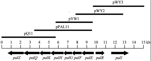
A genomic DNA library prepared from E. rhapontici was screened by using a previously described partial sequence coding for a sucrose isomerase (20) as a probe. To analyze the corresponding gene cluster in more detail, overlapping subfragments spanning a contiguous 15-kb DNA region were isolated using probes generated either by restriction digestion from the previously identified positive clones or by PCR using primers derived from such clones. Sequence analysis revealed nine complete loci within this 15-kb region (Fig. 1). Since they have been implicated in palatinose metabolism, we have named these genes pal. The genes are arranged in the transcriptional order palEFGHKQZ, with palR and palI being divergently oriented upstream of palE. Additionally, palR and palI seem to be individually transcribed. On the basis of sequence homologies, palK, palF, palG, and palE appear to encode a periplasmic-binding protein-dependent transport system whereas palR appears to encode a protein with regulatory properties. The closest homologues to palH, palI, palQ, and palZ found in the database encode proteins involved in breakdown of di- and oligoscaccharides. The proposed functions of the deduced gene products and their closest database homologues are summarized in Table 2. A more detailed analysis of the derived gene products is given below.
FIG. 1.
Molecular organization of the pal gene cluster from E. rhapontici. Arrows indicate the locations of open reading frames and the directions of gene transcription. Gene designations are given below the arrows. The individual clones used to assemble the contiguous DNA region are indicated as bars at the top of the figure.
TABLE 2.
Proposed functions of the deduced gene products and their closest database homologues
| Deduced protein | Closest database homologue (reference) | % of identical amino acids | Protein family (reference) | Sequence features |
|---|---|---|---|---|
| PalI | Bacillus thermoglucosidasius oligo-1,6-glucosidase (31) | 49 | Family 13 of glycosyl hydrolases (14, 16) | Contains invariant amino acids found at the active site of α-amylase enzymes (28) |
| PalR | Rhodococcus opacus ClcR (10) | 12 | LysR type transcriptional regulator | LysR family signature (2) |
| PalE | Sinorhizobium meliloti ThuEa | 39 | MalE family of periplasmic sugar-binding proteins | |
| PalF | Sinorhizobium meliloti ThuFa | 41 | MalFG family of inner membrane permeases | |
| PalG | Sinorhizobium meliloti ThuGa | 58 | MalFG family of inner membrane permeases | Inner membrane permease signature (2) |
| PalK | Salmonella typhimurium MalK (7) | 53 | ABC/ATP-binding cassette proteins | ATP/GTP-binding motif (P-loop) and ABC transporter signature (2) |
| PalH | Bacillus subtilis melibiase (α-galactosidase) (31) | 22 | Family 4 of glycosyl hydrolases (14, 16) | |
| PalQ | Bacillus thermoglucosidasius oligo-1,6-glucosidase (31) | 49 | Family 13 of glycosyl hydrolases (14–16) | Contains invariant amino acids found at the active site of α-amylase enzymes (28) |
| PalZ | Bacillus staerothermophilus exo-α-1,4-glucosidase (29) | 57 | Family 13 of glycosyl hydrolases (14–16) | Contains invariant amino acids found at the active site of α-amylase enzymes (28) |
J. B. Jensen, T. V. Bhuvaneswari, and N. K. Peters, unpublished data.
palK, palF, and palG are putative components of a ATP-dependent inner membrane permease.
The deduced gene products of palF and palG are homologous to members of the MalFG family of inner membrane sugar permeases. Hydrophobicity analysis suggests that both proteins form six membrane-spanning domains, which supports the notion that they are integral membrane proteins. PalK shows strong homology to several members of the ATP-binding cassette family of cytoplasmic ATP-hydrolyzing peripheral membrane proteins. The E. coli PalK homologue MalK is proposed to be involved in protein-protein interactions with MalG, which in turn is the E. coli homologue of PalG (8, 22). Taken together, it is very likely that PalK, PalG, and PalF constitute a system for sugar transport across the inner membrane.
PalE has homology to periplasmic solute-binding proteins.
PalE appears to be a member of the MalE periplasmic solute-binding protein family. It shows the closest homology to ThuE of Sinorhizobium meliloti, a putative trehalose- and maltose-binding protein, which in turn has homology to the maltose- and maltodextrin-binding protein MalE from E. coli (9). The sequence of PalE consists of 1,269 nucleotides; hence, the putative binding protein is composed of 422 residues and has an apparent molecular mass of 48 kDa. Support for the inference that palE is a periplasmic binding protein is provided by a putative N-terminal signal peptide with a predicted cleavage site at position 22 of the polypeptide (21). Furthermore, genes encoding periplasmic solute-binding proteins are located directly upstream of their associated inner membrane permease genes, which holds true for almost all examples found in the literature (3). Thus, its tempting to speculate that palE encodes a periplasmic palatinose- binding protein which would interact with the putative sugar permeases encoded by palF and palG.
PalR, a putative transcriptional regulator, is homologous to DNA binding proteins of the LysR family.
The palR gene product, upstream of palE and divergently oriented, shows a weak but significant homology to regulatory proteins of the LysR family. In response to different coinducers, LysR proteins activate divergent transcription of linked target genes (for an overview, see reference 24). All members of this family of proteins show a high degree of homology in their amino-terminal domains, where the helix-turn-helix DNA-binding region is located. In a proposed consensus sequence, the helix-turn-helix motif is located 23 residues from the amino-terminal end of the polypeptide (13). PalR possesses a sequence homologous to this motif within the 62 amino acid residues from the initial methionine residue.
PalI and PalQ have homology to α-1,6-glucosidases.
The predicted amino acid sequences of PalI and PalQ have homology to many members of family 13 of glycanases (14–16). Typically, members of this family are involved in breakdown of starch and its degradation products (28). Notably, PalI and PalQ have the highest homology to proteins involved in the hydrolysis of 1,6-α-d-glucosidic linkages in isomaltose and dextrins produced from starch and glycogen by α-amylase but have also been demonstrated to cleave palatinose (26). In turn, the two deduced protein sequences share about 70% homology to each other. PalI is most probably located in the cell's periplasmic space since in contains a putative N-terminal signal peptide with a potential cleavage site at position 22 (21).
PalZ has homology to α-1,4-glucosidases.
The closest homologue of the palZ gene product within the database is an α-1,4-glucosidase from Bacillus stearothermophilus (29). Along with PalI and PalQ, PalZ can be grouped into family 13 of glycanases (14–16). The enzyme also contains invariant amino acids found at the active site of α-amylases (28).
PalH has homology to α-galactosidases.
The deduced protein sequence of palH has significant homology to members of family 4 of glycanases (14–16). Interestingly, PalH has its highest homology to an α-galactosidase from Bacillus subtilis, which catalyzes the hydrolysis of melibiose (6-O-α-d-galactopyranosyl-d-glucopyranose) into galactose and glucose. Surprisingly, computer prediction (21) shows that PalH is most probably targeted to the inner membrane of the bacterium. This could argue for an involvement of PalH in uptake rather than in metabolism of sucrose isomers. Further studies are necessary to clarify the role of PalH in metabolism of sucrose isomers in E. rhapontici.
Expression of palI in E. coli reveals its sucrose isomerase activity.
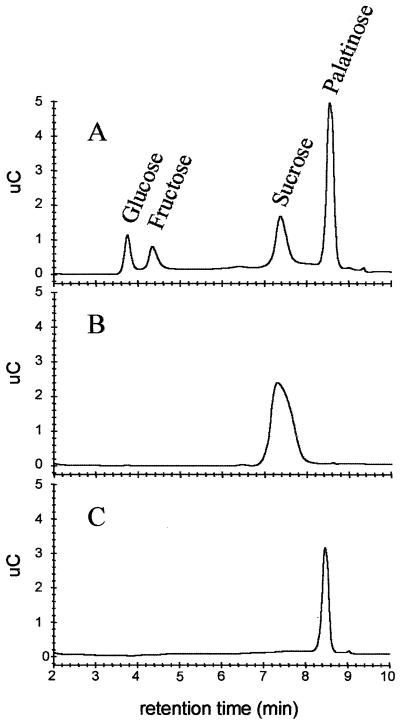
To confirm the nature of the palI product, the protein was expressed in E. coli under the control of an IPTG-inducible promoter. Enzymatic activity was assayed by incubation of a crude cell extract prepared from the expressor strain with sucrose solution and by subsequent sugar analysis via HPLC. Chromatograms indicated the presence of additional peaks in the reaction mixture (Fig. 2A). By comparison to standards, the major novel peak could be assigned to palatinose (Fig. 2C). The occurrence of glucose and fructose as by-products of the reaction has been described previously (5). Cell extracts from the control strain harboring the empty vector created no respective signals (Fig. 2B). This clearly demonstrates the sucrose isomerase activity of the recombinant PalI protein. However, the occurrence of trehalulose as the minor product of the reaction could not be demonstrated in this experiment.
FIG. 2.
(A and B) Composition of a sucrose solution after incubation with protein extract prepared from E. coli harboring the palI expression plasmid (A) in comparison to incubation with extract prepared from cells harboring the empty vector (B). (C) The palatinose peak was assigned from its elution positions and by comparison to a palatinose standard.
The optimum pH for enzyme activity was between 6.0 and 6.5, which is in good agreement with the finding that the enzyme is localized to the periplasmic space of E. rhapontici cells (5). The optimum temperature for isomerase activity was 30°C. It has previously been reported that the reaction temperature affects the composition of the product; i.e., the ratio between palatinose and trehalulose is skewed in the direction of trehalulose as the reaction temperature decreases (30). However, this phenomenon was not investigated in this experiment. From kinetic experiments, the Km for sucrose was 200 mM and the enzyme displayed maximal activity at a substrate concentration of 1.6 M. The Km is lower than the 280 mM reported for the native enzyme (5); however, this could reflect either differences in the detection method or slightly different kinetic properties possessed by the recombinant enzyme. The rather high Km value for sucrose supports the model of the sucrose isomerase reaction as a side reaction of carbon metabolism. If the sucrose uptake system of E. rhapontici has a lower Km than the sucrose isomerase, sucrose consumption is favored over palatinose production. Only a limited amount would be used for the formation of sucrose isomers as a reserve material, which would then be unavailable to competitors.
Characterization of palQ expressed in E. coli.
In a similar experiment to that described above, the palQ gene product was expressed in E. coli and palatinase activity was assayed as the release of free glucose from palatinose. Maximal activity was observed at 30°C and pH 7.0. Kinetic measurements revealed an apparent Km for palatinose of 10 mM, with maximal activity at 90 mM. Activity decreased slightly at substrate concentrations above 100 mM. However, an inhibitory effect of fructose as one of the reaction products could not be observed (data not shown), and due to the measuring principle, the effect of glucose on the reaction was not investigated. Thus, product inhibition effect on palatinase activity by glucose cannot be ruled out at this stage. The palatinase from E. rhapontici showed a strict substrate specificity toward palatinose. No release of glucose from the disaccharides sucrose, maltose, trehalose, and melibiose could be observed (data not shown).
Transcriptional regulation of the pal genes.
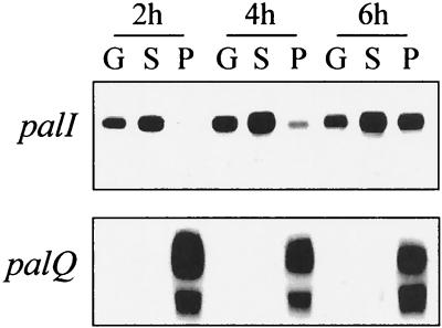
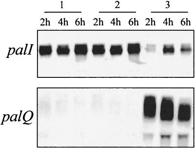
To determine the transcriptional regulation of the pal genes, Northern blot analysis was performed on total RNA of E. rhapontici that had been grown on different carbon sources with the DNA fragment of palI or palQ as a probe. As depicted in Fig. 3, the expression of palI was repressed in the presence of palatinose whereas a strong signal could be observed in the presence of glucose or sucrose. The expression appears to be even higher in the presence of sucrose than it is in the presence of glucose, indicating that sucrose is slightly inducing but is not necessary for expression per se. However, as the palatinose concentration in the medium declined during the prolonged incubation time, the repression of palI transcription was abolished. In turn, palQ expression was induced only in the presence of palatinose. The palQ probe appeared to give two bands on Northern blots. In fact, a ribosomal band interfered with the probe so that a spot of weaker hybridization occurred (Fig. 3). In the presence of sucrose, palatinose was not able to repress palI expression and the palQ transcript was not induced (induction would be a prerequisite for palatinose consumption) (Fig. 4).
FIG. 3.
Transcription of palI and palQ under different conditions of growth. Each well was loaded with 20 μg of total RNA isolated from exponentially growing cells harvested at the time points indicated. M9 medium was supplemented with 0.5% glucose (G), 0.5% sucrose (S), or 0.5% palatinose (P). The entire coding region of palI and palQ was used as a probe.
FIG. 4.
palI and palQ mRNA levels analyzed by Northern blot probing. Cells were harvested from exponentially growing cultures at the time points indicated. Each slot was loaded with 20 μg of total RNA isolated from M9 medium supplemented with 1% sucrose (lanes 1), with 1% sucrose and 1% palatinose (lanes 2), or with 1% palatinose (lanes 3).
This supports the notion that under conditions of excess carbon availability, palatinose is formed as a reserve material but its consumption is postponed until the preferentially metabolized carbon source (e.g., sucrose) is depleted, rather than being metabolized wastefully and unproductively.
Inhibition of a yeast invertase by palatinose.
As depicted in Table 3, palatinose itself is no substrate for an invertase from Saccharomyces cerevisiae. However, the presence of palatinose in the reaction mixture had a strong inhibitory effect on invertase activity in vitro. Concentrations of 20 mM palatinose decreased invertase activity to 76% compared to that in the control reaction utilizing sucrose alone. This proceeded to a residual activity of only 34% of the initial value when the palatinose concentration was increased to 100 mM. Hitherto, the in vivo amount of palatinose accumulation has not been determined. However, since the conversion of sucrose into palatinose can occur very efficiently, with less than 1% sucrose remaining (6), it is conceivable that due to the sometimes high concentration of sucrose in plant tissue, a similarly high concentration of palatinose accumulates. Hence, fungal invertases secreted into the surrounding of the cells would be strongly inhibited if the palatinose concentration is sufficiently high. This would impede carbon utilization for fungi and thus provide E. rhapontici with a mechanism to inhibit the spread of competitive organisms.
TABLE 3.
Inhibition of yeast invertase activity by palatinose
| Sugars | Relative invertase activity [%] ± SD |
|---|---|
| 100 mM sucrose | 100 |
| 100 mM palatinose | 0 |
| 100 mM sucrose–20 mM palatinose | 76 ± 2 |
| 100 mM sucrose–40 mM palatinose | 60 ± 6 |
| 100 mM sucrose–60 mM palatinose | 50 ± 5 |
| 100 mM sucrose–80 mM palatinose | 42 ± 1 |
| 100 mM sucrose–100 mM palatinose | 34 ± 6 |
Taken together, our results provide a plausible model for the adaptive role of sucrose conversion in that sucrose isomers are built during periods of excess carbon availability and sequestered in a form unavailable to competitors such as fungi or the host plant. The affinity of the sucrose isomerase toward sucrose is much too low to readily conclude that sucrose consumption via sucrose conversion is the only possibility for metabolization. Thus, sucrose conversion is only a minor mechanism of sucrose metabolism during periods of sufficient nutrient availability. Induction of the mobilizing enzyme does not occur until the preferred carbon source is depleted, and only then does the metabolism switch from palatinose production to consumption. As a consequence of sucrose depletion in that case, sucrose isomerase expression is halted.
Moreover, palatinose is a potent inhibitor of a yeast invertase, which provides a second strategy to exclude competitors from utilization of the available sucrose.
REFERENCES
- 1.Altschul S F, Madden T L, Schäffer A A, Zhang J, Zhang Z, Miller W, Lipman D J. Gapped BLAST and PSI-BLAST: a new generation of protein database search programs. Nucleic Acids Res. 1997;25:3389–3402. doi: 10.1093/nar/25.17.3389. [DOI] [PMC free article] [PubMed] [Google Scholar]
- 2.Bairoch A. PROSITE: a dictionary of sites and patterns in proteins. Nucleic Acids Res. 1992;20:2013–2018. doi: 10.1093/nar/20.suppl.2013. [DOI] [PMC free article] [PubMed] [Google Scholar]
- 3.Boos W, Lucht J M. Periplasmic binding protein-dependent ABC transporters. In: Neidhardt F C, Curtiss III R, Ingraham J L, Lin E C C, Low K B, Magasanik B, Reznikoff W S, Riley M, Schaechter M, Umbarger H E, editors. Escherichia coli and Salmonella: cellular and molecular biology. 2nd ed. Washington, D.C.: ASM Press; 1996. pp. 1175–1209. [Google Scholar]
- 4.Chaplin M F. Monosaccharides. In: Chaplin M F, Kennedy J F, editors. Carbohydrate analysis: a practical approach. Oxford, United Kingdom: IRL Press; 1986. p. 3. [Google Scholar]
- 5.Cheetham P S J. The extraction and mechanism of a novel isomaltulose-synthesizing enzyme from Erwinia rhapontici. Biochem J. 1984;220:213–220. doi: 10.1042/bj2200213. [DOI] [PMC free article] [PubMed] [Google Scholar]
- 6.Cheetham P S J, Imber C E, Isherwood J. The formation of isomaltulose by immobilized Erwinia rhapontici. Nature. 1982;299:628–631. [Google Scholar]
- 7.Dahl M K, Francoz E, Saurin W, Boos W, Manson M D, Hofnung M. Comparison of sequences from the malB regions of Salmonella typhimurium and Enterobacter aerogenes with Escherichia coli K12: a potential new regulatory site in the interoperonic region. Mol Gen Genet. 1989;218:199–207. doi: 10.1007/BF00331269. [DOI] [PubMed] [Google Scholar]
- 8.Dassa E. Sequence-function relationships in MalG, an inner membrane protein from the maltose transport system in E. coli. Mol Microbiol. 1993;7:39–47. doi: 10.1111/j.1365-2958.1993.tb01095.x. [DOI] [PubMed] [Google Scholar]
- 9.Duplay P, Bedoulle H, Fowler A, Zabin I, Saurin W, Hofnung M. Sequences of the malE gene and of its product, the maltose-binding protein of Escherichia coli K12. J Biol Chem. 1984;259:10606–10613. [PubMed] [Google Scholar]
- 10.Eulberg D, Kourbatova E M, Golovleva L A, Schlomann M. Evolutionary relationship between chlorocatechol catabolic enzymes from Rhodococcus opacus 1CP and their counterparts in proteobacteria: sequence divergence and functional convergence. J Bacteriol. 1998;180:1082–1094. doi: 10.1128/jb.180.5.1082-1094.1998. [DOI] [PMC free article] [PubMed] [Google Scholar]
- 11.Fujii S, Kishihara S, Komoto M, Shimizu J. Isolation and characterization of oligosaccharides produced from sucrose by transglucosylation action of Serratia plymuthica. Nippon Shokuhin Kogyo Gakkaishi. 1983;30:339–344. [Google Scholar]
- 12.Hayano K, Fukui S. Purification and properties of 3-ketosucrose-forming enzyme from the cells of Agrobacterium tumefaciens. J Biol Chem. 1967;242:3665–3672. [PubMed] [Google Scholar]
- 13.Henikoff S, Haughn G W, Calva J M, Wallace J C. A large family of bacterial activator proteins. Proc Natl Acad Sci USA. 1988;85:6602–6606. doi: 10.1073/pnas.85.18.6602. [DOI] [PMC free article] [PubMed] [Google Scholar]
- 14.Henrissat B. A classification of glycosyl hydrolases based on amino acid sequence similarities. Biochem J. 1991;280:309–316. doi: 10.1042/bj2800309. [DOI] [PMC free article] [PubMed] [Google Scholar]
- 15.Henrissat B, Bairoch A. New families in the classification of glycosyl hydrolases based on amino acid sequence similarities. Biochem J. 1993;293:781–788. doi: 10.1042/bj2930781. [DOI] [PMC free article] [PubMed] [Google Scholar]
- 16.Henrissat B, Romeau A. Families, superfamilies and subfamilies of glycosyl hydrolases. Biochem J. 1995;311:350–351. doi: 10.1042/bj3110350. [DOI] [PMC free article] [PubMed] [Google Scholar]
- 17.Herbers K, Mönke G, Badur R, Sonnewald U. A simplified procedure for the subtractive cDNA cloning of photoassimilate-responding genes: isolation of cDNAs encoding a new class of pathogenesis-related proteins. Plant Mol Biol. 1995;29:1027–1038. doi: 10.1007/BF00014975. [DOI] [PMC free article] [PubMed] [Google Scholar]
- 18.Huang J H, Hsu L H, Su Y C. Conversion of sucrose to isomaltulose by Klebsiella planticola CCRC 19112. J Inds Microbiol Biotechnol. 1998;21:22–27. [Google Scholar]
- 19.Lapidus A, Galleron N, Sorokin A, Ehrlich S D. Sequencing and functional annotation of the Bacillus subtilis genes in the 200 kb rrnB-dnaB region. Microbiology. 1997;143:3431–3441. doi: 10.1099/00221287-143-11-3431. [DOI] [PubMed] [Google Scholar]
- 20.Mattes, R., K. Klein, H. Schiweck, M. Kunz, and M. Munir. 28 July 1998. DNA's encoding sucrose isomerase and palatinase. U.S. Patent 5,786,140.
- 21.Nakai K, Kanehisa M. Expert system for predicting protein localization sites in Gram-negative bacteria. Proteins. 1991;11:95–110. doi: 10.1002/prot.340110203. [DOI] [PubMed] [Google Scholar]
- 22.Nikaido H. Maltose transport system of Escherichia coli: an ABC-type transporter. FEBS Lett. 1994;346:55–58. doi: 10.1016/0014-5793(94)00315-7. [DOI] [PubMed] [Google Scholar]
- 23.Sambrook J, Fritsch E F, Maniatis T. Molecular cloning: a laboratory manual. 2nd ed. Cold Spring Harbor, N.Y: Cold Spring Harbor Laboratory Press; 1989. [Google Scholar]
- 24.Schell M A. Molecular biology of the LysR family of transcriptional regulators. Annu Rev Microbiol. 1993;47:597–626. doi: 10.1146/annurev.mi.47.100193.003121. [DOI] [PubMed] [Google Scholar]
- 25.Sonnewald U. Expression of E. coli inorganic pyrophosphatase in transgenic plants alters photoassimilate partitioning. Plant J. 1992;2:571–581. [PubMed] [Google Scholar]
- 26.Stefani A, Janett M, Semenza G. Small intestinal sucrase and isomaltase split the bond between glucosyl-C1 and the glycosyl oxygen. J Biol Chem. 1975;250:7810–7813. [PubMed] [Google Scholar]
- 27.Summers W C. A simple method for extraction of RNA from E. coli utilizing diethyl pyrocarbonate. Anal Biochem. 1970;33:459–463. doi: 10.1016/0003-2697(70)90316-7. [DOI] [PubMed] [Google Scholar]
- 28.Svensson B. Protein engineering in the α-amylase family: catalytic mechanism, substrate specificity, and stability. Plant Mol Biol. 1994;25:141–157. doi: 10.1007/BF00023233. [DOI] [PubMed] [Google Scholar]
- 29.Takii Y, Takahashi K, Yamamoto K, Sogabe Y, Suzuki Y. Bacillus staerothermophilus ATCC12016 α-glucosidase specific for α-1,4 bonds of maltosaccharides and α-glucans shows high amino acid sequence similarities to seven α-d-glucohydrolases with different substrate specificity. Appl Microbiol Biotechnol. 1996;44:629–634. [Google Scholar]
- 30.Véronèse T, Bouchu A, Perlot P. Rapid method for trehalulose production and its purification by preparative high-performance liquid chromatography. Biotechnol Tech. 1999;13:43–48. [Google Scholar]
- 31.Watanabe K, Chishiro K, Kitamura K, Suzuki Y. Proline residues responsible for thermostability occur with high frequency in the loop regions of an extremely thermostable oligo-1,6-glucosidase from Bacillus thermoglucosidasius KP1006. J Biol Chem. 1991;266:24287–24294. [PubMed] [Google Scholar]
- 32.Weidenhagen R, Lorenz S. Palatinose [6-(-α-Glucopyranoside)-fructofuranose], ein neues bakterielles Umwandlungsprodukt der Saccharose. Z. Zuckerindust. 1957;7:533–534. [Google Scholar]
- 33.Whiting P H, Midgley M, Dawes E A. The role of glucose limitation in the regulation of transport of glucose, gluconate and 2-oxogluconate, and of glucose metabolism in Pseudomonas aeruginosa. J Gen Microbiol. 1976;92:304–310. doi: 10.1099/00221287-92-2-304. [DOI] [PubMed] [Google Scholar]


