Abstract
Antler is the fastest growing and ossifying tissue in animals and it is a valuable model for cartilage/bone development. To understand the molecular mechanisms of chondrogenesis and osteogenesis of antlers, the PacBio Sequel II and Illumina sequencing technology were combined and used to investigate the mRNA expression profiles in antler tip, middle, and base at six different developmental stages, i.e., at 15th, 25th, 45th, 65th, 100th and 130th growth days. Consequently, we identified 24,856 genes (FPKM > 0.1), including 8778 novel genes. Besides, principal component analysis (PCA) revealed a significant separation between the growth stage (25th, 45th and 65th days) and ossification stage (100th and 130th days). COL2A1 gene was significantly abundant in the growth stage, whereas S100A7, S100A12, S100A8, and WFDC18 genes were abundant at the ossification stage. Subsequently screened to 14,765 significantly differentially expressed genes (DEGs), WGCNA and GO functional enrichment analyses revealed that genes related to cell division and chondrocyte differentiation were up-regulated, whereas those with steroid hormone-mediated signaling pathways were down-regulated at ossification stages. Additionally, 25 tumor suppressor genes and 11 oncogenes were identified and were predicted to interact with p53. Co-regulation of tumor suppressor genes and oncogenes is responsible for the special growth pattern of antlers. Together, we constructed the most complete sika deer antler transcriptome database so far. The database provides data support for subsequent studies on the molecular mechanism of sika deer antler chondrogenesis and osteogenesis.
Subject terms: Transcriptomics, Gene expression
Introduction
Antlers are a unique feature of males in the Cervidae family which regenerate annually as per their sexual cycles. Except in genera Rangifer, where it is found in both sexes, the antler is typically a secondary feature of males. Antler is a bony outgrowth of the cranial frontal bones and differs from the keratinous horn of the Bovidae in that horns are not discarded but are annually re-grown. Annual antler regeneration originates from pedicle periosteum cells differentiating into adipocytes, chondrocytes, osteocytes, and neuron-like cells in vitro1. Antler bone growth occurs through a combination of modified endochondral ossification within each antler distal tip after cartilage formation, and intramembranous ossification within the shaft which increases antler diameter2. Notably, modified endochondral ossification involves extensive resorption of mineralized cartilage by osteoclast or chondroblast cells, followed by secretion of bone matrix by osteoblasts, and subsequent mineralization3. The rapid antler growth (up to 2 cm/d) is based on the proliferation and differentiation of reserve mesenchymal cells in the antler tip, which eventually evolve into cartilage tissue. Yu et al. reported that the correlation between antlers and osteosarcoma gene expression profile (r = 0.67 ~ 0.78) is higher than that of antlers and normal bone tissue (r = 0.33 ~ 0.47); they speculated the existence of similar patterns of antler growth and tumorigenesis4. Several proto-oncogenes (FOS, FAM83A, and REL) and tumor suppressor genes (PML, TP53 pathway-related, and ADAMTS family members) are positively selected in cervids and highly expressed in the antler.
Antler regrowth is regulated by local and systemic factors and is involved in the reproductive cycle. It is well established that testosterone is a major reproductive hormone regulating antler regrowth5,6. A high androgen level is necessary for the initiation of antler mineralization, whereas low levels are responsible for antler casting and regeneration. It was previously showed that testosterone can be converted into estradiol or dihydrotestosterone, which might be responsible for antler ossification and desiccation as well as the death of the velvet7. Meanwhile, Suttie found a strong positive correlation between antler growth rate and circulating IGF-1 concentration, which interacts with testosterone to stimulate intense antler growth8. Moreover, previous studies revealed a series of growth factors and signaling pathways involved in the regulation of antler regrowth, including IGF-1, parathyroid hormone-related peptide, retinoid acid, melatonin, and Wnt signaling pathway9,10.
These features render the antler a valuable model for studying bone growth and mineralization as well as epimorphic regeneration11. Therefore, several scientists have focused on exploring molecular regulatory mechanisms of regeneration, rapid growth, osteogenesis, and shedding of antlers using modern biological technologies (including transcriptomics, and proteomics)12–14. Furthermore, Xiumei Xing recently assembled a high-quality sika deer whole-genome sequence using combined technologies of SMRT, Illumina sequencing, and Hi-C, which significantly facilitated our work15. Antler growth is a classic "S" type curve, and its gene expression pattern undergoes major changes at different stages. Herein, we collected sika deer antlers during six different development periods, the initial growth period (15th and 25th days), rapid growth period (45th and 65th days), and ossification period (100th and 130th days). Subsequently, a gene expression database was constructed using transcriptomic technology. Meanwhile, we analyzed the gene expression dynamics and screened gene or signaling pathways related to antler chondrogenesis and osteogenesis. Our findings will improve the understanding of the biological processes for antler development at a molecular level. Antlers can complete the growth of cartilage, bone tissue and mineral accumulation in a short time to achieve rapid ossification. Thus, the exploration of the mechanism of rapid growth and ossification of antler provides a reference for the treatment of cartilage- and bone-related disorders, e.g. osteoporosis, fracture healing.
Results
Antler transcriptome sequencing
The RNA obtained from the antlers at growth stages (15, 25, 45, and 65 days) were combined to prepare the SMRTbell library (ANTLER1), and the RNA in ossification stages (100 and 130 days) to prepare another library (ANTLER2). We obtained 31.39 Gb of raw data and 14,495,425 sub-reads. The subread BAM of the offline data was self-corrected to obtain 1,367,298 CCS. After clustering the full-length non-chimeric sequences, 252,913 polished consensuses for ANTLER1 and 300,178 for ANTLER2 were generated (Table 1). Corrected consensus sequences were aligned to the sika deer reference genome using the GMAP software, showing a mapping rate of 86.72% and 88.26% for ANTLER1 and ANTLER2 (Table S2). Finally, 86,321 isoforms were generated, of which 14,402 were aligned to unannotated regions of the reference genome GTF file, corresponding to 6593 non-redundant novel genes (Table S3). The IsoSeq cluster representative sequences were added to the reference genome GTF file construct a new reference genome for Illumina short reads alignment and gene expression analysis.
Table 1.
Data output using the SMRT sequencing platforms.
| Statistics | ANTLER1 | ANTLER2 |
|---|---|---|
| Subreads base (G) | 14.49 | 16.9 |
| Subreads numbers | 6,838,310 | 7,657,115 |
| Circular Consensus Sequence | 625,715 | 74,1583 |
| Full-length non-chimeric reads | 441,644 | 545,108 |
| Consensus reads | 252,913 | 300,178 |
| Mean_length of transcripts (bp) | 3240 | 3247 |
| Min_length of transcripts (bp) | 170 | 189 |
| Max_length of transcripts (bp) | 17,475 | 17,839 |
| N50 of transcripts (bp) | 4223 | 4214 |
| N90 of transcripts (bp) | 2070 | 2020 |
Additionally, 48 samples were analyzed using Illumina technology (Table S1). In total, 2,172,875,780 clean reads were obtained after filtering raw data; the short reads were successfully mapped back to the new reference genome with an average mapping ratio of 90.96%. Genome-mapping results from all the sequenced short-reads data were gathered, assembled using Cufflinks, and compared to known gene models using the Cuffcompare, yielding a total of 2185 novel genes.
Gene functional annotation
In total, 30,227 genes were functionally annotated against seven public databases (Table 2), of which 21,449 were derived from the reference genome, 6593 were novel genes from Iso-Seq, and 2185 were novel genes from short reads. 28,698 gene sequences (94.94%) were successfully aligned to more than one public database, out of which the NT database had the highest annotation ratio at 93.55%; followed by the NR database with 84.46%. A total of 20,909 genes sequences were annotated in the SwissProt database, corresponding to 17,168 entries.
Table 2.
Statistics of BLASTX database searches for gene sequences.
| Databases | Number of genes | Percentage (%) |
|---|---|---|
| NR | 25,530 | 84.46 |
| NT | 28,280 | 93.55 |
| KEGG | 13,383 | 44.27 |
| Swiss-Prot | 20,909 | 69.17 |
| PFAM | 18,329 | 60.63 |
| GO | 18,738 | 61.99 |
| KOG | 8965 | 29.65 |
| In all Databases | 5668 | 18.75 |
| At least one Database | 28,698 | 94.94 |
Gene expression analysis
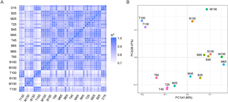
First, we performed correlation and principal component analysis (PCA). The squares of Pearson's correlation coefficient (R2) were all greater than 0.8, implying that the samples had a good biological repetition (Fig. 1A). PCA analysis revealed that antlers at the growth and ossification periods had a clear separation. Moreover, the antler tip was different from the antler's middle and base (Fig. 1B).
Figure 1.
The correlation (A) and principal component analysis (B) for the antler samples. The figures weres generated using the R software (vesion 4.1.3, https://www.r-project.org/).
Further, we characterized the 20 most abundant genes per sample (Table S4), covering 51 genes. Among these, Collagen alpha-1(I) chain (COL1A1), Collagen alpha-2(I) chain (COL1A2), Secreted protein acidic and rich in cysteine (SPARC), and Thymosin beta-10 (TMSB10) were shared by all samples. Cartilage marker genes (COL2A1, COL3A1) were specifically prevalent in the top 20 list of T45 and T65, whereas the abundance of Protein S100-A7 (S100A7), Protein S100-A12 (S100A12), Protein S100-A8 (S100A8), and WAP four-disulfide core domain protein 18 (WFDC18) drastically increased in T100 and T130. Additionally, several high-abundance genes were related to different parts of the antler. For instance, the expression of osteopontin (OPN), Osteocalcin (OCN), and pigment epithelium-derived factor (PEDF) in the antler middle and base was higher than that in the corresponding antler tip. Although at 45th and 65th days were adjacent periods, the FPKM value of OCN of M65 was 20 times that of M45.
Differentially expressed gene analysis
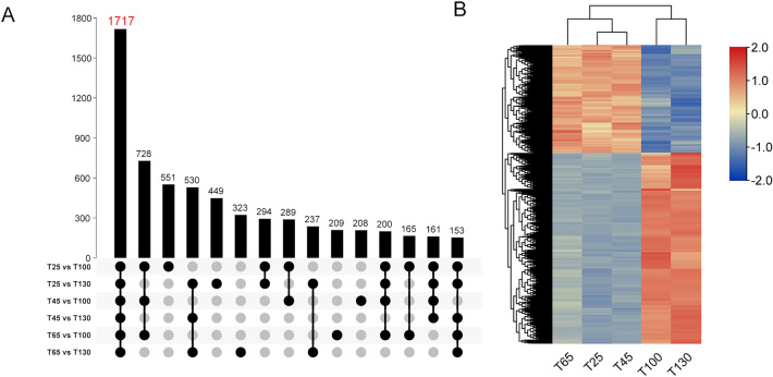
The DESeq2 software was used to screen the significantly differentially expressed genes (DEGs) (p-adjusted < 0.05, |log2(foldchange)|> 1) and 14,765 DEGs were obtained. As shown in Table S5, a small number of DEGs were screened in the groups (T25 vs. T45, T25 vs. T65 and T45 vs. T65), which were 8, 7 and 1 respectively. On the contrary, abundant DEGs were discovered between the growing antler tip and ossification antler tip (T25 vs. T100, T25 vs. T130, T45 vs. T100, T45 vs. T130, T65 vs. T100, T65 vs. T130), among which 1717 DEGs were shared by the 6 groups (Fig. 2A). Based on the expression levels of these genes, the antler tip from the five periods could be divided into two clusters, in which T25, T45 and T65 were in one cluster, and T100 and T130 were in the other cluster. Heat map of gene expression showed that 610 genes were up-regualted and 1107 genes were down-regulated in T25, T45 and T65 (Fig. 2B). Gene function enrichment analysis revealed that up-regulated genes were mainly related to collagen fiber assembly, chondrocyte differentiation, osteoblast differentiation, and glycolysis (Table S6). The down-regulated genes were primarily related to the establishment of the skin barrier, arachidonic acid metabolism, innate immune response, and ovarian steroidogenesis. Additionally, the difference between the antler tip and middle gradually increased with the developing antler, whereas the difference between the antler middle and base was smaller.
Figure 2.
Differentially expressed gene analysis. (A) UpSet Plot for DEGs between the growing antler tip and ossification antler tip. 1717 DEGs were shared by the 6 groups. (B) Heat map of 1717 differentially expressed genes at growth and ossification stages. The UpSet Plot and gene expression heat map were generated using TBtools software (version 1.082, https://github.com/CJ-Chen/TBtools).
WGCNA analysis for DEGs
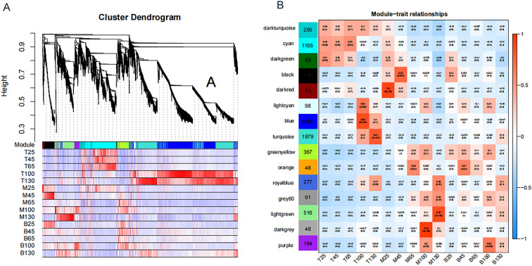
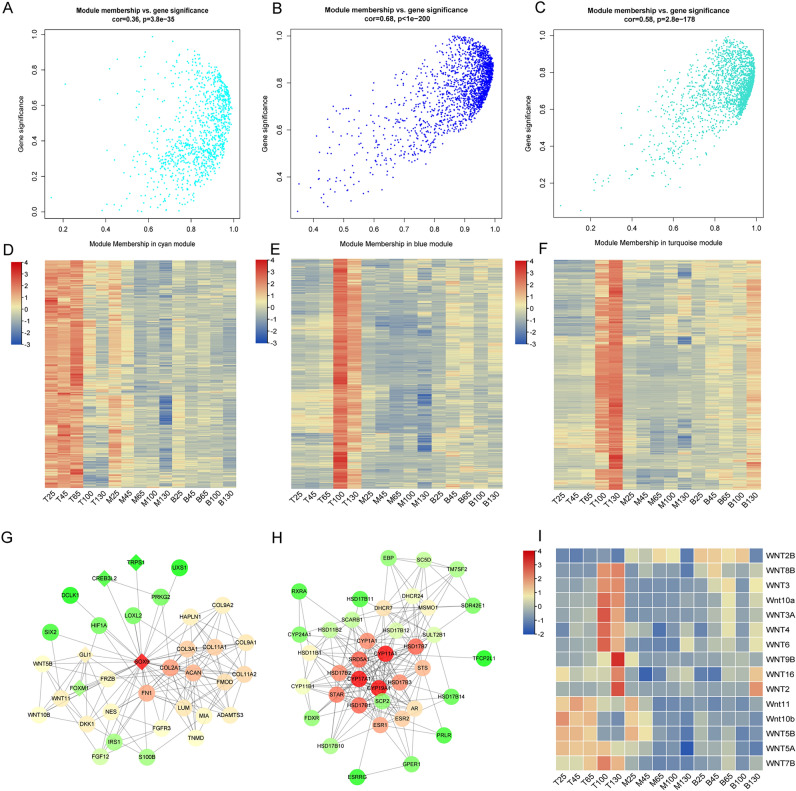
Notably, genes with similar expression patterns participate in similar pathways or have similar functions. Therefore, the weighted gene co-expression network analysis (WGCNA) was used for the modular analysis of DEGs to screen characteristic module genes of different samples (Fig. 3A). Consequently, 7417 DEGs were divided into 15 distinct modules (labeled with different colors in Fig. 3B) and we presented correlation coefficients between the characteristic genes of each module and each different sample. Of note, 3 modules were worthy of special attention, i.e., cyan, blue, and turquoise, which contained 67% of DEGs (Fig. 4A~F). Cyan module comprised 1105 genes, which positively correlated with T25, T45, and T65. Both blue and turquoise modules revealed a positive correlation with the T100 and T130, where the blue module comprised 1910 genes whereas the turquoise module contained 1979 genes.
Figure 3.
Through WGCNA analysis, 7417 DEGs were divided into 15 distinct modules. (A) Gene dendrogram and correlation heat map between gene and module. Red represents positively correlated, blue depicts negatively correlated; (B) Correlation heat map between modules and sample. The figures were generated by R WGCNA package (version 1.71, https://cran.r-project.org/package=WGCNA).
Figure 4.
Rapid growth and ossification-related genes and networks. (A, B, and C) Scatter diagram of module membership and gene significance for cyan, blue and turquoise module were obtained by R WGCNA package (https://cran.r-project.org/package=WGCNA); (D, E, and F) Expression heat maps of genes in cyan, blue and turquoise modules were generated using Tbtools software (version 1.082, https://github.com/CJ-Chen/TBtools); (G and E) Networks of genes associated with rapid growth and ossification were established by the STRING database (version 11.5, https://cn.string-db.org/) and visualized in the Cytoscape software (version 3.4.0, http://www.cytoscape.org/); (I) Heat map of 15 WNT gene expression in different antler samples were generated using Tbtools software.
For further functional enrichment analysis, genes with kME (for modular membership, also known as eigengene-based connectivity) greater than 0.7 were selected as module members. Cyan module contained 874 genes, which were enriched in biological processes including cell division, DNA replication, chondrocyte differentiation, and collagen catabolism (Fig. 4G, Table S7). Blue and Turquoise module genes were enriched in steroid hormone-mediated signaling pathways, retinoic acid receptor signaling pathway, Wnt signaling pathway, the establishment of the skin barrier, ceramide biosynthetic process, proteolysis, and retinol metabolic process (Fig. 4H~I, Table S7). Moreover, the Royalblue module revealed a positive correlation with T130, M130, and B130, and the Lightcyan module showed a positive correlation with T100, M100, and B100. These two modules can be used as the characteristic module genes of velvet antler at 130th and 100th growth days, respectively. Besides, special genes at the 130th antler growth day were enriched in translational initiation, immune response, and negative regulation of tumor necrosis factor production. The 100th-day special genes were related to innate immune response and defense response. Other modules comprised fewer genes or were less relevant to specific antler samples.
Tumor suppressor genes and oncogenes analysis
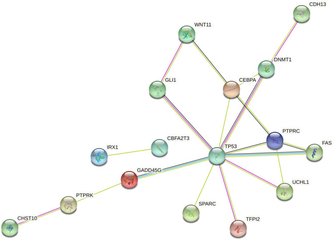
Significantly differentially expressed tumor suppressor genes and oncogenes were searched in Tumor Suppressor Gene Database16 (https://bioinfo.uth.edu/TSGene/index.html) and Oncogene database (http://ongene.bioinfo-minzhao.org/download.html). A total of 777 differentially expressed tumor suppressor genes and 508 oncogenes were screened, which were involved in cell cycle regulation, apoptosis, cell differentiation, etc. A total of 752 were differentially expressed genes in the antler and osteosarcoma, out of which 490 were also differentially expressed at different growth stages of antler; 25 were included in the list of tumor suppressor genes (Table 3), 11 were included in the list of oncogenes (Table 4). Moreover, STRING online software analysis showed that these genes interact with TP53, including GLI1, GADD45G, UCHL1, FAS, DNMT1, and PTPRC (Fig. 5).
Table 3.
25 tumor suppressors associated with antler development.
| Gene symbol | Gene name | FPKM | ||||||||
|---|---|---|---|---|---|---|---|---|---|---|
| T25 | T45 | T65 | T100 | T130 | Osteosarcoma | Osteosarcoma | Roe deer Antler | Sika deer Antler | ||
| PIWIL2 | Piwi-like protein 2 | 7.73 | 7.75 | 7.90 | 4.25 | 3.82 | 0.34 | 1.12 | 7.56 | 6.59 |
| PTPRK | Receptor-type tyrosine-protein phosphatase kappa | 3.86 | 3.99 | 3.83 | 12.43 | 11.53 | 0.18 | 0.09 | 8.15 | 6.76 |
| BTG3 | Protein BTG3 | 56.87 | 57.58 | 52.51 | 41.24 | 37.12 | 0 | 0 | 14.50 | 20.80 |
| TGM3 | Protein-glutamine gamma-glutamyltransferase E | 4.50 | 4.63 | 10.38 | 120.73 | 88.59 | 2.03 | 0 | 14.39 | 16.21 |
| FOXO6 | Forkhead box protein O6 | 1.11 | 1.48 | 2.53 | 14.28 | 8.96 | 0.37 | 0.1 | 5.10 | 4.19 |
| PTPRC | Receptor-type tyrosine-protein phosphatase C | 1.80 | 2.07 | 1.73 | 4.03 | 6.65 | 0 | 0.15 | 7.92 | 6.90 |
| EPHA1 | Ephrin type-A receptor 1 | 2.40 | 2.96 | 6.36 | 68.56 | 58.24 | 3.2 | 0.03 | 15.40 | 11.38 |
| IRX1 | Iroquois-class homeodomain protein IRX-1 | 2.87 | 2.26 | 2.35 | 26.45 | 17.65 | 0.44 | 0.18 | 13.41 | 8.64 |
| PLCD1 | 1-phosphatidylinositol 4,5-bisphosphate phosphodiesterase delta-1 | 88.64 | 132.62 | 98.37 | 39.61 | 50.39 | 9.22 | 11.59 | 64.56 | 67.16 |
| GADD45G | Growth arrest and DNA damage-inducible protein GADD45 gamma | 25.56 | 32.83 | 50.03 | 109.59 | 95.64 | 6.95 | 1.5 | 23.59 | 32.18 |
| CEBPA | CCAAT/enhancer-binding protein alpha | 21.14 | 20.48 | 33.60 | 280.77 | 170.46 | 5.25 | 2.09 | 38.33 | 43.15 |
| GLI1 | Zinc finger protein GLI1 | 10.31 | 9.10 | 17.96 | 6.60 | 1.51 | 1.09 | 0.25 | 3.12 | 3.15 |
| CHST10 | Carbohydrate sulfotransferase 10 | 96.70 | 121.45 | 76.59 | 31.56 | 59.25 | 3.87 | 4.71 | 24.00 | 32.99 |
| FAS | Tumor necrosis factor receptor superfamily member 6 | 2.48 | 3.05 | 3.04 | 6.73 | 11.85 | 8.16 | 12.28 | 1.23 | 0.76 |
| POU2F3 | POU domain, class 2, transcription factor 3 | 2.14 | 3.66 | 3.97 | 66.32 | 45.87 | 0.82 | 0.01 | 3.08 | 3.10 |
| PGRMC2 | Membrane-associated progesterone receptor component 2 | 117.91 | 116.99 | 109.99 | 347.47 | 304.30 | 16.53 | 10.72 | 103.30 | 118.08 |
| GUCY2C | Heat-stable enterotoxin receptor | 0.31 | 0.28 | 0.26 | 1.89 | 1.28 | 0.22 | 0.09 | 1.15 | 0.80 |
| TFPI2 | Tissue factor pathway inhibitor 2 | 43.01 | 31.07 | 40.06 | 102.47 | 260.43 | 1.08 | 0.12 | 50.91 | 60.36 |
| DNMT1 | DNA (cytosine-5)-methyltransferase 1 | 19.32 | 18.12 | 25.10 | 15.49 | 12.21 | 7.08 | 8.25 | 41.24 | 60.53 |
| SPARC | SPARC | 14,375.78 | 14,530.88 | 10,628.45 | 6504.71 | 4483.71 | 1286.8 | 1766.17 | 11,040.98 | 10,407.78 |
| CDH13 | Cadherin-13 | 24.90 | 24.07 | 16.84 | 55.67 | 74.93 | 3.02 | 5.53 | 20.53 | 25.20 |
| ADAMTS18 | A disintegrin and metalloproteinase with thrombospondin motifs 18 | 31.00 | 36.64 | 53.94 | 6.12 | 1.02 | 2.99 | 0.07 | 23.90 | 24.05 |
| CBFA2T3 | Protein CBFA2T3 | 3.30 | 2.71 | 2.82 | 7.61 | 10.14 | 1.27 | 0.26 | 5.20 | 4.30 |
| WNT11 | Protein Wnt-11 | 23.58 | 44.15 | 27.77 | 9.98 | 7.47 | 0.61 | 0.39 | 3.96 | 4.77 |
| UCHL1 | Ubiquitin carboxyl-terminal hydrolase isozyme L1 | 55.84 | 78.46 | 186.74 | 20.74 | 15.65 | 5.82 | 2.91 | 24.44 | 26.96 |
Table 4.
11 oncogenes associated with antler development.
| Gene symbol | Gene name | FPKM | ||||||||
|---|---|---|---|---|---|---|---|---|---|---|
| T25 | T45 | T65 | T100 | T130 | Osteosarcoma | Osteosarcoma | Roe deer Antler | Sika deer Antler | ||
| CNTN2 | Contactin-2 | 0.07 | 0.16 | 0.19 | 2.90 | 0.29 | 0.05 | 0.00 | 0.28 | 0.31 |
| GLI1 | Zinc finger protein GLI1 | 10.31 | 9.10 | 17.96 | 6.60 | 1.51 | 1.09 | 0.25 | 3.12 | 3.15 |
| HMGN5 | High mobility group nucleosome-binding domain-containing protein 5 | 11.66 | 9.31 | 9.87 | 6.01 | 7.64 | 5.55 | 2.11 | 16.49 | 14.74 |
| LMO1 | Rhombotin-1 | 1.77 | 1.38 | 2.91 | 30.18 | 18.24 | 0.51 | 0.14 | 7.60 | 6.28 |
| MYCN | N-myc proto-oncogene protein | 1.89 | 1.58 | 1.84 | 18.04 | 6.28 | 0.51 | 0.10 | 3.90 | 3.82 |
| SBSN | Suprabasin | 26.06 | 31.05 | 66.50 | 793.69 | 982.11 | 0.03 | 0.00 | 103.82 | 113.76 |
| SPARC | SPARC | 14,375.78 | 14,530.88 | 10,628.45 | 6504.71 | 4483.71 | 1286.80 | 1766.17 | 11,040.98 | 10,407.78 |
| TGM3 | Protein-glutamine gamma-glutamyltransferase E | 4.50 | 4.63 | 10.38 | 120.73 | 88.59 | 2.03 | 0.00 | 14.39 | 16.21 |
| TMEM140 | Transmembrane protein 140 | 4.67 | 5.22 | 4.81 | 8.91 | 15.60 | 0.97 | 0.74 | 11.73 | 7.91 |
| UCHL1 | Ubiquitin carboxyl-terminal hydrolase isozyme L1 | 55.84 | 78.46 | 186.74 | 20.74 | 15.65 | 5.82 | 2.91 | 24.44 | 26.96 |
| WNT3 | Proto-oncogene Wnt-3 | 1.11 | 0.93 | 1.90 | 24.10 | 24.99 | 0.03 | 0.00 | 2.75 | 1.76 |
Figure 5.
Network of interaction between tumor suppressor and p53 gene. The network was established by the STRING database (version 11.5, https://cn.string-db.org/) and visualized in the Cytoscape software (version 3.4.0, http://www.cytoscape.org/).
qRT-PCR validation of DEGs
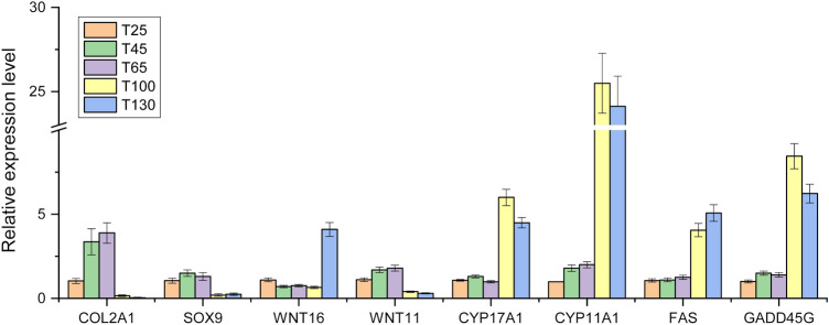
To further validate the results of RNA-seq, 8 DEGs, including Collagen alpha-1(II) chain (COL2A1), Transcription factor SOX-9 (SOX9), Protein Wnt-16 (WNT16), Protein Wnt-11 (WNT11), Steroid 17-alpha-hydroxylase/17,20 lyase (CYP17A1), Cholesterol side-chain cleavage enzyme (CYP11A1), Tumor necrosis factor receptor superfamily member 6 (FAS), Growth arrest and DNA damage-inducible protein GADD45 gamma (GADD45G), were selected to perform qRT-PCR. COL2A1, SOX9 and WNT11 had higher FPKM in the growing stage of antler, while WNT16, CYP17A1, CYP11A1, FAS and GADD45G had higher FPKM in antler ossifitaion stages. As shown in Fig. 6, the relative expression level of the genes obtained by qRT-PCR were basically consistent with FPKM values by Illumina RNA-seq, which confirmed the reliability of the sequencing data.
Figure 6.
The relative expression levels of 8 genes by qRT-PCR. The figure was generated by OriginPro 2018 software (b9.5.0.193, https://www.originlab.com/).
Discussion
The sika deer (Cervus nippon) reference genome was adopted in this work, and the RNA-seq mapping ratio was greater than 89%; this was adequate for subsequent analyses. Additionally, we integrated full-length transcriptome analysis, which yielded an additional 6593 non-redundant novel genes; 96.53% of the sequences were annotated in at least one public database. A total of 1343 sequences were blasted to the SwissProt database, corresponding to 398 non-redundant proteins. Higher FPKM values were found in protein S100-A9, WFDC18, and other genes unidentified in the reference genome. Therefore, the integration of SMRT sequencing further improves the transcriptome expression data of deer antlers.
The dynamic analysis of gene expression showed that the genes related to chondrogenic differentiation had higher FPKM values in the growth stage of antler, this is consistent with the previous proteomics results14, including COL2A1, COL11A2, ACANA, and HAPLN1. COL2A1 is a good marker gene for the analysis of molecular events causing initiation and progressive chondrogenesis17. SOX9 is a crucial transcription factor that regulates cartilage development and activates the above-mentioned chondrocyte markers. Baojin Yao discovered that SOX9 participates in the rapid growth of antlers by controlling multiple cell lineages, including pluripotent mesenchymal cells, proliferating chondrocytes, and hypertrophic chondrocytes18. The comparative genomic analysis of ruminants showed that SOX9 is a highly conserved gene of horned ruminants, involved in the neural crest cell migration pathway and regulates the origin and horn development4. Moreover, a headgear-specific gene, FOXL2, was upregulated in growing antlers and located upstream of the SOX9. This further confirms the important regulatory role of the SOX9 gene in the rapid growth of the antler. Therefore, the rapid growth of antler may be attributed to the chondrogenesis process mediated by SOX9.
In addition to cartilage, bone tissue is also an important component of antler, which is visually demonstrated through the expression of bone matrix proteins (osteopontin and osteocalcin). The former was primarily expressed at the early stage of osteoblast differentiation, and the latter was a late osteoblast differentiation gene promoting bone matrix mineralization19,20. Additionally, S100 family genes appear to contribute to osteogenesis, as S100 genes are expressed at higher levels during the ossification phase, including S100A2, S100A4, S100A7, S100A8, S100A9, S100A12, S100A14, S100A15A, and S100A16. Among that, S100A8 was rich in neutrophils, macrophages, osteoblasts, and osteoclasts. In the presence of Zn2+ and Ca2+, S100A8/A9 protein heterodimer participates in cell migration, arachidonic acid metabolism, and bone marrow cell maturation21. Also, it increases the calcification of the cartilage matrix and bone trabeculae construction22. TLR4, a common receptor of S100A8/A9, was up-regulated in antlers at the 130th growth day, indicating that S100A8/A9 may mediate the rapid ossification process of antlers by binding TLR423,24.
Then, we performed functional enrichment analysis of the DEGs. The results showed that Wnt signaling pathway was an important enrichment pathway, which could mediate distinct effects on the initiation of chondrogenesis and differentiation of chondrocytes25–28. For instance, Wnt5a and Wnt5b promote early chondrogenesis at the same time inhibiting terminal differentiation. In contrast, Wnt4 suppresses the initiation of chondrogenesis and accelerates terminal chondrocyte differentiation. Besides, Wnt3a inhibits chondrogenesis of mesenchymal cells by stabilizing cell–cell adhesion, independent of β-catenin transcriptional activity29. Furthermore, Wnt16 is a vital determinant of cortical bone mass and is majorly derived from osteoblasts. Osteoblast-specific Wnt16 overexpression increases both cortical and trabecular bone mass and structure in mice30. Wergedal discovered that the periosteal bone formation rate and mineral apposition rate were reduced in Wnt16 knockout vs wild-type mice31. Ying Jin revealed that Wnt16 promotes osteoblast differentiation of periosteal-derived cells in vitro and in vivo with a potential application in improving bone regeneration32. Together, it is speculated Wnt16 signaling pathway can regulate ossification rather than chondrogenesis of antler.
Antler is considered to be a secondary sexual characteristic of male deer, and its periodic regeneration is also associated with sex hormone levels. In this study, it is found that several genes related to steroid hormone mediated signaling pathways were up-regulated during the ossification stage of antler, such as androgen receptor (AR), aromatase (CYP19A1), steroid 17-alpha-hydroxylase/17,20 lyase (CYP17A1), sex hormone-binding globulin (SHBG), 3-oxo-5-alpha-steroid 4-dehydrogenase 1 (SRD5A1). Androgen regulates bone growth, fracture healing, and bone mass maintenance in male animals. Its receptor (AR) is expressed in mesenchymal cells, osteoblasts, and osteoclasts. Androgen directly binds androgen receptors to regulate bone development in male animals. 5α-reductase, a protein encoded by SRD5A1, could transform testosterone into 5-alpha-dihydrotestosterone (DHT) with more AR binding capacity. 5α-reductase can also be converted into estrogen under the action of P450 aromatase. Therefore, it can be concluded that testosterone play an important role in the ossification of antler. However, the signaling pathway mediated by testosterone still needs to be further explored.
Antler can achieve orderly and rapid growth, and its cell proliferation rate is 30 times faster than cancer cells. Therefore, many scientists have linked the development of antler with cancer, believing that the growth of antler is similar to bone cancer with normal bone growth33,34. The significant connection of gene expression between antler and osteosarcoma compared to antler and normal bone helps to explain this viewpoint. Nonetheless, unlike cancerous tissues whose rapid growth is unregulated and pathological, antler tissue regeneration is a highly coordinated physiological process and regulated by multiple tumor suppressors and proto-oncogenes. Based on previous studies, we further screened significantly differentially expressed tumor suppressor genes and oncogenes during antler growth. These genes play important roles in both the initiation and progression of cancer by regulating the cell cycle and apoptosis. For instance, PLCD1 suppresses the proliferation, invasion, and migration of esophageal squamous cell carcinoma by suppressing the Wnt/β-catenin signaling pathway35. GLI1 is a terminal effector of the Hh signaling pathway36, which activates numerous downstream proteins, including SHH, PTCH, and cycle-regulating proteins, including proto-oncogene N-MYC and cycliD. DNMT1 is a DNA methyltransferase, whose high expression in tumors is linked to chronic inflammation or persistent viral infection37. Additionally, overexpression of these genes including GLI1, GADD45G, UCHL1, PTPRC, DNMT1, SPARC, TFPI12, and FAS activate transcription of the p53 gene, which causes apoptosis38–40. The p53 signaling pathway regulates cell division and prevents tumor formation. Yu et al. found that p53 cofactor genes including PML, NMT2, and CD2AP, and p53 regulator genes including ELOVL6, S100A8, ISG15, CNOT3, and CCDC69 were expressed in antlers by positive selection of cervid lineage4. Summarily, co-regulation of tumor suppressor genes and oncogenes is responsible for special antler growth patterns.
Conclusion
In conclusion, we combined the PacBio and RNA-seq technologies to analyze the gene expression profiling of antlers during different developmental stages. The results showed that genes associated with chondrogenesis including COL2A1, COL3A1, COL11A2, ACANA, HAPLN1, and SOX9 are closely related to the rapid growth process of antler and androgen may promote antler ossification. Moreover, based on previous studies, we further screened the tumor suppressor genes and oncogenes associated with the specific growth pattern of antler. Therefore, our findings provide a reference for subsequent studies on the molecular mechanism of sika deer antler chondrogenesis and osteogenesis.
Materials and methods
Samples selection and preparation
Nine 4-year-old male sika deer (Cervus nippon hortulorum) was selected from the deer farm of Institute of Special Animal and Plant Sciences, Chinese Academy of Agricultural Sciences. Antlers were harvested on the 15th, 25th, 45th, 65th, 100th, and 130th growth days after casting the previous hard antlers, respectively. Each period had 3 antlers from different deer as repetitions. Therefore, 18 antlers were obtained as samples for this experiment. The deer was first anesthetized with 5% xylazine at 0.5 mg/kg body weight intramuscularly; this was performed according to the ARRIVE guidelines (https://arriveguidelines.org). To remove contaminants from the surface, antlers were washed using PBS buffer. Subsequently, the slices were selected at the tip, middle, and base of antlers, respectively, and ground into powder using the liquid nitrogen grinder. Specifically, the 15-day antler was not segmented and was only used for database construction. All animal experimental protocols were approved and authorized by the Chinese Academy of Agricultural Sciences Animal Care and Use Committee.
RNA extraction
Total RNA was isolated from antlers using the TRIzol reagent (Invitrogen, CA, USA). RNA degradation and contamination were monitored on 1% agarose gels. Purity, concentration, and integrity were assessed using the NanoPhotometer spectrophotometer (IMPLEN, CA, USA), Qubit 2.0 Fluorimeter (Life Technologies, CA, USA), and Bioanalyzer 2100 system (Agilent Technologies, CA, USA). High-quality RNA was used for transcriptome library construction.
Transcriptome library construction and sequencing
The Iso-Seq library was prepared as per the Isoform Sequencing protocol (Iso-Seq) using the Clontech SMARTer PCR cDNA Synthesis Kit (Clontech Laboratories, CA, USA) and the BluePippin Size Selection System protocol as described by Pacific Biosciences (PN 100-092-800-03) and sequenced on a PacBio Sequel II platform. The Illumina library was generated using the NEBNext Ultra™ RNA Library Prep Kit for Illumina (NEB, CA, USA) following the manufacturer’s recommendations and sequenced on an Illumina 2500 platform.
Data processing
Raw Iso-Seq data was processed using the SMRTlink 7.0 software. Circular consensus sequence (CCS) was generated from subread BAM files, parameters: min_length 50, min_passes 1, max_length 15,000. CCS.BAM files were output, which were then classified into full length and non-full length reads using pbclassify.py, ignorepolyA false, minSeqLength 200. Non-full length and full-length fasta files produced were then fed into the cluster step, which does isoform-level clustering (ICE), followed by final Arrow polishing, hq_quiver_min_accuracy 0.99, bin_by_primer false, bin_size_kb 1, qv_trim_5p 100, qv_trim_3p 30. Additional nucleotide errors of SMRT high-quality consensus reads were corrected using the clean Illumina RNA-seq data with the software LoRDEC (vesion 0.7, parameters: k = 23; s = 3).
The GMAP software (version 2017-06-20) was used to align the high-quality polished consensus sequences to the sika deer whole genome sequences (https://bigd.big.ac.cn/gwh; Accession number: GWHANOY00000000) with parameters: no-chimeras; expand-offsets 1; -B 5; -f samse; -n 1. Gene structure analysis was performed using TAPIS (version 1.2.1) pipeline with default parameters. Reads comparable to unannotated regions of the genome were considered novel genes. The short reads were aligned to the reference genome using HISAT software (version 2.0.4) with default parameters.
Gene functional annotation
All newly predicted genes were annotated using public databases, including the NCBI non-redundant nucleotide database (NT), NCBI non-redundant protein database (NR), Swiss-Prot, Cluster of Orthologous Groups of proteins (COG), and Kyoto Encyclopedia of Genes and Genomes (KEGG)41, Protein family (Pfam) and Gene Ontology (GO). Further, searches were performed using the BLASTX algorithm with an E-value threshold of 10−5.
Gene expression analysis
Gene expression analysis was performed for each sample using the HTSeq software (vesion 0.6.1; parameters: -m union) with the model union. Statistical analyses of gene expression between different samples were performed according to FPKM. The Pearson correlation coefficients and principal component analysis were calculated and visualized using the R software (vesion 4.1.3). Differential gene expression analysis was performed using the DESeq2 software with screening criteria of foldchange ≥ 2 and P value < 0.05. All differential expression genes were analyzed using the WGCNA package of R software (FPKM > 0.1, Module Fold of 0.3, min, ModuleSize of 30, power threshold of 8). GO and KEGG enrichment analyses were conducted using the Tbtools software (version 1.082)42. Protein–protein interaction (PPI) network of DEGs was established by the STRING database (version 11.5) and visualized in the Cytoscape software (version 3.4.0). TBtools software (version 1.082) was used to draw UpSet Plot and gene expression heat map of DEGs.
Quantitative RT-PCR analysis
Quantitative RT-PCR (qRT-PCR) assay was performed using the SYBR® Premix Ex Taq™ kit (TaKaRa, Japan) on a Roche LightCycler480 instrument. The sika deer GAPDH gene was selected as an internal control. Primer sequences used in the experiment are listed in Table S8. Three biological replicates and three technical repeats were used for each gene and sample. The relative mRNA expression level was calculated based on the 2−ΔΔCT method and the figure was drawn with OriginPro 2018.
Ethical approval
The deer used in this study were approved and authorized by the Chinese Academy of Agricultural Sciences Animal Care and Use Committee. All experimental procedures were carried out in accordance with the approved guidelines and regulations.
Supplementary Information
Acknowledgements
This study was supported by funds from the Agricultural Science and Technology Innovation Program of China (CAAS-ASTIP-201X-ISAPS) and the Special Economic Animals Sharing Platform in China (TZDW2020).
Author contributions
R.Z. and X.X. conceived and designed the experiments. R.Z. performed the experiments and data analysis and prepared figures. R.Z. Y.D. and X.X. wrote the main manuscript text. All authors reviewed the manuscript.
Data availability
All datasets generated during the current study have been deposited in the NCBI under project number PRJNA831887 (https://www.ncbi.nlm.nih.gov/bioproject/PRJNA831887).
Competing interests
The authors declare no competing interests.
Footnotes
Publisher's note
Springer Nature remains neutral with regard to jurisdictional claims in published maps and institutional affiliations.
Supplementary Information
The online version contains supplementary material available at 10.1038/s41598-022-20244-1.
References
- 1.Li C, Yang F, Sheppard A. Adult stem cells and mammalian epimorphic regeneration-insights from studying annual renewal of deer antlers. Curr. Stem Cell Res. Ther. 2009;4:237–251. doi: 10.2174/157488809789057446. [DOI] [PubMed] [Google Scholar]
- 2.Price JS, Allen S, Faucheux C, Althnaian T, Mount JG. Deer antlers: A zoological curiosity or the key to understanding organ regeneration in mammals? J. Anat. 2005;207:603–618. doi: 10.1111/j.1469-7580.2005.00478.x. [DOI] [PMC free article] [PubMed] [Google Scholar]
- 3.Feleke M, et al. New physiological insights into the phenomena of deer antler: A unique model for skeletal tissue regeneration. J. Orthop. Transl. 2021;27:57–66. doi: 10.1016/j.jot.2020.10.012. [DOI] [PMC free article] [PubMed] [Google Scholar]
- 4.Wang Y, et al. Genetic basis of ruminant headgear and rapid antler regeneration. Science. 2019 doi: 10.1126/science.aav6335. [DOI] [PubMed] [Google Scholar]
- 5.Gaspar-Lopez E, et al. Biometrics, testosterone, cortisol and antler growth cycle in Iberian red deer stags (Cervus elaphus hispanicus) Reprod. Domest. Anim. 2010;45:243–249. doi: 10.1111/j.1439-0531.2008.01271.x. [DOI] [PubMed] [Google Scholar]
- 6.Akhtar RW, et al. Identification of proteins that mediate the role of androgens in antler regeneration using label free proteomics in sika deer (Cervus nippon) Gen. Comp. Endocrinol. 2019;283:113235. doi: 10.1016/j.ygcen.2019.113235. [DOI] [PubMed] [Google Scholar]
- 7.Bubenik GA, et al. Testosterone and estradiol concentrations in serum, velvet skin, and growing antler bone of male white-tailed deer. J. Exp. Zool. A Comp. Exp. Biol. 2005;303:186–192. doi: 10.1002/jez.a.139. [DOI] [PubMed] [Google Scholar]
- 8.Suttie JM, et al. Insulin-like growth factor 1 (IGF-1) antler-stimulating hormone? Endocrinology. 1985;116:846–848. doi: 10.1210/endo-116-2-846. [DOI] [PubMed] [Google Scholar]
- 9.Kierdorf U, Li C, Price JS. Improbable appendages: Deer antler renewal as a unique case of mammalian regeneration. Semin. Cell Dev. Biol. 2009;20:535–542. doi: 10.1016/j.semcdb.2008.11.011. [DOI] [PubMed] [Google Scholar]
- 10.Sun X, et al. Melatonin promotes antler growth by accelerating MT1-mediated mesenchymal cell differentiation and inhibiting VEGF-induced degeneration of chondrocytes. Int. J. Mol. Sci. 2022 doi: 10.3390/ijms23020759. [DOI] [PMC free article] [PubMed] [Google Scholar]
- 11.Landete-Castillejos T, et al. Antlers - Evolution, development, structure, composition, and biomechanics of an outstanding type of bone. Bone. 2019;128:115046. doi: 10.1016/j.bone.2019.115046. [DOI] [PubMed] [Google Scholar]
- 12.Hu P, et al. Full-length transcriptome and microRNA sequencing reveal the specific gene-regulation network of velvet antler in sika deer with extremely different velvet antler weight. Mol. Genet. Genom. 2019;294:431–443. doi: 10.1007/s00438-018-1520-8. [DOI] [PubMed] [Google Scholar]
- 13.Sui Z, et al. Quantitative proteomics analysis of deer antlerogenic periosteal cells reveals potential bioactive factors in velvet antlers. J. Chromatogr. A. 2020;1609:460496. doi: 10.1016/j.chroma.2019.460496. [DOI] [PubMed] [Google Scholar]
- 14.Zhang R, Li Y, Xing X. Comparative antler proteome of sika deer from different developmental stages. Sci. Rep. 2021;11:10484. doi: 10.1038/s41598-021-89829-6. [DOI] [PMC free article] [PubMed] [Google Scholar]
- 15.Xing, X. et al.. The first high-quality reference genome of sika deer provides insights for high-tannin adaptation (2021). [DOI] [PMC free article] [PubMed]
- 16.Zhao M, Sun J, Zhao Z. TSGene: A web resource for tumor suppressor genes. Nucleic Acids Res. 2013;41:D970–976. doi: 10.1093/nar/gks937. [DOI] [PMC free article] [PubMed] [Google Scholar]
- 17.Hering TM, Wirthlin L, Ravindran S, McAlinden A. Changes in type II procollagen isoform expression during chondrogenesis by disruption of an alternative 5' splice site within Col2a1 exon 2. Matrix Biol. 2014;36:51–63. doi: 10.1016/j.matbio.2014.04.004. [DOI] [PMC free article] [PubMed] [Google Scholar]
- 18.Yao B, et al. Sox9 functions as a master regulator of antler growth by controlling multiple cell lineages. DNA Cell Biol. 2018;37:15–22. doi: 10.1089/dna.2017.3885. [DOI] [PubMed] [Google Scholar]
- 19.Paredes R, et al. The Runx2 transcription factor plays a key role in the 1alpha,25-dihydroxy Vitamin D3-dependent upregulation of the rat osteocalcin (OC) gene expression in osteoblastic cells. J. Steroid Biochem. Mol. Biol. 2004;89–90:269–271. doi: 10.1016/j.jsbmb.2004.03.076. [DOI] [PubMed] [Google Scholar]
- 20.Shen Q, Christakos S. The vitamin D receptor, Runx2, and the Notch signaling pathway cooperate in the transcriptional regulation of osteopontin. J. Biol. Chem. 2005;280:40589–40598. doi: 10.1074/jbc.M504166200. [DOI] [PubMed] [Google Scholar]
- 21.Goyette J, Geczy CL. Inflammation-associated S100 proteins: New mechanisms that regulate function. Amino Acids. 2011;41:821–842. doi: 10.1007/s00726-010-0528-0. [DOI] [PubMed] [Google Scholar]
- 22.Zreiqat H, Howlett CR, Gronthos S, Hume D, Geczy CL. S100A8/S100A9 and their association with cartilage and bone. J. Mol. Histol. 2007;38:381–391. doi: 10.1007/s10735-007-9117-2. [DOI] [PubMed] [Google Scholar]
- 23.Luo G, et al. Bone marrow adipocytes enhance osteolytic bone destruction by activating 1q213(S100A7/8/9-IL6R)-TLR4 pathway in lung cancer. J. Cancer Res. Clin. Oncol. 2020;146:2241–2253. doi: 10.1007/s00432-020-03277-9. [DOI] [PMC free article] [PubMed] [Google Scholar]
- 24.Ehrchen JM, Sunderkotter C, Foell D, Vogl T, Roth J. The endogenous Toll-like receptor 4 agonist S100A8/S100A9 (calprotectin) as innate amplifier of infection, autoimmunity, and cancer. J. Leukoc Biol. 2009;86:557–566. doi: 10.1189/jlb.1008647. [DOI] [PubMed] [Google Scholar]
- 25.Ba H, Wang D, Yau TO, Shang Y, Li C. Transcriptomic analysis of different tissue layers in antler growth Center in Sika Deer (Cervus nippon) BMC Genom. 2019;20:173. doi: 10.1186/s12864-019-5560-1. [DOI] [PMC free article] [PubMed] [Google Scholar]
- 26.Andrade AC, Nilsson O, Barnes KM, Baron J. Wnt gene expression in the post-natal growth plate: Regulation with chondrocyte differentiation. Bone. 2007;40:1361–1369. doi: 10.1016/j.bone.2007.01.005. [DOI] [PMC free article] [PubMed] [Google Scholar]
- 27.Church V, Nohno T, Linker C, Marcelle C, Francis-West P. Wnt regulation of chondrocyte differentiation. J. Cell Sci. 2002;115:4809–4818. doi: 10.1242/jcs.00152. [DOI] [PubMed] [Google Scholar]
- 28.French DM, et al. WISP-1 is an osteoblastic regulator expressed during skeletal development and fracture repair. Am. J. Pathol. 2004;165:855–867. doi: 10.1016/S0002-9440(10)63348-2. [DOI] [PMC free article] [PubMed] [Google Scholar]
- 29.Hwang SG, Yu SS, Lee SW, Chun JS. Wnt-3a regulates chondrocyte differentiation via c-Jun/AP-1 pathway. FEBS Lett. 2005;579:4837–4842. doi: 10.1016/j.febslet.2005.07.067. [DOI] [PubMed] [Google Scholar]
- 30.Alam I, et al. Osteoblast-specific overexpression of human WNT16 increases both cortical and trabecular bone mass and structure in mice. Endocrinology. 2016;157:722–736. doi: 10.1210/en.2015-1281. [DOI] [PMC free article] [PubMed] [Google Scholar]
- 31.Wergedal JE, Kesavan C, Brommage R, Das S, Mohan S. Role of WNT16 in the regulation of periosteal bone formation in female mice. Endocrinology. 2015;156:1023–1032. doi: 10.1210/en.2014-1702. [DOI] [PMC free article] [PubMed] [Google Scholar]
- 32.Jin Y, Sun X, Pei F, Zhao Z, Mao J. Wnt16 signaling promotes osteoblast differentiation of periosteal derived cells in vitro and in vivo. PeerJ. 2020;8:e10374. doi: 10.7717/peerj.10374. [DOI] [PMC free article] [PubMed] [Google Scholar]
- 33.Price J, Allen S. Exploring the mechanisms regulating regeneration of deer antlers. Philos. Trans. R. Soc. Lond. Ser. B Biol. Sci. 2004;359:809–822. doi: 10.1098/rstb.2004.1471. [DOI] [PMC free article] [PubMed] [Google Scholar]
- 34.Wang DT, Chu WH, Sun HM, Ba HX, Li CY. Expression and functional analysis of tumor-related factor S100A4 in antler stem cells. J. Histochem. Cytochem. 2017;65:579–591. doi: 10.1369/0022155417727263. [DOI] [PMC free article] [PubMed] [Google Scholar]
- 35.He X, et al. PLCD1 suppressed cellular proliferation, invasion, and migration via inhibition of wnt/beta-catenin signaling pathway in esophageal squamous cell carcinoma. Dig. Dis. Sci. 2021;66:442–451. doi: 10.1007/s10620-020-06218-1. [DOI] [PubMed] [Google Scholar]
- 36.Lau BW, et al. Hedgehog/GLI1 activation leads to leukemic transformation of myelodysplastic syndrome in vivo and GLI1 inhibition results in antitumor activity. Oncogene. 2019;38:687–698. doi: 10.1038/s41388-018-0431-9. [DOI] [PMC free article] [PubMed] [Google Scholar]
- 37.Kanai Y, Hirohashi S. Alterations of DNA methylation associated with abnormalities of DNA methyltransferases in human cancers during transition from a precancerous to a malignant state. Carcinogenesis. 2007;28:2434–2442. doi: 10.1093/carcin/bgm206. [DOI] [PubMed] [Google Scholar]
- 38.Yoon JW, et al. p53 modulates the activity of the GLI1 oncogene through interactions with the shared coactivator TAF9. DNA Repair (Amst) 2015;34:9–17. doi: 10.1016/j.dnarep.2015.06.006. [DOI] [PMC free article] [PubMed] [Google Scholar]
- 39.Cai Q, et al. Effects of expression of p53 and Gadd45 on osmotic tolerance of renal inner medullary cells. Am. J. Physiol. Renal. Physiol. 2006;291:F341–349. doi: 10.1152/ajprenal.00518.2005. [DOI] [PubMed] [Google Scholar]
- 40.Xiang T, et al. The ubiquitin peptidase UCHL1 induces G0/G1 cell cycle arrest and apoptosis through stabilizing p53 and is frequently silenced in breast cancer. PLoS ONE. 2012;7:e29783. doi: 10.1371/journal.pone.0029783. [DOI] [PMC free article] [PubMed] [Google Scholar]
- 41.Kanehisa M, et al. KEGG for linking genomes to life and the environment. Nucleic Acids Res. 2008;36:D480–484. doi: 10.1093/nar/gkm882. [DOI] [PMC free article] [PubMed] [Google Scholar]
- 42.Chen C, et al. TBtools: An integrative toolkit developed for interactive analyses of big biological data. Mol. Plant. 2020;13:1194–1202. doi: 10.1016/j.molp.2020.06.009. [DOI] [PubMed] [Google Scholar]
Associated Data
This section collects any data citations, data availability statements, or supplementary materials included in this article.
Supplementary Materials
Data Availability Statement
All datasets generated during the current study have been deposited in the NCBI under project number PRJNA831887 (https://www.ncbi.nlm.nih.gov/bioproject/PRJNA831887).


