Abstract
Background
Chronic low-level inflammation is thought to play a role in many age-related diseases and to contribute to multimorbidity and to the disability related to this condition. In this framework, inflamma-miRs, an important subset of miRNA able to regulate inflammation molecules, appear to be key players. This study aimed to evaluate plasma levels of the inflamma-miR-181a in relation to age, parameters of health status (clinical, physical, and cognitive) and indices of multimorbidity in a cohort of 244 subjects aged 65- 97.
Methods
MiR-181a was isolated from plasma according to standardized procedures and its expression levels measured by qPCR. Correlation tests and multivariate regression analyses were applied on gender-stratified groups.
Results
MiR-181a levels resulted increased in old men, and significantly correlated with worsened blood parameters of inflammation (such as low levels of albumin and bilirubin and high lymphocyte content), particularly in females. Furthermore, we found miR-181a positively correlated with the overall multimorbidity burden, measured by CIRS Comorbidity Score, in both genders.
Conclusions
These data support a role of miR-181a in age-related chronic inflammation and in the development of multimorbidity in older adults and indicate that the routes by which this miRNA influence health status are likely to be gender specific. Based on our results, we suggest that miR-181a is a promising biomarker of health status of the older population.
Keywords: Aging, miRNA, Multimorbidity, Inflammation, miR-181a, Inflamma-miRs
Highlights
Levels of the inflamma-miR-181a correlate with multimorbidity burden in older people.
MiR-181a levels correlate with blood inflammation markers in a gender-specific manner.
MiR-181a is positively correlated with age in males but not in females.
The paths by which miR-181a can influence health status likely differ between genders.
Background
The continuous rise of older individuals in western societies is leading to a significant increase of the prevalence of age-related diseases, such as cardiovascular diseases, diabetes, dementia, and cancer. Alongside this, there has been a significant increase in the prevalence of multimorbidity, i.e., the coexistence of two or more health conditions in an individual, and comorbidity, i.e., the presence of one or more additional conditions concurrent with a primary condition, resulting in a higher proportion of subjects at higher risk of disability, functional loss, and death [1–4]. Currently, inflammaging, the upregulation of the inflammatory response occurring in normal aging process and that determines a chronic, low-grade pro-inflammatory state, is considered one of the central pathogenetic mechanisms at the basis of most diseases and comorbidities accompanying aging [5].
MicroRNAs (miRNAs) are small, non-coding endogenous RNA molecules that regulate gene expression either by mRNA cleavage/destabilization or inhibition of translation [6]. The high capacity of miRNAs to target large networks of mRNAs makes them master epigenetic regulators, able to fine-tune almost all cellular processes [7]. Multiple miRNAs have been demonstrated to regulate canonical aging signalling pathways, and increasingly recognized as important modulators of the processes associated with age-related decline [8–10]. As summarised in several reviews, the dysregulated expression of many of them has been linked to multiple chronic diseases of aging [11–15]. A crucial role in the induction of aging phenotypes seems to be played by a relatively small number of miRNAs involved in the regulation of immune response and inflammatory processes, termed inflamma-miRs [16, 17]. Diverse patterns of dysregulated expression profiles of inflamma-miRs (miR-21, miR-34, miR-126, miR-146a, and miR-155, to name a few) have been identified in pathological conditions [18–20], thus making them attractive biomarkers of the quality of aging.
Mir-181a, a member of the miR-181 family, is increasingly regarded as an inflamma-miR [21] because of its ability to modulate the expression of important anti-inflammatory (TGFβ and IL-10) and pro-inflammatory (IL-6, TNFα, IL-1) cytokines [22, 23], and key components of NF-κB signalling [24]. It also exerts immune modulatory functions mainly by regulating T cell differentiation and proliferation [25–28]. Recent research has also classified miR-181a as a mitochondrial miRNA (“mitomiR”) because its activity in controlling the expression of genes at the crossroads of mitochondrial function, response to inflammation and cellular senescence [21, 29].
The levels of this mediator of inflammation have been correlated with increasing number of chronic diseases, including coronary artery disease [30], neurodegenerative diseases [31] and cancer [31, 32]. Moreover, studies in mice support its role in the loss of muscle mass and function with age [33]. These findings highlight the potential of miR-181a as biomarker of aging and gauge of individual decline. To date, however, few studies have assessed the changes of miR-181a expression levels with age and the relationship with development of negative health outcomes associated to normal aging.
Also, gender differences remain largely unexplored, although this may be a clinically relevant factor considering that the differential expression of miRNAs between the sexes has been found an important underlying mechanism for gender-biased disease outcome [34].
Thus, the aim of this study was to investigate age-and sex-related changes in miR-181a levels in a cohort of subjects in the age-range 65–97 years and to explore the relationship with parameters (clinical, physical, and cognitive) related to the health status and with indices of multimorbidity.
Methods
Study participants
Samples used in this study have been collected from elderly nursing homes located in the Calabria region (southern Italy) as part of a larger study examining the quality of aging in Calabria. The study population included 244 subjects with age (range 65–97 years, mean age 82.45 ± 7.11 years) of which 160 were females (range 65–97 years, 83.18 ± 7.12) and 84 males (range 65–97 years, mean age 81.07 ± 6.91 years). A descriptive analysis of this segment of the population, comprising also indicators of their health and functional status, was provided in De Rango et al., 2011 [35]. According to this survey, for its characteristics the studied cohort is representative of the elderly Calabrian population of the same age range. All study participants underwent a multidimensional geriatric assessment designed to evaluate cognitive status, functional abilities, physical health, and social aspects. Data were collected through a structured questionnaire administered by a trained operator. Subjects with physical or mental severe impairments were excluded from the sampling. A peripheral blood sample was collected for each participant for clinical and laboratory examination, and an informed consent was signed. The study was performed following the Strengthening the Reporting of OBservational studies in Epidemiology (STROBE) statement [36].
Geriatric assessment
Physical performance
Muscle strength was assessed as hand grip strength (HGS) using a handheld dynamometer (SMEDLEY’s dynamometer TTM) while the subject was sitting with the arm close to his/her body. The test was repeated three times with the stronger hand and the maximum of these values was considered.
Gait speed was measured at the usual pace over 4-m (m). Timing began when subjects started foot movement and stopped when one foot contacted the ground after completely crossing the 4 m mark. Gait speed was measured using distance in meters and time in seconds (m/s). The best time of two attempts was recorded.
Functional activity
Disability was measured using the Activities of Daily Living (ADL) score [37]. The score is given counting the number of activities (bathing, dressing, toileting, transfer from bed to chair, and feeding) in which the participant is dependent or independent at the time of the visit. ADL scores were dichotomized as one if the subject was not independent in all five items and zero otherwise.
Cognitive performance
The mini mental state examination (MMSE) tool was used to assess cognitive function, evaluating orientation, episodic memory, attention, language, and construction functions [38]. The MMSE scores (from 0 to 30) were normalized for age and educational status [39]. A MMSE score < 24 was used to diagnose cognitive impairment.
Multimorbidity
Multimorbidity was evaluated using the modified Cumulative Illness Rating Scale (CIRS), a scoring system that measures the burden of chronic medical illnesses by considering 14 items, corresponding to different physiological systems (cardiac, vascular, respiratory, otorhinolaryngology, upper gastrointestinal, lower gastrointestinal, hepatic and pancreatic, renal, genitourinary, musculoskeletal and dermatologic, neurology, endocrinology, metabolic, breast, and psychiatric) [40, 41]. The score for each organ system was based on disease severity ranging from 0 (no problem) to 4 (severe impairment in function), resulting in a total score ranging from 0 to 56. Higher scores indicate higher morbidity burden. A comorbidity index was computed by counting the number of items ranking three or four in disease severity.
Biochemical measurements
Samples from the venous blood were withdrawn after an overnight fast of 12 h in the morning.
Biochemical measurements were performed at the Italian National Research Centre on Ageing (Cosenza) using standard protocols. The same blood sample was used for miRNA analysis.
RNA extraction and miRNA quantification
Plasma for miRNAs analysis was separated by centrifugation at 1800 g for 10 min at room temperature, collected in RNase-free tubes and further centrifuged at 1200 × g for 20 min at 10 °C to completely remove contaminant cells. All samples were kept at − 80 °C until analysis.
MiR-181a was isolated from 200 μL of plasma using miRNeasy® Serum/Plasma kit (Qiagen, Hilden, Germany) according to the manufacturer’s instructions. Arabidopsis thaliana miR-159a (assay ID 000,338) was used as a spike-in control throughout the workflow. RNA yield was quantified on the Qubit 2.0 Fluorometer (Life Technologies, Milan, Italy) and it was around 30–50 ng/mL each sample. Of this, 5 μL were converted in cDNA using TaqMan® microRNA Reverse Transcription Kit (Life Technologies) and stem–loop specific RT primers for miR-181a (hsa-miR-181a-5p assay, ID 000,480). Small nuclear (snRNA) U6 was used as endogenous control (assay ID 001,973). Afterward, quantitative real-time PCR was performed on a QuantStudio3™ Real-Time PCR System (Applied Biosystems, Milan, Italy) with automatic baseline setting. All reactions were run in triplicate. The relative expression levels of the miRNA in comparison with the normalizer were then calculated using the comparative threshold (Ct) method 2−ΔCt [42].
Statistical analysis
Categorical variables are reported as percentages, while continuous variables are reported as means and standard deviations (SD). The Kolmogorov–Smirnov test was used to evaluate the normality of data distribution. Differences in anthropometric and clinical parameters between groups were determined by independent-samples t-test for normal distribution data, the Mann–Whitney U test for skewed data or the Chi-Square test for categorical variables. Pearson’s and Spearman’s correlation tests were used to determine the relationship between parametric and non-parametric variables, respectively. Effect sizes (Cohen's d) were computed using group means with considering the dispersion of the group mean values. Partial correlation was used to test the strength of associations while controlling for age. Multivariate regression analyses were also used to control for potentially confounding factors. Statistical comparisons among groups were performed by using one-way ANOVA followed by post hoc Tukey’s test. All statistical data were analyzed by the SPSS software version 27.0 (SPSS, Inc., Chicago, IL, USA). P < 0.05 was considered statistically significant.
Results
Table 1 summarizes the anthropometric, clinical, and biochemical characteristics of the study subjects, both in the whole sample and in subjects divided according to sex. As Table 1 shows, several characteristics were differently distributed between females and males, but no significant differences in miR-181a mean values were found between the two genders.
Table 1.
Demographic and selected clinical characteristics
| Variables |
Entire Cohort (N = 244) |
Female (N = 160) |
Male (N = 84) |
p-value |
Effect Size (Female vs Male) |
95% CI |
|---|---|---|---|---|---|---|
| Age (yrs) | 82.45 (7.11) | 83.18 (7.12) | 81.07 (6.91) | 0.026 | -0.299 | -0.565- -0.034 |
| BMI (Kg/m2) | 25.77 (6.06) | 25.53 (6.61) | 26.32 (4.51) | ns | - | - |
| Glucose (mg/dL) | 99.16 (29.93) | 99.29 (31.95) | 98.91 (25.91) | ns | - | - |
| HbA1C (%) | 6.08 (1.56) | 6.15 (1.63) | 5.95 (1.41) | ns | - | - |
| Total protein (g/dL) | 6.58 (0.59) | 6.57 (0.59) | 6.59 (0.60) | ns | ||
| Albumin (g/dL) | 54.61 (8.03) | 53.44 (7.67) | 56.62 (8.29) | 0.010 | 0.403 | 0.137–0.670 |
| Total cholesterol (mg/dL) | 159.16 (40.44) | 163.86 (40.42) | 150.50 (39.32) | 0.024 | -0.334 | -0.599- -0.068 |
| LDL cholesterol (mg/dL) | 88.75 (32.79) | 90.76 (32.53) | 85.04 (33.17) | ns | - | - |
| HDL cholesterol (mg/dL) | 49.69 (14.17) | 51.39 (14.62) | 46.56 (12.81) | 0.021 | -0.344 | -0.610- -0.079 |
| Triglycerides (mg/dL) | 116.74 | 122.65 (71.97) | 105.84 (44.21) | ns | - | - |
| Creatinine (mg/dL) | 1.12 (0.43) | 1.05 (0.43) | 1.24 (0.40) | < 0.0001 | 0.452 | 0.185–0.720 |
| Total bilirubin (mg/dL) | 0.70 (0.36) | 0.67 (0.34) | 0.75 (0.38) | ns | - | - |
| Uric acid (mg/dL) | 4.64 (1.39) | 4.58 (1.40) | 4.78 (1.38) | ns | - | - |
| Ferritin (ng/mL) | 142.50 (130.75) | 126.07 (126.43) | 170.34 (134.08) | 0.004 | 0.343 | 0.077–0.609 |
| C-Reactive Protein (mg/L) | 14.63 (20.90) | 15.30 (22.60) | 13.17 (17.07) | ns | - | - |
| RBC (× 1012/L) | 4.26 (0.65) | 4.20 (0.60) | 4.37 (0.73) | 0.017 | 0.263 | 0.003–0.528 |
| WBC (× 109/L) | 6.74 (2.09) | 6.91 (2.28) | 6.41 (1.64) | ns | - | - |
| Lymphocytes % | 24.64 (10.58) | 26.07 (10.67) | 22.06 (9.99) | 0.012 | -0.384 | -0.650- -0.118 |
| Neutrophils % | 56.57 (16.05) | 56.88 (14.48) | 55.99 (18.63) | ns | - | - |
| Monocytes % | 10.31 (5.18) | 10.22 (4.88) | 10.48 (5.70) | ns | - | - |
| Basophils % | 0.83 (0.53) | 0.83 (0.53) | 0.81 (0.52) | ns | - | - |
| Eosinophils % | 2.55 (2.13) | 2.50 (2.13) | 2.65 (2.15) | ns | - | - |
| Platelets (× 109/L) | 244.48 (107.28) | 262.19 (117.33) | 212.73 (77.43) | < 0.0001 | -0.469 | -0.737—-0.202 |
| HGS (Kg) | 17.54 (10.49) | 12.53 (5.98) | 24.91 (11.36) | < 0.0001 | 1.504 | 1.208—1.80 |
| GS Gait Speed (m/s) | 0.64 (0.28) | 0.55 (0.22) | 0.78(0.32) | 0.001 | 0.889 | 0.613—1.165 |
| MMSE | 18.01 (5.64) | 17.52 (5.87) | 18.91 (5.12) | ns | - | - |
| ADL dependence (> 1) | 65.5% | 71.8% | 53.2% | 0.008 | 0.351 | - |
| CIRS-TS | 17.71 (12.57) | 17.86 (12.65) | 17.38 (12.47) | ns | - | - |
| CIRS- CI | 2.54 (2.90) | 2.62 (3.01) | 2.37 (2.81) | ns | - | - |
| Mir-181a (log 2− ΔCt) | -0.28 (1.18) | -0.26 (1.22) | -0.30 (1.09) | ns | - | - |
Continuous variables are expressed as mean and standard deviations (SD), while categorical variables are expressed as percentage (%). P value from t-test or Mann–Whitney depending on continuous data distribution and from chi-squared test of association for categorical variables
Abbreviations: HGS Hand Grip Strength, GS Gait Speed, LDL Low-density lipoprotein, HDL High-density lipoprotein, RBC Red Blood Cells, WBC White Blood Cells, MMSE Mini Mental State Examination, ADL Activities of Daily Living, CIRS-TS Cumulative Illness Rating Scale (CIRS)-Total Score, CIRS-CI, Cumulative Illness Rating Scale (CIRS)-Comorbidity Index, ns not significant
Correlation analysis between miR-181a levels and age
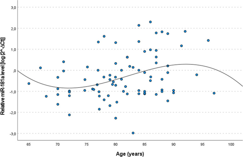
First, we investigated changes of miR-181a expression occurring upon age. In the entire cohort, no significant correlation with age was observed (r = 0.057, p = 0.347). To explore potential sex-specific age-related changes the analysis was conducted separately for women and men, revealing a difference between the two sexes: in males there was a statistically significant increase of miR-181a levels with increasing age (r = 0.295, p = 0.006) but not in females (r = -0.057, p = 0.437) This result was confirmed by linear regression analysis [β = 0.312 (95% CI = 0.102–0.522), p = 0.004 in males and β = -0.008 (95% CI = -0.164–0.169, p = 0.922 in females]. To better delineate the age-related changes, polynomial regression analysis showed that cubic curve fits the data better than a linear curve (linear regression r2 = 0.098 vs cubic regression r2 = 0.116), indicating a major increase after the age of 80 years (Fig. 1).
Fig. 1.
Scatter plot and fitted cubic curve of plasma miR‐181a expression levels as a function of age in male subjects. Data are reported as log 2− ΔCt normalized to U6 expression
Given the sex-specific effect of age on miR-181a expression, we conducted the subsequent analyses with parameters reported in Table 1 separately in the two sexes, after controlling for age.
Correlation analysis between miR-181a levels and indices of physical, cognitive performance and multimorbidity
We first evaluated the relationship between the miR-181a levels and physical and cognitive abilities measured by geriatric assessments, including Hand Grip (HG), Gait Speed (GS), Activity of Daily Living (ADL), and Mini Mental State Examination (MMSE). No correlation was found between miR-181a levels and these geriatric parameters.
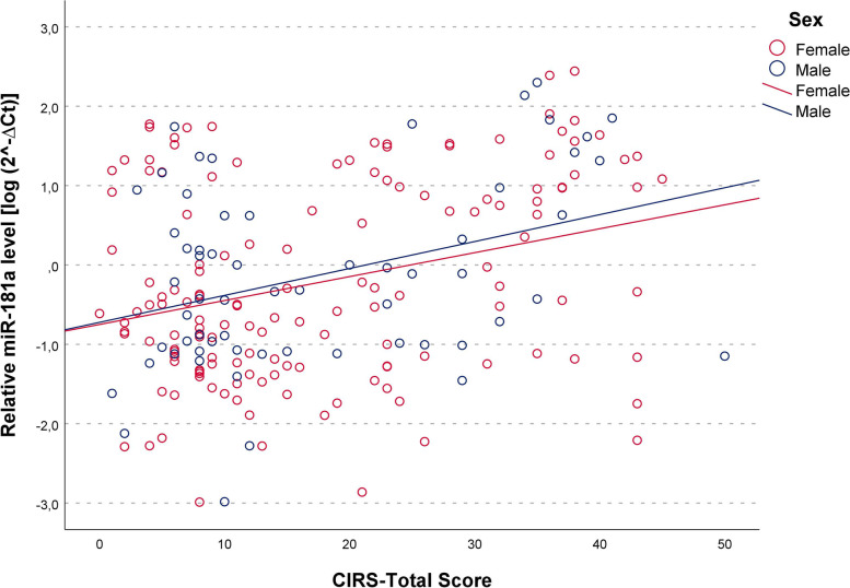
We then sought to examine the relationship between levels of miR-181a and multimorbidity burden by using the Cumulative Illness Rating Scale for Geriatrics (CIRS-total score) index. We found this index positively correlated with age of subjects (r = 0.268, p = 0.001 and r = 0.277, p = 0.03 for female and males respectively). Partial correlation analysis adjusted for age showed that circulating levels of miR-181a were significantly higher in subjects with a higher multimorbidity burden (that is, higher CIRS total score) in both sexes (rpartial = 0.307, p < 0.001 and rpartial = 0.330, p = 0.016, in women and men respectively). Linear regression analysis confirmed the positive correlation between miR-181a expression and CIRS after the adjustment of age [β = 0.330 (95% CI = 0.176–0.503), p < 0.001 and β = 0.299 (95% CI = 0.046–0.602), p = 0.023, respectively for females and males]. The scatter plots and linear regressions relative to these tests are presented in Fig. 2. Similarly, evaluation of the CIRS-Comorbidity Index (CIRS-CI), computed by counting the number of items for which moderate to severe pathology was reported, yielded positive correlations [rpartial = 0.292, p = 0.001 and β = 0.289 (95% CI = 0.127–0.483), p = 0.001] in females and [rpartial = 0.306, p = 0.026 and β = 0.302 (95% CI = 0.043–0.639) p = 0.026] in males, again reflecting higher miR-181a levels among persons with greater multimorbidity.
Fig. 2.
Correlation between plasma miR‐181a levels and multimorbidity. Scatter plots illustrate the relationship between plasma miR‐181a levels and the Cumulative Illness Rating Scale (CIRS) total score in male and female subjects. Data are reported as log 2− Δ.Ct normalized to U6 expression. Regression lines are displayed for males (blue, rpartial = 0.306, p = 0.026) and females (red, rpartial = 0.306, p = 0.026)
Correlation analysis between miR-181a levels and clinical markers
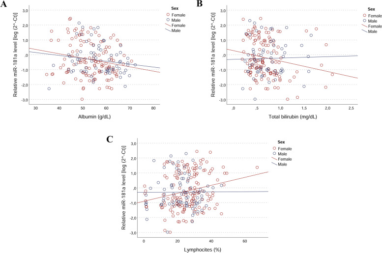
The association of miR-181a with the health status of analysed samples prompted us to investigate potential associations with clinical measurements. Again, sex-specific associations between miR-181a levels and blood biomarkers of generalized inflammation were found. More precisely, in women miR-181a expression was negatively correlated with Albumin [rpartial = -0.187; p = 0.041 and β = -0.188 (95% CI = -0.354—-0.008, p = 0.041] and total bilirubin [rpartial = -0.199; p = 0.024 and β = -0.199 (95% CI = -0.383—-0.028, p = 0.024] levels, and positively correlated with lymphocytes percentage [rpartial = 0.213; p = 0.004 and β = 0.237 (95% CI = 0.097—0.401, p = 0.004]. Scatter plots and linear regressions are shown in Fig. 3 (A, B and C). In males, miR-181a levels tended to be positively correlated with neutrophils percentage, but this correlation did not reach statistical significance at nominal level.
Fig. 3.
Correlations between plasma miR‐181a levels and biochemical variables in male and female subjects. Scatter plots illustrate the relationship between plasma miR‐181a levels and levels of (A) Albumin ( p = 0.041 in females); (B) Total Bilirubin (p = 0.024 in females); (C) Lymphocytes (p = 0.01 in females). Data are reported as log 2− ΔCt normalized to U6 expression
Discussion
In the present study, circulating levels of miR-181a, widely regarded as an inflamma-miR [16, 21], have been analysed in a cohort of elderly subjects aged from 65 to 97 years. We found that in both genders, but more significantly in females, circulating miR-181a levels positively correlated with the burden of multimorbidity when assessed either by the CIRS total score or by the CIRS-Comorbidity Index, which are reliable measures of multimorbidity and poor health status in geriatric patients [41, 43]. Multimorbidity is recognized as the most common chronic condition, determining a progressive increase of susceptibility to the occurrence of functional impairment and disability [44]. Factors affecting its development are complex and manifold [45], but chronic inflammation has consistently been found as one of its main risk factors [46].
The positive correlation of miR-181a with the burden of multimorbidity which we report, considering the role of miR-181a as a modulator of inflammatory responses, suggests that higher expression of miR-181a may be associated with a higher inflammatory state.
Interestingly, increased levels of miR-181a have been reported in inflammatory- and oxidative stress-related conditions, such as after treatment with lipopolysaccharide and hydrogen peroxide [47, 48]. Also, the overexpression of this miRNA has been seen in diverse age-related illnesses, including neurodegenerative and cancer diseases, although contrasting results have been found in the direction of the effect of miR-181a on these diseases [30–32, 49, 50]. These divergent results can be reconciled by considering that miR-181a can play a dual role as an anti- or a pro-inflammatory miRNA, likely depending on the cellular and physiological context.
Of note, we found miR-181a levels negatively correlated with the levels of albumin and bilirubin and positively correlated with lymphocytes in women, while levels in men only showed a trend toward a positive correlation with neutrophils. Although these findings must be interpreted with caution as some of the correlations only approach statistical significance, they may provide some hints about the relationship among miR-181a, inflammation and health status. First, they are compatible with our previous suggestion that the upregulation of miRNA-181a expression correlates with inflammatory status, given that low levels of albumin and bilirubin and high levels lymphocytes and neutrophils are characteristic features of inflammation [51–54]. Second, sex-specific correlations may reflect specific differences in immune-inflammatory responses between men and women with age. For example, men older than 65 years display higher innate and lower adaptive immune function than women [55, 56]. Being lymphocytes and neutrophils among the main cell types involved in the adaptative and innate response, respectively, the association of miR-181a levels with lymphocytes in females and neutrophils in males could mirror these differences.
The finding that miR-181a is positively correlated with age in males but not in females further supports this view. Studies investigating age associated changes of miR-181a expression in vitro and in vivo models yielded contrasting results, with some reports showing a downregulation [57, 58] and others an upregulation [29, 59]. As to humans, two studies found miR-181a downregulated in older individuals compared to younger ones [22, 60]. However, the subjects analysed in the above cited studies were younger than those included in our study (< 70 years). In addition, sex-stratified analyses were not performed in those studies, thus making it challenging to compare results and draw a conclusion. On the other hand, mir-181a has been found up-regulated in centenarians [61, 62]. These evidences, together with ours, suggest that changes in miR-181a levels with age follow nonlinear trajectories, characterized by a decrease in the expression levels from young to old age, and by an increase from old to very old age. This reasoning is supported by literature reports of nonlinear age-related changes and sex-dependent differences in levels of various miRNAs, including miR-181a [34, 63, 64], as well as non-linear alterations in plasma proteome with age [65].
Conclusion
Data herein support the hypothesis that miR-181a acts as an inflamma-miRNA playing important roles in the aging process. We were able to identify miR-181a as multimorbidity-associated miRNA. Its potential use as biomarker of health status has to consider that it might have different effects at different ages, as well as reported for other factors [66, 67]. Importantly, our results highlighted sex-specific correlations of miR-181a with risk factors for negative outcomes, suggesting that the routes by which this miRNA can influence health status are different between genders. From a more general point of view, this supports the growing belief that for better understanding the underpinnings of the gender differences in aging, as well as in age-related diseases, gender-stratified analyses should be performed.
Our conclusions should be viewed in the light of some limitations. First, the correlations we found are of moderate effect, partially due to the relatively small size of the analysed sample; thus, a larger sample would have probably allowed us to better document the validity of our statistical associations. Therefore, larger future studies and across a wider age range are recommended. Second, the results here presented are limited to revealing associative, rather than causal, relations between miR-181a expression and multimorbidity at old age. Therefore, investigations on its targets and regulators would provide more insights about the signalling pathways underlying the observed associations and evaluate its potential as biomarker of multimorbidity risk.
Notwithstanding these limitations, data here presented may lay the groundwork for a more complete research study in the future.
Acknowledgements
The work has been made possible by the collaboration with Gruppo Baffa (Sadel Spa, Sadel San Teodoro srl, Sadel CS srl, Casa di Cura Madonna dello Scoglio, AGI srl, Casa di Cura Villa del Rosario srl, Savelli Hospital srl, Casa di Cura Villa Ermelinda)
Abbreviations
- miRNA
MicroRNA
- qPCR
Quantitative Polymerase Chain Reaction
- CIRS
Cumulative Illness Rating Scale
- inflamma-miRs
Inflammatory microRNAs
- TGFβ
Transforming Growth Factor beta
- IL-10
Interleukin 10
- IL-6
Interleukin 6
- TNFα
Tumor necrosis factor alfa
- IL-1
Interleukin 1
- HGS
Hand Grip Strength
- ADL
Activities of Daily Living
- MMSE
Mini Mental State Examination
- cDNA
Complementary DNA
- SD
Standard Deviation
- BMI
Body Mass Index
- HbA1C
Haemoglobin A1C
- GS
Gait Speed
- LDL
Low-density lipoprotein
- HDL
High-density lipoprotein
- RBC
Red Blood Cells
- WBC
White Blood Cells
- CIRS-TS
Cumulative Illness Rating Scale (CIRS)-Total Score
- CIRS-CI
Cumulative Illness Rating Scale (CIRS)-Comorbidity Index
Authors’ contributions
Francesca Iannone and Paolina Crocco: Methodology- Data curation-. Review & editing. Serena Dato: Writing—review & editing. Giuseppe Passarino: Writing review & editing. Giuseppina Rose: Conceptualization, Writing—original draft, review & editing. The author(s) read and approved the final manuscript.
Funding
This research was supported by “SI.F.I.PA.CRO.DE.–Sviluppo e industrializzazione farmaci innovativi per terapia molecolare personalizzata PA.CRO.DE.” PON ARS01_00568 granted by MIUR (Ministry of Education, University and Research) Italy to G.P.
Availability of data and materials
The data that support the findings of this study are available from the corresponding author upon reasonable request.
Declarations
Ethics approval and consent to participate
The study was conducted in accordance with the Declaration of Helsinki, and the protocol was approved by the local Ethical Committee (Comitato Etico Regione Calabria-Sezione Area Nord) on 2017–10-31 (code n. 25/2017). A written informed consent was obtained from all participants included in the study.
Consent for publication
Not applicable.
Competing interests
No conflict of interest to declare.
Footnotes
Publisher’s Note
Springer Nature remains neutral with regard to jurisdictional claims in published maps and institutional affiliations.
Francesca Iannone and Paolina Crocco contributed equally to this work.
Contributor Information
Francesca Iannone, Email: francesca.iannone@unical.it.
Paolina Crocco, Email: paolina.crocco@unical.it.
Serena Dato, Email: serena.dato@unical.it.
Giuseppe Passarino, Email: giuseppe.passarino@unical.it.
Giuseppina Rose, Email: pina.rose@unical.it.
References
- 1.Barnett K, Mercer SW, Norbury M, Watt G, Wyke S, Guthrie B. Epidemiology of multimorbidity and implications for health care, research, and medical education: a cross-sectional study. Lancet. 2012;380(9836):37–43. doi: 10.1016/S0140-6736(12)60240-2. [DOI] [PubMed] [Google Scholar]
- 2.Nojiri S, Itoh H, Kasai T, Fujibayashi K, Saito T, Hiratsuka Y, Okuzawa A, Naito T, Yokoyama K, Daida H. Comorbidity status in hospitalized elderly in Japan: analysis from national database of health insurance claims and specific health checkups. Sci Rep. 2019;9(1):20237. doi: 10.1038/s41598-019-56534-4. [DOI] [PMC free article] [PubMed] [Google Scholar]
- 3.Rizzuto D, Melis R, Angleman S, Qiu C, Marengoni A. Effect of chronic diseases and multimorbidity on survival and functioning in elderly adults. J Am Geriatr Soc. 2017;65(5):1056–1060. doi: 10.1111/jgs.14868. [DOI] [PubMed] [Google Scholar]
- 4.Atella V, Piano Mortari A, Kopinska J, Belotti F, Lapi F, Cricelli C, Fontana L. Trends in age-related disease burden and healthcare utilization. Aging Cell. 2019;18(1):e12861. doi: 10.1111/acel.12861. [DOI] [PMC free article] [PubMed] [Google Scholar]
- 5.Franceschi C, Garagnani P, Parini P, Giuliani C, Santoro A. Inflammaging: a new immune-metabolic viewpoint for age-related diseases. Nat Rev Endocrinol. 2018;14(10):576–590. doi: 10.1038/s41574-018-0059-4. [DOI] [PubMed] [Google Scholar]
- 6.He L, Hannon GJ. MicroRNAs: small RNAs with a big role in gene regulation. Nat Rev Genet. 2004;5:522–531. doi: 10.1038/nrg1379. [DOI] [PubMed] [Google Scholar]
- 7.Bartel DP. MicroRNAs: target recognition and regulatory functions. Cell. 2009;136:215–233. doi: 10.1016/j.cell.2009.01.002. [DOI] [PMC free article] [PubMed] [Google Scholar]
- 8.Inukai S, Slack F. MicroRNAs and the genetic network in aging. J Mol Biol. 2013;425(19):3601–3608. doi: 10.1016/j.jmb.2013.01.023. [DOI] [PMC free article] [PubMed] [Google Scholar]
- 9.Huan T, Chen G, Liu C, Bhattacharya A, Rong J, Chen BH, Seshadri S, Tanriverdi K, Freedman JE, Larson MG, Murabito JM, Levy D. Age-associated microRNA expression in human peripheral blood is associated with all-cause mortality and age-related traits. Aging Cell. 2018;17(1):e12687. doi: 10.1111/acel.12687. [DOI] [PMC free article] [PubMed] [Google Scholar]
- 10.Kinser HE, Pincus Z. MicroRNAs as modulators of longevity and the aging process. Hum Genet. 2020;139:291–308. doi: 10.1007/s00439-019-02046-0. [DOI] [PMC free article] [PubMed] [Google Scholar]
- 11.Saeidimehr S, Ebrahimi A, Saki N, Goodarzi P, Rahim F. MicroRNA-based linkage between aging and cancer: from epigenetics view point. Cell J. 2016;18(2):117–126. doi: 10.22074/cellj.2016.4303. [DOI] [PMC free article] [PubMed] [Google Scholar]
- 12.de Lucia C, Komici K, Borghetti G, Femminella GD, Bencivenga L, Cannavo A, Corbi G, Ferrara N, Houser SR, Koch WJ, Rengo G. microRNA in cardiovascular aging and age-related cardiovascular diseases. Front Med. 2017;4:74. doi: 10.3389/fmed.2017.00074. [DOI] [PMC free article] [PubMed] [Google Scholar]
- 13.Verjans R, van Bilsen M, Schroen B. MiRNA deregulation in cardiac aging and associated disorders. Int Rev Cell Mol Biol. 2017;334:207–263. doi: 10.1016/bs.ircmb.2017.03.004. [DOI] [PubMed] [Google Scholar]
- 14.Quinlan S, Kenny A, Medina M, Engel T, Jimenez-Mateos EM. MicroRNAs in neurodegenerative diseases. Int Rev Cell Mol Biol. 2017;334:309–343. doi: 10.1016/bs.ircmb.2017.04.002. [DOI] [PubMed] [Google Scholar]
- 15.Wang M, Qin L, Tang B. MicroRNAs in Alzheimer's Disease. Front Genet. 2019;10:153. doi: 10.3389/fgene.2019.00153. [DOI] [PMC free article] [PubMed] [Google Scholar]
- 16.Olivieri F, Rippo MR, Procopio AD, Fazioli F. Circulating inflamma-miRs in aging and age-related diseases. Front Genet. 2013;4:121. doi: 10.3389/fgene.2013.00121. [DOI] [PMC free article] [PubMed] [Google Scholar]
- 17.Cătană CS, Calin GA, Berindan-Neagoe I. Inflamma-miRs in aging and breast cancer: are they reliable players? Front Med. 2015;2:85. doi: 10.3389/fmed.2015.00085. [DOI] [PMC free article] [PubMed] [Google Scholar]
- 18.Schroen B, Heymans S. Small but smart–microRNAs in the centre of inflammatory processes during cardiovascular diseases, the metabolic syndrome, and ageing. Cardiovasc Res. 2012;93(4):605–613. doi: 10.1093/cvr/cvr268. [DOI] [PubMed] [Google Scholar]
- 19.Olivieri F, Spazzafumo L, Bonafè M, Recchioni R, Prattichizzo F, Marcheselli F, Micolucci L, Mensà E, Giuliani A, Santini G, Gobbi M, Lazzarini R, Boemi M, Testa R, Antonicelli R, Procopio AD, Bonfigli AR. MiR-21-5p and miR-126a-3p levels in plasma and circulating angiogenic cells: relationship with type 2 diabetes complications. Oncotarget. 2015;6(34):35372–35382. doi: 10.18632/oncotarget.6164. [DOI] [PMC free article] [PubMed] [Google Scholar]
- 20.Prattichizzo F, Giuliani A, Ceka A, Rippo MR, Bonfigli AR, Testa R, Procopio AD, Olivieri F. Epigenetic mechanisms of endothelial dysfunction in type 2 diabetes. Clin Epigenet. 2015;7(1):56. doi: 10.1186/s13148-015-0090-4. [DOI] [PMC free article] [PubMed] [Google Scholar]
- 21.Rippo MR, Olivieri F, Monsurrò V, Prattichizzo F, Albertini MC, Procopio AD. MitomiRs in human inflamm-aging: a hypothesis involving miR-181a, miR-34a and miR-146a. Exp Gerontol. 2014;56:154–163. doi: 10.1016/j.exger.2014.03.002. [DOI] [PubMed] [Google Scholar]
- 22.Hooten NN, Fitzpatrick M, Wood WH, De S, Ejiogu N, Zhang Y, Mattison JA, Becker KG, Zonderman AB, Evans MK. Age-related changes in microRNA levels in serum. Aging (Albany NY) 2013;5:725–740. doi: 10.18632/aging.100603. [DOI] [PMC free article] [PubMed] [Google Scholar]
- 23.Xie W, Li M, Xu N, Lv Q, Huang N, He J, Zhang Y. MiR-181a regulates inflammation responses in monocytes and macrophages. PLoS ONE. 2013;8(3):e58639. doi: 10.1371/journal.pone.0058639. [DOI] [PMC free article] [PubMed] [Google Scholar]
- 24.Kozloski GA, Jiang X, Bhatt S, Ruiz J, Vega F, Shaknovich R, Melnick A, Lossos IS. miR-181a negatively regulates NF-κB signaling and affects activated B-cell-like diffuse large B-cell lymphoma pathogenesis. Blood. 2016;127(23):2856–2866. doi: 10.1182/blood-2015-11-680462. [DOI] [PubMed] [Google Scholar]
- 25.Li QJ, Chau J, Ebert PJ, Sylvester G, Min H, Liu G, Braich R, Manoharan M, Soutschek J, Skare P, Klein LO, Davis MM, Chen CZ. miR-181a is an intrinsic modulator of T cell sensitivity and selection. Cell. 2007;129(1):147–161. doi: 10.1016/j.cell.2007.03.008. [DOI] [PubMed] [Google Scholar]
- 26.Ye Z, Li G, Kim C, Hu B, Jadhav RR, Weyand CM, Goronzy JJ. Regulation of miR-181a expression in T cell aging. Nat Commun. 2018;9(1):3060. doi: 10.1038/s41467-018-05552-3. [DOI] [PMC free article] [PubMed] [Google Scholar]
- 27.Grewers Z, Krueger A. MicroRNA miR-181—A Rheostat for TCR signaling in thymic selection and peripheral T-cell function. Int J Mol. Sci. 2020;21:6200. doi: 10.3390/ijms21176200. [DOI] [PMC free article] [PubMed] [Google Scholar]
- 28.Kim C, Ye Z, Weyand CM, Goronzy JJ. miR-181a-regulated pathways in T-cell differentiation and aging. Immun Ageing. 2021;18(1):28. doi: 10.1186/s12979-021-00240-1. [DOI] [PMC free article] [PubMed] [Google Scholar]
- 29.Giuliani A, Cirilli I, Prattichizzo F, Mensa E, Fulgenzi G, Sabbatinelli J, Graciotti L, Olivieri F, Procopio AD, Tiano L, Rippo MR. The mitomiR/Bcl-2 axis affects mitochondrial function and autophagic vacuole formation in senescent endothelial cells. Aging (Albany NY) 2018;10:2855–2873. doi: 10.18632/aging.101591. [DOI] [PMC free article] [PubMed] [Google Scholar]
- 30.Hulsmans M, Sinnaeve P, Van der Schueren B, Mathieu C, Janssens S, Holvoet P. Decreased miR-181a expression in monocytes of obese patients is associated with the occurrence of metabolic syndrome and coronary artery disease. J Clin Endocrinol Metab. 2012;97(7):E1213–E1218. doi: 10.1210/jc.2012-1008. [DOI] [PubMed] [Google Scholar]
- 31.Indrieri A, Carrella S, Carotenuto P, Banfi S, Franco B. The Pervasive role of the miR-181 family in development, neurodegeneration, and cancer. Int J Mol Sci. 2020;21(6):2092. doi: 10.3390/ijms21062092. [DOI] [PMC free article] [PubMed] [Google Scholar]
- 32.Yan X, Gao M, Zhang P, Ouyang G, Mu Q, Xu K. MiR-181a functions as an oncogene by regulating CCND1 in multiple myeloma. Oncol Lett. 2020;20(1):758–764. doi: 10.3892/ol.2020.11579. [DOI] [PMC free article] [PubMed] [Google Scholar]
- 33.Borja-Gonzalez M, Casas-Martinez JC, McDonagh B, Goljanek-Whysall K. Aging science talks: the role of miR-181a in age-related loss of muscle mass and function. Transl Med. 2020;4:81–85. doi: 10.1016/j.tma.2020.07.001. [DOI] [PMC free article] [PubMed] [Google Scholar]
- 34.Sheinerman K, Tsivinsky V, Mathur A, Kessler D, Shaz B, Umansky S. Age- and sex-dependent changes in levels of circulating brain-enriched microRNAs during normal aging. Aging (Albany NY) 2018;10(10):3017–3041. doi: 10.18632/aging.101613. [DOI] [PMC free article] [PubMed] [Google Scholar]
- 35.De Rango F, Montesanto A, Berardelli M, Mazzei B, Mari V, Lattanzio F, Corsonello A, Passarino G. To grow old in Southern Italy: a comprehensive Description of the old and oldest old in Calabria. Gerontology. 2011;57:327–334. doi: 10.1159/000316941. [DOI] [PubMed] [Google Scholar]
- 36.von Elm E, Altman DG, Egger M, Pocock SJ, Gøtzsche PC, Vandenbroucke JP, for the STROBE initiative The strengthening the reporting of observational studies in epidemiology (STROBE) statement: guidelines for reporting observational studies. Lancet. 2007;370(9596):1453–1457. doi: 10.1016/S0140-6736(07)61602-X. [DOI] [PubMed] [Google Scholar]
- 37.Katz S, Downs TD, Cash HR, Grotz RC. Progress in development of the index of ADL. Gerontologist. 1970;10(1):20–30. doi: 10.1093/geront/10.1_Part_1.20. [DOI] [PubMed] [Google Scholar]
- 38.Folstein MF, Folstein SE, McHugh PR. "Mini-mental state". A practical method for grading the cognitive state of patients for the clinician. J. Psychiatr Res. 1975;12(3):189–198. doi: 10.1016/0022-3956(75)90026-6. [DOI] [PubMed] [Google Scholar]
- 39.Grigoletto F, Zappalà G, Anderson DW, Lebowitz BD. Norms for the mini-mental state examination in a healthy population. Neurology. 1999;53(2):315–320. doi: 10.1212/WNL.53.2.315. [DOI] [PubMed] [Google Scholar]
- 40.de Groot V, Beckerman H, Lankhorst GJ, Bouter LM. How to measure comorbidity. a critical review of available methods. J Clin. Epidemiol. 2003;56:221–229. doi: 10.1016/S0895-4356(02)00585-1. [DOI] [PubMed] [Google Scholar]
- 41.Salvi F, Miller MD, Grilli A, Giorgi R, Towers AL, Morichi V, Spazzafumo L, Mancinelli L, Espinosa E, Rappelli A, Dessì-Fulgheri P. A manual of guidelines to score the modified cumulative illness rating scale and its validation in acute hospitalized elderly patients. J Am Geriatr Soc. 2008;56(10):1926–1931. doi: 10.1111/j.1532-5415.2008.01935.x. [DOI] [PubMed] [Google Scholar]
- 42.Schmittgen TD, Livak KJ. Analyzing real-time PCR data by the comparative C(T) method. Nat Protoc. 2008;3(6):1101–1108. doi: 10.1038/nprot.2008.73. [DOI] [PubMed] [Google Scholar]
- 43.Parmelee PA, Thuras PD, Katz IR, Lawton MP. Validation of the Cumulative Illness Rating Scale in a geriatric residential population. J Am Geriatr Soc. 1995;43(2):130–137. doi: 10.1111/j.1532-5415.1995.tb06377.x. [DOI] [PubMed] [Google Scholar]
- 44.Fabbri E, Zoli M, Gonzalez-Freire M, Salive ME, Studenski SA, Ferrucci L. Aging and multimorbidity: new tasks, priorities, and frontiers for integrated Gerontological and clinical research. J Am Med Dir Assoc. 2015;16(8):640–647. doi: 10.1016/j.jamda.2015.03.013. [DOI] [PMC free article] [PubMed] [Google Scholar]
- 45.Kadambi S, Abdallah M, Loh KP. Multimorbidity, function, and cognition in aging. Clin Geriatr Med. 2020;36(4):569–584. doi: 10.1016/j.cger.2020.06.002. [DOI] [PMC free article] [PubMed] [Google Scholar]
- 46.Friedman E, Shorey C. Inflammation in multimorbidity and disability: an integrative review. Health Psychol. 2019;38(9):791–801. doi: 10.1037/hea0000749. [DOI] [PMC free article] [PubMed] [Google Scholar]
- 47.Wang L, Huang H, Fan Y, Kong B, Hu H, Hu K, Guo J, Mei Y, Liu WL. Effects of downregulation of microRNA-181a on H2O2-induced H9c2 cell apoptosis via the mitochondrial apoptotic pathway. Oxid Med Cell Longev. 2014;2014:960362. doi: 10.1155/2014/960362. [DOI] [PMC free article] [PubMed] [Google Scholar]
- 48.Liu HY, Yu LF, Zhou TG, Wang YD, Sun DH, Chen HR, Hou YF. Lipopolysaccharide-stimulated bone marrow mesenchymal stem cells-derived exosomes inhibit H2O2-induced cardiomyocyte inflammation and oxidative stress via regulating miR-181a-5p/ATF2 axis. Eur Rev Med Pharmacol Sci. 2020;24(19):10069–10077. doi: 10.26355/eurrev_202010_23224. [DOI] [PubMed] [Google Scholar]
- 49.Song X, Xue Y, Cai H. Down-Regulation of miR-181a-5p Prevents Cerebral Ischemic Injury by Upregulating En2 and Activating Wnt/β-catenin Pathway. J Stroke Cerebrovasc Dis. 2021;30(3):105485. doi: 10.1016/j.jstrokecerebrovasdis.2020.105485. [DOI] [PubMed] [Google Scholar]
- 50.Xiang Y, Tian Q, Guan L, Niu SS. The dual role of miR-186 in cancers: Oncomir battling with tumor suppressor miRNA. Front Oncol. 2020;10:233. doi: 10.3389/fonc.2020.00233. [DOI] [PMC free article] [PubMed] [Google Scholar]
- 51.Vogel ME, Zucker SD. Bilirubin acts as an endogenous regulator of inflammation by disrupting adhesion molecule-mediated leukocyte migration. Inflamm Cell Signal. 2016;3(1):e1178. doi: 10.14800/ics.1178. [DOI] [PMC free article] [PubMed] [Google Scholar]
- 52.Meeuwsen J, Wesseling M, Hoefer IE, de Jager S. Prognostic value of circulating inflammatory cells in patients with stable and acute coronary artery disease. Front Cardiovasc Med. 2017;4:44. doi: 10.3389/fcvm.2017.00044. [DOI] [PMC free article] [PubMed] [Google Scholar]
- 53.Soeters PB, Wolfe RR, Shenkin A. Hypoalbuminemia: pathogenesis and clinical significance. JPEN J Parenter Enter Nutr. 2019;43(2):181–193. doi: 10.1002/jpen.1451. [DOI] [PMC free article] [PubMed] [Google Scholar]
- 54.Liew PX, Kubes P. The neutrophil's role during health and disease. Physiol Rev. 2019;99(2):1223–1248. doi: 10.1152/physrev.00012.2018. [DOI] [PubMed] [Google Scholar]
- 55.Márquez EJ, Chung CH, Marches R, Rossi RJ, Nehar-Belaid D, Eroglu A, Mellert DJ, Kuchel GA, Banchereau J, Ucar D. Sexual-dimorphism in human immune system aging. Nat Commun. 2020;11(1):751. doi: 10.1038/s41467-020-14396-9. [DOI] [PMC free article] [PubMed] [Google Scholar]
- 56.Hägg S, Jylhävä J. Sex differences in biological aging with a focus on human studies. eLife. 2021;10:e63425. doi: 10.7554/eLife.63425. [DOI] [PMC free article] [PubMed] [Google Scholar]
- 57.Li G, Yu M, Lee WW, Tsang M, Krishnan E, Weyand CM, Goronzy JJ. Decline in miR-181a expression with age impairs T cell receptor sensitivity by increasing DUSP6 activity. Nat Med. 2012;18(10):1518–1524. doi: 10.1038/nm.2963. [DOI] [PMC free article] [PubMed] [Google Scholar]
- 58.Tugay K, Guay C, Marques AC, Allagnat F, Locke JM, Harries LW, Rutter GA, Regazzi R. Role of microRNAs in the age-associated decline of pancreatic beta cell function in rat islets. Diabetologia. 2016;59(1):161–169. doi: 10.1007/s00125-015-3783-5. [DOI] [PMC free article] [PubMed] [Google Scholar]
- 59.Mancini M, Saintigny G, Mahé C, Annicchiarico-Petruzzelli M, Melino G, Candi E. MicroRNA-152 and -181a participate in human dermal fibroblasts senescence acting on cell adhesion and remodeling of the extra-cellular matrix. Aging (Albany NY) 2012;4(11):843–853. doi: 10.18632/aging.100508. [DOI] [PMC free article] [PubMed] [Google Scholar]
- 60.Xu Y, Xu L, Chen C, Zhang Y, Zeng C, Jin Z, Chen S, Li B, Zha X, Yin Z, Li Y. Age-related immune profile of the T cell receptor repertoire, Thymic recent output function, and miRNAs. BioMed Res Int. 2020;2020:5910823. doi: 10.1155/2020/5910823. [DOI] [PMC free article] [PubMed] [Google Scholar]
- 61.Gombar S, Jung HJ, Dong F, Calder B, Atzmon G, Barzilai N, Tian XL, Pothof J, Hoeijmakers JHJ, Campisi J, Vijg J, Suh Y. Comprehensive microRNA profiling in B-cells of human centenarians by massively parallel sequencing. BMC Genomics. 2012;13:353. doi: 10.1186/1471-2164-13-353. [DOI] [PMC free article] [PubMed] [Google Scholar]
- 62.Serna E, Gambini J, Borras C, Abdelaziz KM, Belenguer A, Sanchis P, Avellana JA, Rodriguez-Mañas L, Viña J. Centenarians, but not octogenarians, up-regulate the expression of microRNAs. Sci Rep. 2012;2:961. doi: 10.1038/srep00961. [DOI] [PMC free article] [PubMed] [Google Scholar]
- 63.Olivieri F, Capri M, Bonafè M, Morsiani C, Jung HJ, Spazzafumo L, Viña J, Suh Y. Circulating miRNAs and miRNA shuttles as biomarkers: Perspective trajectories of healthy and unhealthy aging. Mech Aging Dev. 2017;165(Pt B):162–170. doi: 10.1016/j.mad.2016.12.004. [DOI] [PMC free article] [PubMed] [Google Scholar]
- 64.Mensà E, Giuliani A, Matacchione G, Gurău F, Bonfigli AR, Romagnoli F, De Luca M, Sabbatinelli J, Olivieri F. Circulating miR-146a in healthy aging and type 2 diabetes: age- and gender-specific trajectories. Mech Ageing Dev. 2019;180:1–10. doi: 10.1016/j.mad.2019.03.001. [DOI] [PubMed] [Google Scholar]
- 65.Lehallier B, Gate D, Schaum N, Nanasi T, Lee SE, Yousef H, Moran Losada P, Berdnik D, Keller A, Verghese J, Sathyan S, Franceschi C, Milman S, Barzilai N, Wyss-Coray T. Undulating changes in human plasma proteome profiles across the lifespan. Nat Med. 2019;25(12):1843–1850. doi: 10.1038/s41591-019-0673-2. [DOI] [PMC free article] [PubMed] [Google Scholar]
- 66.Rose G, Crocco P, De Rango F, Corsonello A, Lattanzio F, De Luca M, Passarino G. Metabolism and successful aging: Polymorphic variation of syndecan-4 (SDC4) gene associate with longevity and lipid profile in healthy elderly Italian subjects. Mech Ageing Dev. 2015;150:27–33. doi: 10.1016/j.mad.2015.08.003. [DOI] [PubMed] [Google Scholar]
- 67.Karlsson IK, Lehto K, Gatz M, Reynolds CA, Dahl Aslan AK. Age-dependent effects of body mass index across the adult life span on the risk of dementia: a cohort study with a genetic approach. BMC Med. 2020;18(1):131. doi: 10.1186/s12916-020-01600-2. [DOI] [PMC free article] [PubMed] [Google Scholar]
Associated Data
This section collects any data citations, data availability statements, or supplementary materials included in this article.
Data Availability Statement
The data that support the findings of this study are available from the corresponding author upon reasonable request.


