



1. Introduction
Optimization of diffusion weighted imaging (DWI) is important in the setting of prostate MRI due to its wide use for cancer detection and emerging role for treatment planning [1–13]. Per the Prostate Imaging Reporting & Data System (PI-RADS), currently on version 2.1, peripheral zone lesions (where the majority of prostate cancers occur) are evaluated primarily on DWI and apparent diffusion coefficient (ADC) maps [2]. Additionally, the degree of diffusion restriction present, measured both qualitatively and quantitatively, has been correlated with Gleason score and tumor aggressiveness [9–18]. Therefore, optimizing the DWI sequence to minimize artifacts (e.g., distortion) and to provide reproducible quantitative parameters is paramount to maximizing its clinical utility.
The advancement of DWI applications in the body has relied upon the development and refinement of single-shot echo planar imaging (ssEPI), which allows for rapid data acquisition and robustness against patient motion [19, 20]. However, the long readout times in ssEPI lead to image distortions in the presence of susceptibility-related field inhomogeneity (e.g., from air in the rectum or aerated lung next to liver). In the prostate this distortion can lead to limitations in diagnostic performance and even result in a non-diagnostic DWI acquisition if severe distortion is present that cannot be remedied [2].
Reduced-distortion DWI techniques, such as reduced field of view (rFOV) [21, 22] and multi-shot EPI (msEPI) [23–25], can partially mitigate the presence and severity of susceptibility related distortion. While both rFOV and msEPI are designed to reduce image distortion, they work in different ways: msEPI shortens the echo train while maintaining the phase encoding direction, whereas rFOV keeps the echo train length the same but modifies the phase encoding direction so that field heterogeneities near the rectum do not create distortions into the prostate. Prostate MRI utilizing these techniques have demonstrated more reproducible quantitative diffusion measurements relative to ssEPI, in phantom experiments as well as in volunteers [26], and additional early work evaluating rFOV also demonstrated reduced distortion in the setting of prostate cancer [27, 28]. However, the performance of reduced-distortion DWI sequences, particularly msEPI, in prostate cancer patients has not been systematically evaluated.
Therefore, the purpose of this work is to evaluate image distortion and ADC quantification for rFOV and msEPI DWI, using ssEPI as the widely available and clinically utilized reference standard.
2. Materials and Methods
Institutional Review Board approval was obtained and written informed consent was obtained from all subjects. All data were handled in concordance with U.S. HIPAA guidelines. All subjects referred for clinical MRI of the prostate were eligible for enrollment and consecutive individuals were recruited from July 2018 – January 2020. Common clinical indications for prostate MRI included: (1) staging evaluation in the setting of known prostate cancer, (2) elevated prostate specific antigen (PSA), and (3) post-treatment monitoring or active surveillance.
2.1. MRI acquisition/reconstruction:
MRI was performed at 3.0T (Discovery 750w or Signa Premier, GE Healthcare). Three oblique axial DWI sequences were acquired: 1) ssEPI DWI (reference standard used in routine clinical practice), 2) rFOV DWI, and 3) msEPI DWI. Detailed DWI acquisition parameters are shown in Table 1. The acquisitions were designed to have the same number of signal averages which resulted in a longer scan time for msEPI. The rFOV DWI images were reconstructed with complex averaging using the field-of-view optimized and constrained undistorted single-shot (FOCUS) method [29] and images of msEPI DWI were reconstructed with the phase-corrected multiplexed sensitivity encoding (MUSE) method [23]. The ssEPI DWI images were reconstructed using standard DW-EPI reconstruction with parallel imaging acceleration. ADC maps for each DWI sequence were calculated as follows
In addition, high-resolution, oblique axial T2-weighted (T2W) images were obtained (field of view = 26 × 26 cm; in-plane resolution = 0.7 × 1.0 mm; slice thickness = 2.4 mm; echo time = 109.8 ms; repetition time = 3000 ms) during the clinical prostate MRI protocol. These T2-weighted images were used as an anatomical reference for drawing regions of interest as well as a reference volume for distortion assessment (see details below).
Table 1.
DWI Acquisition Parameters
| Scanner | GE Premier Gmax=70mT/m; SRmax=200mT/m |
GE 750W Gmax=33mT/m; SRmax=120mT/m |
|||||
|---|---|---|---|---|---|---|---|
| Coil | 32-channel Torso coil | Small 30-channel coil | |||||
| Sequence | ssEPI | rFOV | msEPI | ssEPI | rFOV | msEPI | |
| FOV (cm2) | 28×28 | 28×14 | 28×28 | 28×28 | 28×14 | 28×28 | |
| Resolution | 2mm×2mm×4.8mm | ||||||
| Phase encoding direction | A/P | R/L | A/P | A/P | R/L | A/P | |
| Echo Train Length | 48 | 48 | 32 | 48 | 48 | 32 | |
| Partial Fourier | 75% | 75% | None | 75% | 75% | None | |
| Parallel Imaging | 2 | N/A | 2 | 2 | N/A | 2 | |
| Diffusion Direction | All in 1 | ||||||
| b-values (# Averages) | 100(6), 800(12) | 100(6), 800(12) | |||||
| TE (ms) | 55.5 | 55.9 | 56.0 | 70.6 | 65.5 | 71.0 | |
| TR (s) | 4 | ||||||
| Scan time | 1min 16sec | 1min 16sec | 2min 32sec | 1min 16sec | 1min 16sec | 2min 32sec | |
2.2. Quantitative distortion assessment:
Whole prostate contouring was completed by Y.Z. and Z.W. under the supervision of J.S., a board-certified radiologist with 5 years of experience in prostate MRI, utilizing contouring software implemented in Matlab R2019a, Mathworks. Contouring was completed individually for the T2W images as well as the b=100 s/mm2 images of all three DWI sequences. Contours from the T2W images were used as the reference volume and distortion vectors of each slice were calculated by matching the contours of diffusion images (i.e. distorted image volume) to the reference volume [30]. The norm of the distortion vector measures the geometric displacement (mm) induced by distortion over a certain direction (angle φ in polar coordinate system). The final metric dr.m.s. (in mm) was calculated using the root-mean-square value of the norms of distortion vectors over all angles and slices to quantify the overall distortion level of the entire prostate. The algorithm was implemented in Matlab R2019a following the method proposed by Gill AB, et al. [30].
2.3. Quantitative assessment of biopsy correlated lesions:
ROI contouring:
Freehand ROI contouring was performed in Matlab using in-house software, by a radiologist with 6 years of clinical prostate MRI experience, for all patients who had a biopsy of a lesion described on the clinical prostate MRI. This included patients who (1) received MRI-US Fusion biopsy (UroNav, Phillips Healthcare) or (2) MRI-US cognitive biopsy (BK Medical). First, a prostate cancer ROI was drawn on the ADC maps (ssEPI, rFOV, msEPI) for each lesion targeted for biopsy. Subsequently, a non-cancerous ROI was placed on the same slice in the contralateral prostate gland, with maintenance of zonal anatomy and ROI size, for matched quantitative comparison. The oblique axial T2W MRI were utilized for anatomic reference.
Comparative assessment:
Mean, median, standard deviation, and the 20th and 80th percentile of the ADC histograms were calculated and compared across the three acquired sequences. In particular, the presence of a statistically significant difference between matched cancerous and non-cancerous ROIs was evaluated independently for each of the 3 DWI sequences. The calculated mean ADC from cancerous and non-cancerous prostate tissue was also compared separately for differences in quantification from the 3 DWI sequences with each pair of DWI sequences tested separately.
2.4. Image quality assessment:
Three board-certified radiologists (4, 5, 11 years of clinical prostate MRI experience, respectively) completed independent, blinded review of the three DWI sequences (b-value = 800 s/mm2) with the oblique axial T2W images and ADC maps available for reference. The three DWI sequences from a given patient were viewed separately and non-consecutively with the order shuffled regarding DWI sequence presentation. All DWI sequences were cropped to the same field of view of the pelvis/prostate to mitigate bias. Image resolution, capsule demarcation, zonal anatomy, overall image quality, and qualitative SNR were evaluated on a 5-point Likert scale (1, poor; 2, below average; 3, average; 4, above average; 5, excellent). Image distortion/artifacts were graded on their level and influence (Supplementary Table). The scores from all three readers were averaged for comparison between DWI acquisitions.
Wilcoxon signed-rank test and Student’s t-test were used as appropriate to test for statistical significance (p-value ≤ 0.05 = statistically significant).
3. Results
Thirty patients were recruited. Five patients were excluded due to incorrect DWI acquisition parameters resulting in a final study group of 25 patients. Average age was 63.9 years (range, 44–76) and average serum PSA was 6.55 ng/mL (range, 1.13–10.84 ng/mL). The study group included 10 patients undergoing staging prior to treatment, 10 patients without biopsy proven cancer but persistently elevated PSA, 4 patients undergoing active surveillance, and one patient with biochemical recurrence after high intensity focused ultrasound treatment. For sub-group analysis, 15 patients had proven prostate cancer that correlated with a suspicious lesion on MRI.
3.1. Quantitative distortion
There was a small but statistically significant reduction in the average calculated distortion (dr.m.s.) for rFOV compared to ssEPI (3.2 ± 0.2 mm versus 3.4 ± 0.3 mm, respectively; p = 0.033) as well as for msEPI compared to ssEPI (3.0 ± 0.2 mm versus 3.4 ± 0.3 mm, respectively; p = 0.003). The difference between rFOV and msEPI was not statistically significant (p = 0.078). Figure 1 demonstrates an example of reduced image distortion by rFOV and msEPI DWI compared to ssEPI.
Figure 1. Reduced distortion using the rFOV and msEPI techniques.
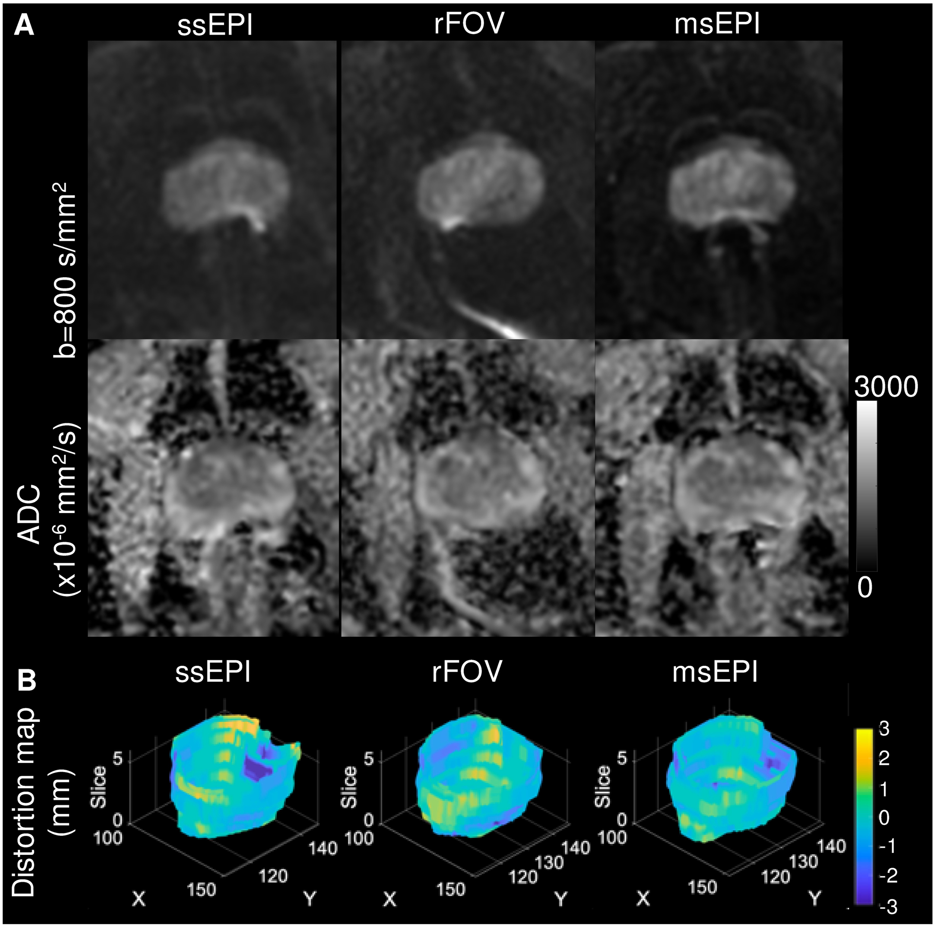
(A) On the ssEPI images, rectal gas results in artifactual signal build-up (white arrow) and contour distortion (dashed arrow) of the posterior prostate. The artifact and distortion are reduced in both the rFOV and, to a greater extent, msEPI images. B. Distortion plots from the same patient demonstrate a small reduction in distortion for both rFOV and msEPI graphically and in a reduction in dr.m.s. (in mm) for rFOV (1.9) and msEPI (1.8) compared to ssEPI (2.2).
3.2. Quantitative ADC results:
Prostate cancer histogram parameters were similar for ssEPI, rFOV, and msEPI DWI (Table 2; all p-values >0.05). When the difference between cancerous and non-cancerous prostate ROIs were compared for each sequence, mean ADC was significantly lower for cancer compared to non-cancerous prostate for the clinical and research DWI sequences (Figure 2A). An illustrative example is presented in Figure 3. When the calculated mean ADC for cancerous and non-cancerous ROIs were compared separately between the three sequences the rFOV non-cancerous prostate mean ADC was significantly lower than from msEPI (p<0.05; Figure 2B).
Table 2.
Histogram parameters for cancerous regions of interest
| ADC parameters (x10−6 mm2/s) | ssEPI | rFOV | msEPI |
|---|---|---|---|
| Standard deviation | 138.1 ± 33.2 | 139.8 ± 30.2 | 156.8 ± 45.4 |
| Mean | 985.1 ± 178 | 1019 ± 144 | 997.4 ± 186 |
| 20th percentile | 859.0 ± 180 | 889.9 ± 137 | 856.7 ± 205 |
| Median | 975.2 ± 172 | 995.7 ± 144 | 992.3 ± 184.1 |
| 80th percentile | 1099 ± 178 | 1139 ± 158 | 1133 ± 182 |
Data are mean ± standard deviation
ADC, apparent diffusion coefficient
Figure 2. Comparison of mean apparent diffusion coefficient values for cancer and non-cancerous prostate regions.
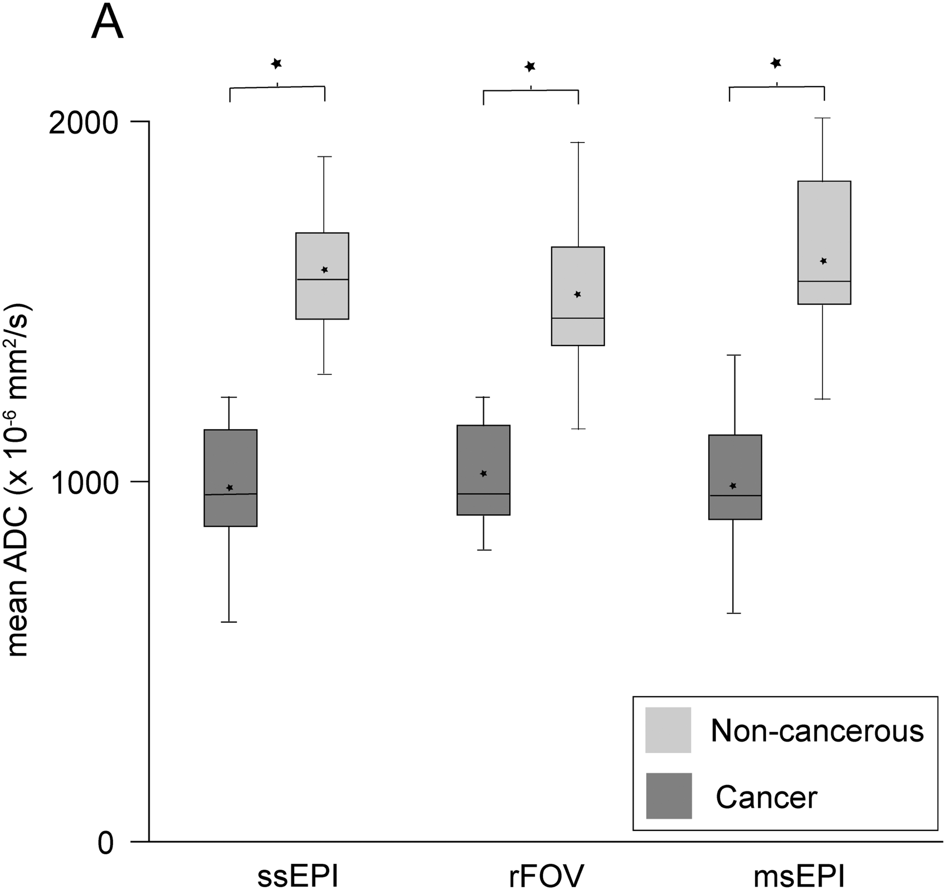
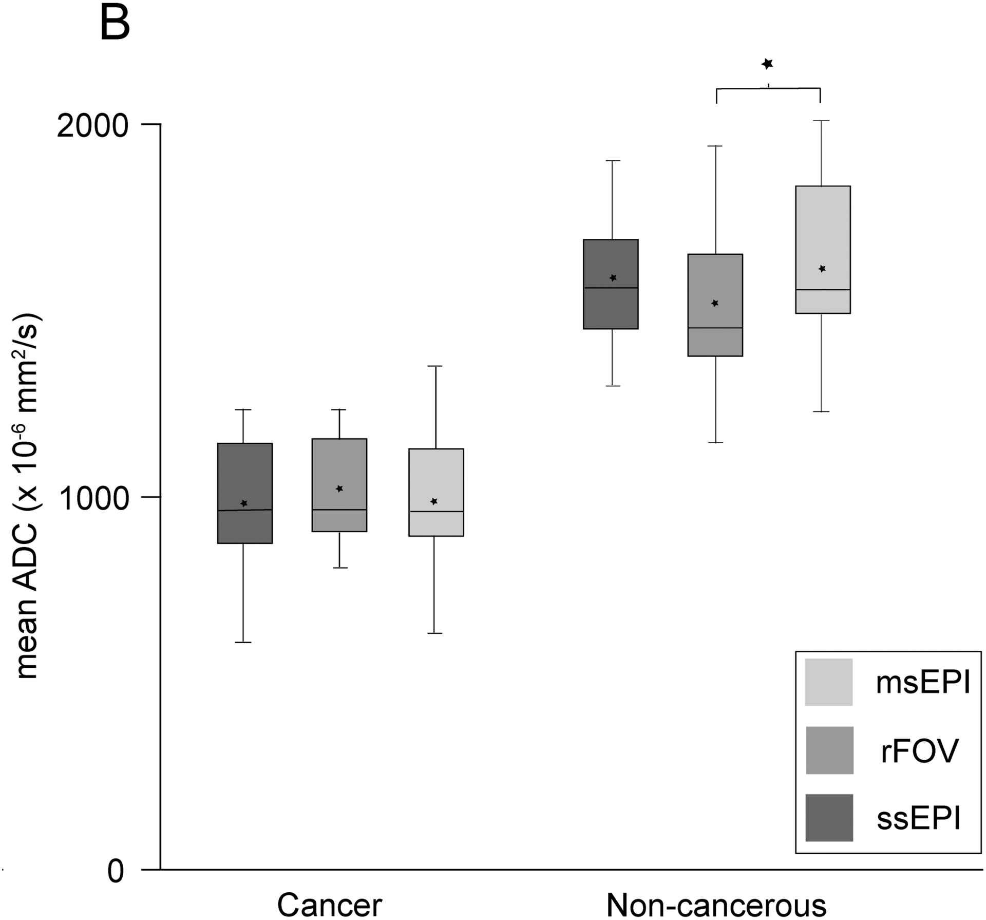
2A) Boxplots grouped and compared according to diffusion acquisition sequence demonstrate that mean ADC was significantly lower for cancer regions compared to non-cancerous prostate for all DWI sequences (*p<0.0001). 2B) Boxplots grouped and compared according to tissue type demonstrate that mean ADC was not significantly different for all comparison except between the non-cancerous prostate values for rFOV compared to msEPI (*p<0.01). Box and whisker plots demonstrate the median (middle line), mean (star), upper and lower quartiles (top and bottom box margins), and minimum/maximum (whiskers). ADC, apparent diffusion coefficient; msEPI, multi-shot echo planar imaging; NC, non-cancerous; rFOV, reduced field-of-view; ssEPI, single-shot echo planar imaging;
Figure 3. Similar apparent diffusion coefficient (ADC) quantification in a 60-year old male with elevated serum prostate specific antigen (4.46 ng/mL).
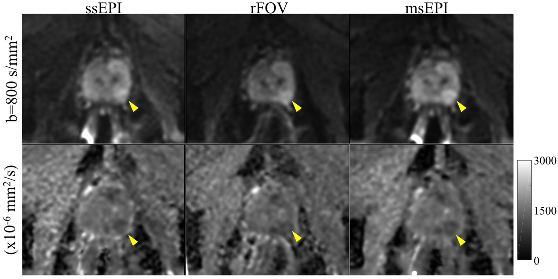
Left peripheral zone PI-RADS 4 lesion (yellow arrowheads) is seen on all three DWI sequences, including the b=800 s/mm2 series (top row) and calculated ADC map (bottom row). The mean ADC value for the lesion was 1075.8, 1098.8, and 1067.5 ×10−6 mm2/s on ssEPI, rFOV, and msEPI, respectively. The patient subsequently underwent radical prostatectomy and was found to have Gleason 3+4 prostate cancer.
3.3. Image quality results:
The DWI acquisition utilizing multi-shot EPI was rated higher in overall image quality, influence of distortion/artifacts, and prostate zonal anatomy compared to both ssEPI and rFOV (p<0.05). Multi-shot EPI also demonstrated significantly improved scores for level of DWI distortion/artifact, compared to ssEPI, and higher qualitative SNR scores, compared to rFOV (p<0.05). Image quality assessment and reader scores are summarized in Table 3.
Table 3.
Multi-reader qualitative assessment of image quality, DWI artifacts, and distortion
| Mean reader score | |||
|---|---|---|---|
| ssEPI | rFOV | msEPI | |
| Resolution | 3.24 ± 0.29 | 3.35 ± 0.31 | 3.47 ± 0.37 |
| Capsule demarcation | 2.91 ± 0.51 | 3.10 ± 0.44* | 3.17 ± 0.46* |
| Zonal anatomy | 2.46 ± 0.52 | 2.52 ± 0.56 | 2.78 ± 0.55**t |
| Qualitative SNR | 3.41 ± 0.32 | 3.39 ± 0.28 | 3.57 ± 0.39t |
| Overall Image Quality | 2.90 ± 0.59 | 3.03 ± 0.46 | 3.28 ± 0.57**t |
| Level of Distortion/Artifacts | 3.04 ± 0.68 | 3.16 ± 0.70 | 3.35 ± 0.66* |
| Influence of Distortion/Artifacts | 3.13 ± 0.80 | 3.23 ± 0.74 | 3.52 ± 0.64**t |
p-value < 0.05 between rFOV or msEPI and ssEPI
p-value < 0.01 between rFOV or msEPI and ssEPI
p-value < 0.05 between msEPI and rFOV
4. Discussion
We found that reduced-distortion DWI methods, based on reduced field of view (rFOV) and multi-shot techniques (msEPI), significantly reduced quantitative distortion of the prostate, maintained similar ADC quantification, and demonstrated evidence of similar to slightly improved image quality, compared to standard single-shot EPI.
DWI of the prostate utilizing ssEPI can suffer severe image distortion due to susceptibility-related field inhomogeneities, often due to rectal gas. Prior work in phantoms, normal volunteers, and limited assessment in prostate cancer patients has shown that two different distortion reduction techniques, rFOV and msEPI, can reduce distortion and ghosting [23, 26–28]. In the current work, we expanded on these results and found a small but significant reduction in the tested distortion metric, dr.m.s., for both rFOV and msEPI compared to ssEPI. This distortion metric had been previously tested in the work by Gill et al. where it was shown to successfully distinguish between three defined groups of patients: ‘non-distorted’, ‘distorted’, and ‘severely distorted due to the effect of a metallic prosthesis’ [30]. In a subsequent study, dr.m.s. was also shown to be correlated with the degree of rectal distension present [31]. While the degree of improvement shown in the current study may seem slight, clinically significant prostate cancer is most frequently encountered in the peripheral zone adjacent to the rectum; hence, any reduction in image distortion may improve cancer detection.
It is also important to consider how a novel DWI sequence might affect quantitative imaging, especially ADC. In the current study, prostate cancer ADC histogram parameters were similar for ssEPI, rFOV, and msEPI DWI and each sequence maintained a significantly lower mean ADC for prostate cancer compared to non-cancerous prostate. Prior works investigating the effect of rFOV on ADC values have demonstrated mixed results [26–28, 32–35]; however, the majority support a non-significant difference between ADC values from rFOV and ssEPI [26, 28, 32, 34, 35]. Interestingly, in the current study mean ADC for non-cancerous prostate was lower for rFOV, compared to the other sequences; however, the underlying cause might be due in part to an overall lower SNR for the rFOV images, leading to noise floor effects at the higher b value, which in turn result in ADC underestimation [36–38]. Indeed, this result is in good agreement with prior results in phantoms and normal volunteers [26].
Regarding qualitative reader analysis, there was a trend of improving average reader scores with the reduced distortion sequences, especially with msEPI, and this difference was statistically significant for many parameters. Taken categorically, it meant a shift in the perceived level of distortion from an average assessment of intermediate (score 3) toward low distortion (score 4) as well as a shift in perceived effect of distortion from an average assessment of satisfactory quality (score 3) toward good quality with hardly any artifacts (score 4). This matches prior work evaluating rFOV DWI in a cohort of 43 patients with prostate MRIs that also demonstrated significantly improved subjective image quality compared to a conventional ssEPI sequence [35]. That study also demonstrated a measured preference for the rFOV sequence when readers were given a choice in the head-to-head comparison [35]. A similar improvement in subjective image quality was also seen for 1 of 2 readers in a study of 49 patients undergoing prostate MR prior to planned MR/US-fusion biopsy; however, there was not a significant difference in reader sensitivity, specificity, or accuracy for GS 3+4 or higher tumors when comparing the two sequences [39]. A novel feature of the current work was evaluating msEPI compared to rFOV, which demonstrated improved average reader scores for msEPI regarding distortion influence, overall image quality, qualitative SNR, and zonal anatomy. The two sequences utilize slightly different techniques to reduce distortion as rFOV reduces distortion by limiting the size of the FOV while msEPI shortens the echo train length. Therefore, msEPI has the capability to maintain a higher resolution diffusion-weighted image without sacrificing scan coverage; however, this comes at the expense of a longer scan time and increased post-processing. Further research evaluating the effect of reduced distortion DWI sequences on the clinical assessment of prostate cancer is warranted, particularly if the proposed sequence would involve an increase in scan time or post-processing as is the case with msEPI.
The current study has several limitations. While the prospective design was a strength, the study cohort was small which could lead to bias. Validation of our findings in a larger cohort might be beneficial. Second, qualitative image assessment can be limited by the subjectivity of reader opinion; however, image quality, as determined subjectively by the interpreting radiologist, is critical to the diagnosis of prostate cancer. The study attempted to standardize the subjective image quality interpretation by using defined categories regarding the level and effect of artifacts as well as by averaging the scores of all three readers prior to statistical analysis. Furthermore, whenever possible quantitative analysis was utilized including an assessment of quantitative prostate distortion and through analysis of multiple ADC quantitative parameters. Third, the use of biopsy results for pathologic comparison can be limited by sampling bias, which is especially true for cognitive MRI-US biopsy. Finally, there were some DWI acquisition parameters that might have affected individual sequences. A standardized higher b-value of b=800 s/mm2 was utilized for ssEPI, msEPI, and rFOV which may optimize the accuracy of the calculated ADC maps; however, qualitative DWI interpretation often now includes the use of higher b-value images (i.e. b=1000 or even 1500 s/mm2). The number of averages was kept consistent between the three acquisitions which resulted in a scan time for msEPI that was double the other sequences.
In conclusion, rFOV and msEPI DWI methods reduced quantitative distortion of the prostate, maintained similar ADC quantification, and demonstrated evidence of slight qualitative improvement, compared to standard single-shot EPI.
Supplementary Material
Acknowledgements
The authors wish to acknowledge GE Healthcare who provides research support to the University of Wisconsin.
Abbreviations
- msEPI
multi shot echo planar imaging
- MUSE
multiplexed sensitivity encoding
- PSA
prostate specific antigen
- rFOV
reduced field of view
- ROI
region of interest
- ssEPI
single shot echo planar imaging
References
- [1].Stabile A, Giganti F, Rosenkrantz AB, Taneja SS, Villeirs G, Gill IS, et al. Multiparametric MRI for prostate cancer diagnosis: current status and future directions. Nat Rev Urol 2020;17:41–61. doi: 10.1038/s41585-019-0212-4. [DOI] [PubMed] [Google Scholar]
- [2].Turkbey B, Rosenkrantz AB, Haider MA, Padhani AR, Villeirs G, Macura KJ, et al. Prostate Imaging Reporting and Data System Version 2.1: 2019 Update of Prostate Imaging Reporting and Data System Version 2. Eur Urol 2019;76:340–351. doi: 10.1016/j.eururo.2019.02.033 [DOI] [PubMed] [Google Scholar]
- [3].Fütterer JJ, Briganti A, De Visschere P, Emberton M, Giannarini G, Kirkham A, et al. Can Clinically Significant Prostate Cancer Be Detected with Multiparametric Magnetic Resonance Imaging? A Systematic Review of the Literature. Eur Urol 2015;68:1045–1053. doi: 10.1016/j.eururo.2015.01.013. [DOI] [PubMed] [Google Scholar]
- [4].Jung SI, Donati OF, Vargas HA, Goldman D, Hricak H, Akin O. Transition zone prostate cancer: incremental value of diffusion-weighted endorectal MR imaging in tumor detection and assessment of aggressiveness. Radiology 2013;269:493–503. doi: 10.1148/radiol.13130029. [DOI] [PubMed] [Google Scholar]
- [5].Park SY, Oh YT, Jung DC, Cho NH, Choi YD, Rha KH, et al. Prediction of biochemical recurrence after radical prostatectomy with PI-RADS version 2 in prostate cancers: initial results. Eur Radiol 2016;26:2502–2509. doi: 10.1007/s00330-015-4077-5. [DOI] [PubMed] [Google Scholar]
- [6].Lawrence EM, Tang SY, Barrett T, Koo B, Goldman DA, Warren AY, et al. Prostate cancer: performance characteristics of combined T₂W and DW-MRI scoring in the setting of template transperineal re-biopsy using MR-TRUS fusion. Eur Radiol 2014;24:1497–1505. doi: 10.1007/s00330-014-3159-0. [DOI] [PubMed] [Google Scholar]
- [7].Barrett T, Lawrence EM, Priest AN, Warren AY, Gnanapragasam VJ, Gallagher FA, et al. Repeatability of diffusion-weighted MRI of the prostate using whole lesion ADC values, skew and histogram analysis. Eur J Radiol 2019;110:22–29. doi: 10.1016/j.ejrad.2018.11.014. [DOI] [PubMed] [Google Scholar]
- [8].Woodfield CA, Tung GA, Grand DJ, Pezzullo JA, Machan JT, Renzulli JF. Diffusion-weighted MRI of peripheral zone prostate cancer: comparison of tumor apparent diffusion coefficient with Gleason score and percentage of tumor on core biopsy. AJR Am J Roentgenol 2010;194:W316–322. doi: 10.2214/AJR.09.2651. [DOI] [PubMed] [Google Scholar]
- [9].Fukunaga T, Tamada T, Kanomata N, Takeuchi M, Ueda Y, Miyaji Y. Quantitative diffusion-weighted imaging and dynamic contrast-enhanced MR imaging for assessment of tumor aggressiveness in prostate cancer at 3T. Magn Reson Imaging 2021; 83:152–159. doi: 10.1016/j.mri.2021.08.009. [DOI] [PubMed] [Google Scholar]
- [10].Salami SS, Ben-Levi E, Yaskiv O, Turkbey B, Villani R, Rastinehad AR. Risk stratification of prostate cancer utilizing apparent diffusion coefficient value and lesion volume on multiparametric MRI. J Magn Reson Imaging 2017; 45:610–616. doi: 10.1002/jmri.25363. [DOI] [PubMed] [Google Scholar]
- [11].Mazzone E, Stabile A, Pellegrino F, Basile G, Cignoli D, Cirulli GO, et al. Positive Predictive Value of Prostate Imaging Reporting and Data System Version 2 for the Detection of Clinically Significant Prostate Cancer: A Systematic Review and Meta-analysis. Eur Urol Oncol 2020;4:697–713. doi: 10.1016/j.euo.2020.12.004. [DOI] [PubMed] [Google Scholar]
- [12].Lawrence EM, Gallagher FA, Barrett T, Warren AY, Priest AN, Goldman DA, et al. Preoperative 3-T diffusion-weighted MRI for the qualitative and quantitative assessment of extracapsular extension in patients with intermediate- or high-risk prostate cancer. AJR Am J Roentgenol 2014;203:W280–286. doi: 10.2214/AJR.13.11754. [DOI] [PubMed] [Google Scholar]
- [13].Vargas HA, Akin O, Franiel T, Mazaheri Y, Zheng J, Moskowitz C, et al. Diffusion-weighted endorectal MR imaging at 3 T for prostate cancer: tumor detection and assessment of aggressiveness. Radiology 2011;259:775–784. doi: 10.1148/radiol.11102066. [DOI] [PMC free article] [PubMed] [Google Scholar]
- [14].Tamada T, Prabhu V, Li J, Babb JS, Taneja SS, Rosenkrantz AB. Prostate Cancer: Diffusion-weighted MR Imaging for Detection and Assessment of Aggressiveness-Comparison between Conventional and Kurtosis Models. Radiology 2017;284:100–108. doi: 10.1148/radiol.2017162321. [DOI] [PubMed] [Google Scholar]
- [15].McGarry SD, Brehler M, Bukowy JD, et al. (2021) Multi-Site Concordance of Diffusion-Weighted Imaging Quantification for Assessing Prostate Cancer Aggressiveness. J Magn Reson Imaging. DOI: 10.1002/jmri.27983. [DOI] [PMC free article] [PubMed] [Google Scholar]
- [16].Li C, Chen M, Wang J, Wang X, Zhang W, Zhang C. Apparent diffusion coefficient values are superior to transrectal ultrasound-guided prostate biopsy for the assessment of prostate cancer aggressiveness. Acta Radiol 2017;58:232–239. doi: 10.1177/0284185116639764. [DOI] [PubMed] [Google Scholar]
- [17].Henderson DR, de Souza NM, Thomas K, Riches SF, Morgan VA, Sohaib SA, et al. Nine-year Follow-up for a Study of Diffusion-weighted Magnetic Resonance Imaging in a Prospective Prostate Cancer Active Surveillance Cohort. Eur Urol 2016;69:1028–1033. doi: 10.1016/j.eururo.2015.10.010. [DOI] [PubMed] [Google Scholar]
- [18].Jie C, Rongbo L, Ping T. The value of diffusion-weighted imaging in the detection of prostate cancer: a meta-analysis. Eur Radiol 2014;24:1929–1941. doi: 10.1007/s00330-014-3201-2. [DOI] [PMC free article] [PubMed] [Google Scholar]
- [19].Turner R, Le Bihan D, Chesnick AS. Echo-planar imaging of diffusion and perfusion. Magn Reson Med 1991;19:247–253. doi: 10.1002/mrm.1910190210. [DOI] [PubMed] [Google Scholar]
- [20].Bammer R, Auer M, Keeling SL, Augustin M, Stables LA, Prokesch RW, et al. Diffusion tensor imaging using single-shot SENSE-EPI. Magn Reson Med 2002;48:128–136. doi: 10.1002/mrm.10184. [DOI] [PubMed] [Google Scholar]
- [21].Zhao L, Madore B, Panych LP. Reduced field-of-view MRI with two-dimensional spatially-selective RF excitation and UNFOLD. Magn Reson Med 2005;53:1118–1125. doi: 10.1002/mrm.20458. [DOI] [PubMed] [Google Scholar]
- [22].Wilm BJ, Svensson J, Henning A, Pruessmann KP, Boesiger P, Kollias SS. Reduced field-of-view MRI using outer volume suppression for spinal cord diffusion imaging. Magn Reson Med 2007;57:625–630. doi: 10.1002/mrm.21167. [DOI] [PubMed] [Google Scholar]
- [23].Chen NK, Guidon A, Chang HC, Song AW. A robust multi-shot scan strategy for high-resolution diffusion weighted MRI enabled by multiplexed sensitivity-encoding (MUSE). Neuroimage 2013;72:41–47. doi: 10.1016/j.neuroimage.2013.01.038. [DOI] [PMC free article] [PubMed] [Google Scholar]
- [24].Holdsworth SJ, Skare S, Newbould RD, Guzmann R, Blevins NH, Bammer R. Readout-segmented EPI for rapid high resolution diffusion imaging at 3 T. Eur J Radiol 2008;65:36–46. doi: 10.1016/j.ejrad.2007.09.016. [DOI] [PMC free article] [PubMed] [Google Scholar]
- [25].Yeom KW, Holdsworth SJ, Van AT, Iv M, Skare S, Lober RM, et al. Comparison of readout-segmented echo-planar imaging (EPI) and single-shot EPI in clinical application of diffusion-weighted imaging of the pediatric brain. AJR Am J Roentgenol 2013;200:W437–443. doi: 10.2214/AJR.12.9854. [DOI] [PubMed] [Google Scholar]
- [26].Zhang Y, Holmes J, Rabanillo I, Guidon A, Wells S, Hernando D. Quantitative diffusion MRI using reduced field-of-view and multi-shot acquisition techniques: Validation in phantoms and prostate imaging. Magn Reson Imaging 2018;51:173–181. doi: 10.1016/j.mri.2018.04.006. [DOI] [PMC free article] [PubMed] [Google Scholar]
- [27].Korn N, Kurhanewicz J, Banerjee S, Starobinets O, Saritas E, Noworolski S. Reduced-FOV excitation decreases susceptibility artifact in diffusion-weighted MRI with endorectal coil for prostate cancer detection. Magn Reson Imaging 2015;33:56–62. doi: 10.1016/j.mri.2014.08.040. [DOI] [PMC free article] [PubMed] [Google Scholar]
- [28].Feng Z, Min X, Sah VK, Li L, Cai J, Deng M, et al. Comparison of field-of-view (FOV) optimized and constrained undistorted single shot (FOCUS) with conventional DWI for the evaluation of prostate cancer. Clin Imaging 2015;39:851–855. doi: 10.1016/j.clinimag.2015.03.004. [DOI] [PubMed] [Google Scholar]
- [29].Saritas EU, Cunningham CH, Lee JH, Han ET, Nishimura DG. DWI of the spinal cord with reduced FOV single-shot EPI. Magn Reson Med 2008;60:468–473. doi: 10.1002/mrm.21640. [DOI] [PubMed] [Google Scholar]
- [30].Gill AB, Czarniecki M, Gallagher FA, Barrett T. A method for mapping and quantifying whole organ diffusion-weighted image distortion in MR imaging of the prostate. Sci Rep 2017;7:12727. doi: 10.1038/s41598-017-13097-6. [DOI] [PMC free article] [PubMed] [Google Scholar]
- [31].Caglic I, Hansen NL, Slough RA, Patterson AJ, Barrett T. Evaluating the effect of rectal distension on prostate multiparametric MRI image quality. Eur J Radiol 2017;90:174–180. doi: 10.1016/j.ejrad.2017.02.029. [DOI] [PubMed] [Google Scholar]
- [32].Ma C, Li YJ, Pan CS, Wang H, Wang J, Chen S, et al. High resolution diffusion weighted magnetic resonance imaging of the pancreas using reduced field of view single-shot echo-planar imaging at 3 T. Magn Reson Imaging 2014;32:125–131. doi: 10.1016/j.mri.2013.10.005. [DOI] [PubMed] [Google Scholar]
- [33].Singer L, Wilmes LJ, Saritas EU, Shankaranarayanan A, Proctor E, Wisner DJ, et al. High-resolution diffusion-weighted magnetic resonance imaging in patients with locally advanced breast cancer. Acad Radiol 2012;19:526–534. doi: 10.1016/j.acra.2011.11.003. [DOI] [PMC free article] [PubMed] [Google Scholar]
- [34].Zaharchuk G, Saritas EU, Andre JB, Chin CT, Rosenberg J, Brosnan TJ, et al. Reduced field-of-view diffusion imaging of the human spinal cord: comparison with conventional single-shot echo-planar imaging. AJNR Am J Neuroradiol 2011;32:813–820. doi: 10.3174/ajnr.A2418. [DOI] [PMC free article] [PubMed] [Google Scholar]
- [35].Warndahl BA, Borisch EA, Kawashima A, Riederer SJ, Froemming AT. Conventional vs. reduced field of view diffusion weighted imaging of the prostate: Comparison of image quality, correlation with histology, and inter-reader agreement. Magn Reson Imaging 2018;47:67–76. doi: 10.1016/j.mri.2017.10.011. [DOI] [PubMed] [Google Scholar]
- [36].Prah DE, Paulson ES, Nencka AS, Schmainda KM. A simple method for rectified noise floor suppression: Phase-corrected real data reconstruction with application to diffusion-weighted imaging. Magn Reson Med 2010;64:418–429. doi: 10.1002/mrm.22407. [DOI] [PMC free article] [PubMed] [Google Scholar]
- [37].Iima M, Partridge SC, Le Bihan D. Six DWI questions you always wanted to know but were afraid to ask: clinical relevance for breast diffusion MRI. Eur Radiol 2020;30:2561–2570. doi: 10.1007/s00330-019-06648-0. [DOI] [PubMed] [Google Scholar]
- [38].Kristoffersen A. Optimal estimation of the diffusion coefficient from non-averaged and averaged noisy magnitude data. J Magn Reson 2007;187:293–305. doi: 10.1016/j.jmr.2007.05.004. [DOI] [PubMed] [Google Scholar]
- [39].Tamada T, Ream JM, Doshi AM, Taneja SS, Rosenkrantz AB. Reduced Field-of-View Diffusion-Weighted Magnetic Resonance Imaging of the Prostate at 3 Tesla: Comparison With Standard Echo-Planar Imaging Technique for Image Quality and Tumor Assessment. J Comput Assist Tomogr 2017;41:949–956. doi: 10.1097/RCT.0000000000000634. [DOI] [PubMed] [Google Scholar]
Associated Data
This section collects any data citations, data availability statements, or supplementary materials included in this article.