Abstract
We herein report a rare case of leiomyoma of the urethra. A young woman with no history of malignancy was referred to our hospital because of a 1-year history of frequent urination, urgency, and dysuria. Postoperative pathologic examination confirmed the diagnosis of urethral leiomyoma. After tumor resection, the patient was followed up for 2 months in the outpatient department and developed no obvious postoperative complications such as urinary incontinence.
Keywords: Leiomyoma, leiomyoma of the urethra, case report, enhanced computed tomography, tumor resection, immunohistochemistry
Introduction
Leiomyoma is a rare benign tumor that arises from smooth muscle cells, most commonly in the genital organs and especially in the uterus. Extrauterine leiomyomas are rare; among them, urethral leiomyoma is an extremely rare type of tumor that arises from smooth muscle cells of mesenchymal tissue. The posterior wall of the proximal urethra is most commonly affected 1 ; very few reports have described posterior urethral leiomyoma. We herein report a rare case of retrourethral leiomyoma and review the pathological features of the urethral smooth muscle and the relevant knowledge of the clinical treatment.
Case report
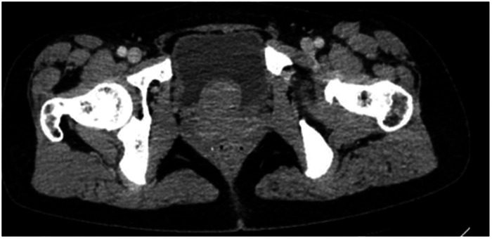
A young woman was admitted to our hospital because of a 1-year history of frequent urination, urgency, and dysuria. She reported that the symptoms became aggravated during menstruation, but she had no other abnormalities such as hematuria or fever. Pelvic enhanced computed tomography revealed a smooth mass, as shown in Figure 1.
Figure 1.
Contrast-enhanced computed tomography showed a smooth mass with a clear boundary in the bladder.
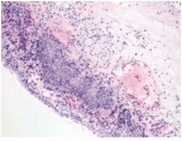
The patient underwent transurethral resection of the urethral mass under general anesthesia. The tumor had a smooth surface and was located between the posterior urethra and the bladder neck, and the resected mass was completely covered by the urethral mucosa. The catheter was removed 3 days after the operation, at which time the patient was urinating well and had no obvious urinary incontinence. Postoperative re-examination showed no obvious symptoms of frequent urination, urgent urination, dysuria, or hematuria. The initial postoperative pathology report considered a “somatocellular tumor,” and the results in the second report were as follows: CD34 (−), CD117 (−), smooth muscle actin (+), desmin (+), Ki-67 (+, 3%), calponin (+), h-caldesmon (+), anaplastic lymphoma kinase (D5F3) (−), estrogen receptor (+), and progesterone receptor (+); these results supported genital tract leiomyoma (Figures 2 and 3).
Figure 2.
Microscopic appearance of the neoplasm with hematoxylin and eosin staining.
Figure 3.
Microscopic appearance of the neoplasm with hematoxylin and eosin staining.
The reporting of this study conforms to the CARE guidelines. 2
Discussion
Urethral leiomyoma is an extremely rare tumor, and Buttner 3 described the first case in 1894. To date, fewer than 45 cases have been described in the literature. 4 About 25% of cases of urethral leiomyoma are asymptomatic, and patients generally complain of symptoms such as a urethral mass, hematuria, frequent urination, urgency, and vaginal bleeding. Because of the different sites of urethral leiomyoma occurrence and different tumor sizes, the clinical symptoms vary. According to the literature, tumors located at the 12- or 6-o’clock position cause symptoms of dysuria, whereas laterally located leiomyomas are more likely to cause irritant symptoms. 5 In our patient, the urethral leiomyoma was located between the urethra and bladder neck and caused significant symptoms of frequent urination, urgency, and dysuria, confirming the findings in previous cases. Urethral leiomyomas are more common in women aged 30 to 40 years, and the mean age at presentation is 41 years. 6 The etiology of urethral leiomyoma may be related to increased estrogen levels. According to some reports, the size of the tumor may decrease with menopause or after delivery, indicating that the disease is hormone-dependent.7–9
Leiomyoma is a benign tumor of the urethral smooth muscle. Although it is more common in the genitourinary tract and gastrointestinal tract, its incidence in the skin is low, and it is rarely seen in deep tissues. In general, soft tissue leiomyoma rarely causes morbidity. 10 Generally, the surface mucosa of urethral leiomyoma is smooth and firm. According to the amount of smooth muscle and fibrous tissue contained within the tumor, the resected tumor may be pink or milky white. The tumor tissue is composed of well-differentiated smooth muscle cells. In contrast, female reproductive tract malignancies are often squamous cells or transitional cells. The leiomyoma cells are triangular in shape, contain abundant cytoplasm and show clear borders, and are clustered into fascicles; mitosis is rare. Leiomyoma must be differentiated from leiomyosarcoma. The presence or absence of anaplasia and identification of mitotic phases are the main criteria used for differentiation.
In this case, pathological biopsy showed that the tumor consisted of interdigitating smooth muscle fiber bundles. No mitosis or nuclei atypia was seen. Immunohistochemical examination showed that the tumor was positive for actin and desmin. The proliferation index (Ki-67) was approximately <3%. These features confirmed the diagnosis of urethral leiomyoma. Surgical resection is the only effective treatment for urethral leiomyoma, and different treatments are adopted according to where the tumor is located within the urethra. Local tumor resection is performed for tumors located in the external urethral meatus, transvaginal tumor resection can be considered for tumors located in the posterior urethral wall, and transurethral resection is feasible for tumors located in the urethra. 11 If the mass is large and close to the bladder neck, it can be removed by transabdominal surgery. Transurethral resection should be performed to avoid injury to the urethral sphincter and prevent urinary incontinence. To date, no reports have described malignant transformation of the urethral smooth muscle; however, local recurrence is possible. Only one patient developed two relapses and thus underwent three surgical procedures.6,12 According to the relevant literature, urethral leiomyoma can also be found in a very small number of men, but postoperative malignant transformation rarely occurs. 5 Our patient showed no obvious symptoms of urinary incontinence during the 1-month postoperative follow-up, and re-examination by B-mode ultrasound showed no obvious tumor recurrence.
Conclusions
Urethral leiomyoma is an extremely rare benign tumor, and fewer than 45 cases have been described in the literature to date. We have herein reported a case of posterior urethral leiomyoma, the location of which differed from the common location of urethral leiomyoma; however, the clinical presentation was roughly similar. Surgical resection is the only treatment for urethral leiomyoma that can ensure long-term disease-free survival. 6 However, the choice of surgical method and the surgeon’s technical skills may affect the risk of postoperative recurrence. This case report supplements the accumulated cases of urethral leiomyomatosis reported to date, but it does not discuss the underlying etiology of cancer formation within the urethral smooth muscle. Because this was only a single case, a retrospective study could not be carried out. Multicenter case studies and long-term follow-up data are needed to further guide clinical decision-making for leiomyoma.
Declaration of conflicting interest: The authors declare that there is no conflict of interest.
Funding: The authors received no financial support for the research, authorship, and/or publication of this article.
Informed consent: The patient provided written informed consent for treatment and for publication of this case for scientific purposes.
ORCID iDs: Yang Liu https://orcid.org/0000-0002-8785-277X
XueSong Yang https://orcid.org/0000-0001-6164-3788
Ethics
Ethics committee approval was not required because of the nature of this study (case report).
References
- 1.Lee MC, Lee SD, Kuo HT, et al. Obstructive leiomyoma of the female urethra: report of a case. J Urol 1995; 153: 420–421. [DOI] [PubMed] [Google Scholar]
- 2.Gagnier JJ, Kienle G, Altman DG, et al. ; CARE Group. The CARE guidelines: consensus-based clinical case reporting guideline development. Headache 2013; 53: 1541–1547. [DOI] [PubMed] [Google Scholar]
- 3.Buttner C. A case of myoma of the female urethra. Z Geburshe Gynak 1894; 28: 135. [Google Scholar]
- 4.Popov SV, Orlov IN, Chernysheva DY, et al. Urethral leiomyoma: a rare neoplasm. Urol Ann 2021; 13: 194–197. [DOI] [PMC free article] [PubMed] [Google Scholar]
- 5.Bai SW, Jung HJ, Jeon MJ, et al. Leiomyomas of the female urethra and bladder: a report of five cases and review of the literature. Int Urogynecol J Pelvic Floor Dysfunct 2007; 18: 913–917. [DOI] [PubMed] [Google Scholar]
- 6.Jiménez Navarro M, Ballesta Martínez B, Rodríguez Talavera J, et al. Recurrence of urethral leiomyoma: a case report. Urol Case Rep 2019; 26: 100968. Published 2019 Jul 16. [DOI] [PMC free article] [PubMed] [Google Scholar]
- 7.Strang A, Lisson SW, Petrou SP. Ureteral endometriosis and coexistent urethral leiomyoma in a postmenopausal woman. Int Braz J Urol 2004; 30: 496–498. [DOI] [PubMed] [Google Scholar]
- 8.Rivière P, Bodin R, Bernard G, et al. Leiomyoma of the female urethra. Prog Urol 2004; 14: 1196–1198. [PubMed] [Google Scholar]
- 9.Merrell RW, Brown HE. Recurrent urethral leiomyoma presenting as stress incontinence. Urology 1981; 17: 588–589. [DOI] [PubMed] [Google Scholar]
- 10.Dell’Atti L, Galosi AB. Female urethra adenocarcinoma. Clin Genitourin Cancer 2018; 16: e263–e267. [DOI] [PubMed] [Google Scholar]
- 11.Pahwa M, Saifee Y, Pahwa AR, et al. Leiomyoma of the female urethra-a rare tumor: case report and review of the literature. Case Rep Urol 2012; 2012: 280816. [DOI] [PMC free article] [PubMed] [Google Scholar]
- 12.Deka PM, Rajeev TP. Leiomyoma of the female urethra: a case report. Urol Int 2003; 71: 224–225. [DOI] [PubMed] [Google Scholar]


