



Abstract
Musculoskeletal pain is an aversive experience that exists within a variety of conditions and can result in significant impairment for individuals. Gaining greater understanding of the factors related to pain vulnerability and resilience to musculoskeletal pain may help target at-risk individuals for early intervention. This analysis builds on our previous work identifying regions where greater gray matter density was associated with lower pain following standardized, exercise induced musculoskeletal injury. Here we sought to examine the relationship between baseline resting state functional connectivity in a priori regions and networks, and delayed onset muscle soreness (DOMS) pain intensity following a single session of eccentric exercise in healthy adults. Participants completed a baseline functional MRI scan and a high intensity trunk exercise protocol in the erector spinae. Pain intensity ratings were collected 48-hours later. Resting state functional connectivity from four seed regions and 3 networks were separately regressed on pain intensity scores. Results revealed that connectivity between left middle frontal gyrus, the left occipital gyrus and cerebellar network seeds and clusters associated with discriminative, emotional, and cognitive aspects of pain were associated with lower post-DOMS pain. Results suggest resilience to clinically relevant pain is associated with aspects of regional and network neural coherence. Investigations of pain modulatory capacity that integrate multimodal neuroimaging metrics are called for.
Keywords: Delayed onset muscle soreness, pain resilience, resting state functional connectivity, pain modulation, musculoskeletal pain
Pain is an aversive sensory, cognitive, and emotional experience that occurs in a variety of conditions and imposes a substantial individual and public health burden. For nearly three decades, musculoskeletal pain has remained a leading cause of years lived with disability globally.22 In the United States, an estimated 20.4% of adults experience chronic pain that is associated with $560B USD/year of total healthcare costs.15 Chronic musculoskeletal pain is especially prevalent, with 19.6% of Americans aged 20 to 59 reporting chronic low back pain,31 and is commonly characterized by lower physical activity, reduced mobility, and cognitive impairment.6 Targeting individuals who are at high risk for developing musculoskeletal pain with effective, early treatments for acute and sub-acute pain could prevent chronic pain by preventing maladaptive neural plasticity associated with negative health consequences.19,43 Gaining greater understanding of the factors related to pain vulnerability and resilience to musculoskeletal pain may help target at risk individuals for early intervention. One approach to identifying factors that predict who will go on to experience chronic pain after injury is to characterize predictors of the experience of experimentally induced but clinically relevant musculoskeletal pain in healthy individuals.
Typical laboratory-based acute pain induction paradigms (eg, cold pressor, heat pain) are limited in their ability to characterize musculoskeletal pain mechanisms because they do not produce the functional impairment or resulting modification of activities of daily living common to musculoskeletal pain conditions. Our group has previously developed and validated a delayed-onset muscle soreness (DOMS) induction paradigm that produces clinically relevant but inherently time-limited pain that peaks within 24 to 48 hours postinduction and resolves within approximately one week.5,16,17,40 DOMS is an eccentric exercise-induced muscle injury approach that produces clinically relevant musculoskeletal pain, typically lasts for several days, vs. seconds or minutes with other laboratory-based induction methods (e.g., heat pain or cold pressor). Additionally, DOMS provides ecological validity as a pain model because it produces pain, participant report of disability/movement restriction,5 and initiation of self-care procedures.
This analysis builds on our previous work identifying regions where gray matter density (GMD) was lower in several cortical regions in individuals who experienced clinically relevant musculoskeletal DOMS-related pain compared to those who did not.8 In this current study, we examined the relationship between resting state functional connectivity (rsFC) and musculoskeletal pain intensity related to DOMS following a single session of eccentric exercise. The previously identified GMD regions were included as seed regions of interest (ROIs) in the current analysis, with the hypothesis that rsFC of these seed ROIs would be significantly associated with pain intensity after the induction of DOMS. We did not predict a specific directionality of this association given that we based our hypothesis on results regarding the correlation of GMD and self-report. Furthermore, although we have detected associations between functional connectivity and pain-related self-report, the directionality of these associations was quite varied.43 The sensorimotor network was also included based on our previous work that found this network was associated with reductions in pain ratings in during repeated inductions of DOMS.43 The default mode and cerebellar network were also included based on previous studies associating these networks with musculoskeletal pain.1,18,27
Methods
Participants
Healthy adults between the ages of 18 and 40 years were recruited for this study. The study was approved by the University of Florida Institutional Review Board. All participants provided informed consent prior to data collection. The current report represents a secondary analysis from a larger randomized clinical trial (NCT01406847). Participants included in this analysis from the larger trial were those who completed baseline rsfMRI scans and the DOMS exercise intervention. These participants were also included in our prior work regarding associations between GMD and DOMS-related pain.8
Study Timeline
Participants completed baseline testing, which consisted of a resting state functional MRI scan, quantitative pain testing and a high intensity trunk exercise to induce the delayed onset muscle soreness (DOMS). Forty-eight hours later, pain intensity scores were collected.
Screening Procedure
During screening sessions, participants completed a standard demographic and health history questionnaire. Responses on the questionnaire were used to determine study eligibility. Exclusion criteria included: Engagement exercise programs involving trunk extensors (eg, deadlifting, Olympic weightlifting) in the previous 6 months; any report of pain in the back or legs in the past 3 months; presence of chronic medical conditions affecting pain perception (eg, diabetes, high blood pressure, major psychiatric disorder including major depression, headaches, kidney dysfunction, muscle damage, or fibromyalgia); history of injury or surgery to the lumbar spine, renal malfunction, cardiac condition, high blood pressure, osteoporosis, or liver dysfunction; consumption of agents (eg, caffeine, alcohol, theophylline, tranquilizers, antidepressants) that may affect pain perception or hydration status from 24 hours prior to and until conclusion of participation; engagement in intervention for symptoms induced by exercise during study participation; recent illness; and any contraindication to MRI (ie, pacemakers, metal implants which are not MRI compatible (eg, aneurysm clip), pregnancy and severe claustrophobia).
Standardized Exercise-Induced Pain Protocol
All subjects completed a warm-up by riding a stationary bicycle (Monark 828E, Monark Exercise AB, Vansbro, Sweden) at 1 kilopond (KP) for 5 minutes. Participants then performed repeated bouts of dynamic exercise to the point of volitional fatigue using a MedX lumber extension exercise machine (MedX Holdings, Inc. Ocala, FL) following a standard protocol. Each participant completed a baseline isometric test of trunk extension torque using a MedX lumbar extension exercise machine. Isometric testing was performed from the participants’ maximal seated trunk flexion and repeated every 12° of trunk extension until maximal seated trunk extension was obtained. Values at each testing position were summed to give a total torque for trunk extension. Previous research has established the repeatability of isometric torque production in participants without pain24 and in groups of patients with LBP.38 After 30 seconds of rest, the subject performed as many repetitions as possible using a weight load equal to 90% of the peak torque from the isometric test, with each repetition performed through the full available range of motion (ROM) in a slow, controlled matter. The subject performed the concentric portion of the repetition for two seconds, paused at full contraction for one second, then completed the eccentric portion over a 4-second period, to a total of seven seconds per repetition. Repetitions were repeated until the patient was unable to move the weight load through a full ROM. After the completion of the set, the isometric torque test was performed again. Participants repeated the sequence of dynamic exercise and static testing until total measured torque decreased to 50% of the baseline total isometric torque. Following the exercise, participants were instructed to avoid taking medication or any other intervention to reduce their pain in the lumbar spine.
Post-Induction Pain Assessment
After 48-hours from pain induction, participants completed a laboratory assessment of their pain using 100 mm visual analogue scales (VAS) anchored from “no pain sensation” to “most intense pain sensation imaginable.” Current pain intensity, worst pain intensity over the previous two days, and pain intensity during the trunk movement (ie, flexion, extension, and right and left lateral bonding) were assessed during this session. The present analysis used only current pain intensity 48-hours after pain induction.
MRI Acquisition
MRI data were acquired with a 3T Philips Achieva scanner equipped with a 32-channel head coil. Resting state functional data were collected in the transaxial orientation using an EPI sequence (XYZ dimension = 80 * 80 * 3; field of view [RL (right-to-left direction), AP (anterior-to-posterior direction), FH (foot-to-head direction) − mm] 240, 240, 114; slice thickness [mm] = 3; gap thickness = 0; voxel dimension [mm] = 3 * 3 * 3; repetition time [milliseconds] = 2000). Acquisition time was 5 minutes and 42 seconds. During resting state fMRI collection, participants were instructed to keep their eyes open and fixated on a cross, remain as still as possible, and to do their best to remain awake. Resting state data were collected before the DOMS induction paradigm.
High-resolution structural brain images were collected using a 3-dimensional (3D) T1-weighted magnetization-prepared rapid gradient-echo (MP-RAGE) sequence with a field-of-view (FOV) = 240 mm (FH) × 240 mm (AP) × 170 mm (RL), voxel wise resolution=1 mm3, TR = 8.1 ms, TE = 3.7ms, FA = 8°. Acquisition time was 7 minutes 56 seconds.
Regions and Networks of Interest
First-level analyses were conducted to the assess BOLD signal connectivity among multiple large, a priori designated clusters that have been previously identified in a sample including these participants by Boissoneault et al8 to be associated with gray matter density with musculoskeletal pain, including the left medial frontal gyrus, left middle occipital gyrus, left middle temporal gyrus, left inferior frontal gyrus and right superior frontal gyrus (Fig 1). Finally, three resting state functional networks, including default mode, sensorimotor and cerebellar1,34 have also been shown to be associated with musculoskeletal pain and were included in our analysis.
Figure 1.

Clusters from Boissoneault et al8 where pain resilient participants had significantly higher GMD than pain susceptible participants in standard MNI space (pFWE<.05). Red: Left medial frontal gyrus; Green: Left middle occipital gyrus; Violet: Left middle temporal gyrus; Blue: Left inferior frontal gyrus; Yellow: Left inferior frontal gyrus; Teal: Right superior frontal gyrus.
fMRI Data Processing
SPM12 (Wellcome Trust Centre for Neuroimaging, London, UK) and the CONN toolbox v18b46 was used to preprocess fMRI data. Steps included slice-time correction, realignment, registration, normalization to MNI space, spatial smoothing (8mm FWHM kernel) and signal artifact reduction using the Artifact Detection Toolbox (ART; http://www.nitrc.org/projects/artifact_detect). Outliers were those where mean global signals exceeded 3 standard deviations, translation exceeded 0.5 mm, or rotation exceeded 0.02 radians from the previous image.12 Component-based noise correction for physiological and other noise source reduction,4 as implemented in the CONN toolbox, was applied during the first-level processing. Regression was used to reduce the influence of 5 principal components each from signal within the CSF and deep cerebral white matter, all 6 movement parameters and their first-order derivatives.
Seed-to-Voxel Functional Connectivity Regression Analysis
GLM was used to examine significant BOLD signal correlation with respect to time between each ROI derived from Boissoneault et al8 and the whole brain (Between-subjects contrast: subject, DOMS pain intensity [0 1]). The average signal within each ROI was used in functional connectivity analyses. The resulting correlation coefficients were converted to Z-scores using Fisher’s r-to-Z transformation. Whole-brain analyses were conducted to identify significant clusters (pheight < .001, uncorrected; pcluster < .05, FDR) where DOMS-related pain severity and seed-to-voxel FC were significantly associated.47 The association between connectivity strength and DOMS-related pain severity was based on average signal within ROIs and significant clusters (vs the average signal within ROIs and the peak voxel of significant clusters).
Network Group Independent Components Analysis
Group-level independent component analysis (ICA) was performed to assess three resting-state networks: default mode, sensorimotor and cerebellar. ICA is a technique that derives distinct sources of variance which are orthogonal in time course between components and correlated with spontaneous fluctuations in voxels within each component. The ICA technique used in this study was performed using the methodology described by Calhoun et al.11 This procedure results in maps of regression coefficients that represent functional connectivity between the IC network and every whole-brain voxel. We identified 20 unique IC components. The IC components with the highest correlation were corresponded to each of the networks of interest were determined using a spatial match-to-template approach. The default mode and sensorimotor network masks used were derived from Yeo et al.45 CONN’s integrated cerebellar network mask was also used. The ICs that corresponded to networks of interest were then independently verified through visual inspection by two researchers (NB and VS). Whole-brain analyses were conducted to identify brain regions where functional connectivity with the spatial extent of each network component was predicted by DOMS-related pain intensity (Between-subjects contrast: subject, DOMS pain intensity [0 1]); pheight < .001, uncorrected; pcluster < .05, FDR).
Results
Participants
A total of 46 subjects completed the baseline MRI scan and DOMS protocol. The average age of the sample was 23.11 (SD = 5.57, range = 18–39). The majority of the sample was female (60.9%), non-Hispanic (82.6%) and identified as White (45.7%), followed by Asian (34.8%), and Black (2.2%) or more than one race (2.2%). Most participants’ highest degree of education was a bachelor’s degree (54.3%) or high school (34.8%), followed by master’s degree (6.5%) and doctorate (2.2%). Participants reported an average pain intensity 48-hours post-DOMS induction as 13.78 (SD = 17.00), and the average 48-hour post-DOMS pain intensity as 24.83 (SD = 25.75).
Seed-to-Voxel Functional Connectivity Regression
Seed-to-voxel functional connectivity was assessed with the regions where gray matter density was associated with musculoskeletal pain.8 The regions included the left middle frontal gyrus, left middle occipital gyrus, and left inferior frontal gyrus. Results revealed several significant clusters where greater connectivity with left middle frontal gyrus (Fig 2) and left middle occipital gyrus (Fig 3) was associated with lower DOMS pain intensity (pFDR< .05). No significant clusters were detected for the left inferior frontal gyrus, left middle temporal gyrus and right superior frontal gyrus seeds. See Table 1 for more details.
Figure 2.
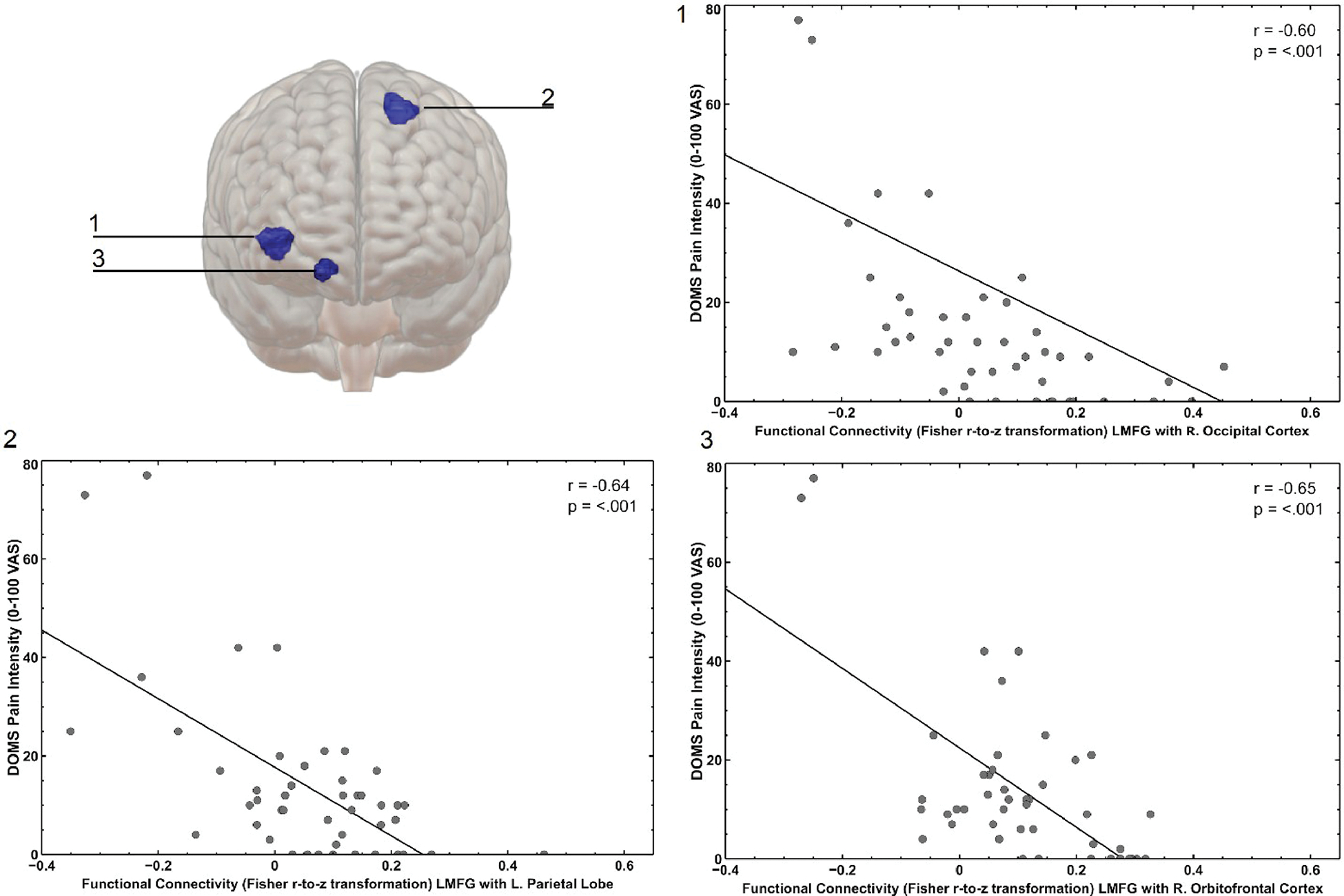
(Upper Left) Anterior view showing the location and spatial extent of clusters (1) (coordinates: 36, −76, 0) including the right lateral occipital cortex, (2) (coordinates: −20, −52, 58) including the left superior parietal lobule and the left lateral occipital cortex, (3) (coordinates: 16, 14, − 16) including the right orbitofrontal cortex, where connectivity with the left middle frontal gyrus was associated with lower DOMS pain severity. (Right and Lower Left) Scatterplots 1 to 3 demonstrating the significant correlation between pain severity and functional connectivity of left middle frontal gyrus with each significant cluster.
Figure 3.

(Left) Left mid-sagittal view showing the location and spatial extent of a cluster (coordinates: −12, −68, −16), including the left cerebellum and left lingual gyrus, where connectivity with the left middle occipital gyrus was associated with lower DOMS pain severity. (Right) Scatterplot demonstrating the significant correlation between pain severity and functional connectivity of left middle occipital gyrus with this cluster.
Table 1.
Cluster Coordinates and Regions Where Functional Connectivity was Associated With Delayed Onset Muscle Soreness Pain Intensity
| Seed Region | Cluster MNI coordinate | Empty Cell | Empty Cell | Empty Cell | Empty Cell | Empty Cell | Empty Cell | |||
|---|---|---|---|---|---|---|---|---|---|---|
| Empty Cell | X | Y | Z | k | Cluster Regions | Voxels in Region | Coverage (%) | Connectivity M(SD) | Correlation With DOMS Pain Intensity (r) | FDR-corrected p-value |
|
| ||||||||||
| Left middle frontal gyrus | 36 | −76 | 0 | 245 | R. lateral occipital cortex | 152 | 7 | .04 (0.17) | −.60 | .00001 |
| Unlabeled or less than 1% coverage | 93 | |||||||||
| −20 | −52 | 58 | 174 | L. superior parietal lobule | 102 | 7 | .05 (0.16) | −.64 | .000141 | |
| L. lateral occipital cortex | 59 | 1 | ||||||||
| Unlabeled or less than 1% coverage | 13 | |||||||||
| 16 | 14 | −16 | 61 | R. orbitofrontal cortex | 43 | 3 | .11 (0.14) | −.65 | .037 | |
| Unlabeled or less than 1% coverage | 18 | |||||||||
| Left middle occipital gyrus | −12 | −68 | −16 | 101 | L. Cerebellum 6 | 72 | 6 | −.02 (0.18) | −.57 | .00696 |
| L. lingual gyrus | 25 | 2 | ||||||||
| Unlabeled or less than 1% coverage | 4 | |||||||||
Independent Component Analysis
Whole-brain analyses were conducted on the ICs with the highest correlation to the DMN (r = .36), CN (r = .48) and SMN (r = .42) templates (Fig 4). Results indicated greater connectivity in another cluster with the sensorimotor network was associated with greater DOMS pain intensity. This cluster included the posterior right supramarginal gyrus, anterior right supramarginal gyrus, right angular gyrus, and the right parietal operculum cortex (Fig 5). Greater connectivity in the CN with a cluster that included the left postcentral gyrus, left precentral gyrus, right postcentral gyrus, and right precentral gyrus (Fig 6); rsFC with this cluster was associated with lower DOMS pain intensity. No significant clusters were found for the default mode network IC. See Table 2 for more details.
Figure 4.

Right and superior views showing the spatial extent of the independent components analysis derived networks: (blue) cerebellar, (red) sensorimotor, and (green) default mode.
Figure 5.

(Left) Right mid-sagittal view showing the location and spatial extent of a cluster (coordinates: 66, −44, 36), including the right posterior and anterior supramarginal gyrus, right angular gyrus and the right parietal operculum cortex, where connectivity with the ICA-derived sensorimotor network was associated with greater DOMS pain severity. (Right) Scatterplot demonstrating the significant correlation between pain severity and functional connectivity of the sensorimotor network with this cluster.
Figure 6.

(Left) Superior view showing the location and spatial extent of a cluster (coordinates: −20, −34, 62), including the left postcentral gyrus and the left and right precentral gyrus, where connectivity with the cerebellar network was associated with lower DOMS pain severity. (Right) Scatterplot demonstrating the significant correlation between pain severity and functional connectivity of the cerebellar network with this cluster.
Table 2.
Cluster Coordinates and Regions Where Functional Connectivity With ICA Networks was Associated With Delayed Onset Muscle Soreness Pain Intensity
| Seed Region | Cluster MNI Coordinate | Empty Cell | Empty Cell | Empty Cell | Empty Cell | Empty Cell | Empty Cell | |||
|---|---|---|---|---|---|---|---|---|---|---|
| Empty Cell | X | Y | Z | k | Cluster Regions | Voxels in Region | Coverage (%) | Connectivity M (SD) | Correlation With DOMS Pain Intensity (r) | FDR-Corrected p-value |
|
| ||||||||||
| Sensorimotor Network | 66 | −44 | 36 | 323 | R. supramarginal gyrus (posterior) | 165 | 13 | −2.49 (1.60) | .61 | .000002 |
| R. supramarginal gyrus (anterior) | 40 | 5 | ||||||||
| R. angular gyrus | 34 | 2 | ||||||||
| R. parietal operculum cortex | 9 | 2 | ||||||||
| Cerebellar Network | −20 | −34 | 62 | 251 | L. postcentral gyrus | 139 | 4 | .005 (1.70) | −0.65 | .000007 |
| L. precentral gyrus | 50 | 1 | ||||||||
| R. precentral gyrus | 2 | 0 | ||||||||
| Not labeled | 43 | |||||||||
Discussion
Overall, we found that DOMS pain intensity was associated with functional connectivity between regions and networks previously identified to be associated with musculoskeletal pain. These included the left middle frontal gyrus and left middle occipital gyrus, where we have previously identified greater gray matter density in people who did not report musculoskeletal pain following DOMS induction, and the ICA-derived sensorimotor and cerebellar networks.
Among the regions with previously identified greater gray matter density associations with lower DOMS-related pain, we found that lower levels of DOMS-related pain predicted greater connectivity of the left MFG with 3 clusters including right lateral occipital cortex, left superior parietal lobule, and right orbitofrontal cortex. The left MFG (BA 6), which includes both premotor cortex and the supplementary motor area, has previously been associated with processing both painful and nonpainful mechanical stimulation.30 This result suggests efficient communication between brain regions involved in motor coordination and the detected clusters, which have been previously implicated with activation in response to acute pain stimulation, pain self-reinforcing placebo mechanisms, and spontaneous pain seen in postherpetic neuralgia patients, may confer resilience to DOMS-related pain.3,14,29,36 We also found associations with connectivity between the left MOG and a cluster located in the left cerebellum and left lingual gyrus, where greater levels of DOMS-related pain predicted lower connectivity. While the MOG is commonly associated with visual processing and object recognition, previous research has found higher levels of painful stimuli to be associated with BOLD signal decreases in the left MOG.36 Potential implications of this finding are further explored below in our discussion of cerebellum network analysis results.
There are several potential explanations as to why some regions previously identified to be associated with gray matter density and lower DOMS-related pain also showed functional associations, but others did not. First, gray matter density may reflect multiple characteristics of tissue contained within a given voxel, including water content (which may fluctuate due to hydration status and/or cerebral blood flow), dendritic arborization, and neuronal density. Each of these characteristics may contribute to voxel-wise gray matter density in regionally specific ways.21 Notably, previous work has identified lesser correspondence among ICA-derived structural and functional components among cortical structures compared to those of the basal ganglia while other findings suggest that sustained practice of cognitive tasks may result in divergent structural and functional effects.26 Second, this study focused on associations of resting state functional connectivity between ROIs and the whole brain with DOMS-related pain. However, there are numerous other ways that a given region may contribute to a functional process, including as the center of a hub or as part of a distributed network.10 It is possible that alternative analytic approaches could elucidate functional roles for other structural ROIs. Our reliance on resting state data for this analysis may limit our ability to identify the association between functional metrics in these regions and the musculoskeletal pain experience.
We also assessed three ICA-derived resting state functional networks. Greater resting state functional connectivity of the cerebellar network with the left postcentral gyrus, left precentral gyrus, and right precentral gyrus was associated with lower reported pain intensity after DOMS induction. However, greater DOMS pain predicted greater connectivity between the SMN and the anterior and posterior supramarginal gyrus, right angular gyrus, and right parietal operculum cortex. Previous findings with similar proximity have shown this area to be associated with pain anticipation and activation in response to pain versus warm stimuli.35,41 This was the only cluster we noted in our analysis where greater connectivity with a seed region or network was associated with greater levels of pain. However, this finding is consistent with the literature as the sensorimotor network is a key component of normal pain processing,28 and suggests that greater connectivity between SMN structures, which are involved in discrimination and localization of pain, and those mediating pain-related expectations, attention, and salience of pain-related stimuli may predispose individuals to experience greater DOMS-related pain.49 Additionally, SMN connectivity with medial frontal structures has been associated with trainability of pain response (ie, reductions in pain intensity over repeated pain exposures that may reflect improved endogenous pain modulatory capacity).8,37
We also found that lower levels of DOMS-related pain were predictive of greater connectivity between the cerebellar network and bilateral pre- and postcentral gyri, as well as greater connectivity between left middle occipital gyrus and cerebellar lobule VI. A mechanistic role for the cerebellum in susceptibility to DOMS-related pain is consistent with both its canonical role in motor processing and its increasingly recognized contributions to sensorimotor integration, cognition, and emotion processing.37 Indeed, previous literature has identified the cerebellum to be an important region for central pain processing and endogenous pain modulation25,33 and structural alterations of the cerebellum have been associated with chronic musculoskeletal conditions.39 The cerebellum is thought to modulate activity (consistent with the Universal Cerebellar Transform25 from both the primary somatosensory and motor cortices via well-characterized neuroanatomic connections through the pons and inferior olive.33 Our finding of an association between lobule VI connectivity and DOMS-related pain intensity is consistent with previous evidence showing overlapping activity in that area during pain and motor processing, as well as significant functional connectivity with the left middle occipital gyrus.32 While evidence suggests occipital structures may be functionally and structurally connected with antinociceptive regions and are commonly reported in functional imaging studies of pain response, limited evidence is available to clarify their role(s) in pain modulation, making this finding difficult to interpret.13
Interestingly, while functional connectivity of the cerebellar network and sensorimotor regions was associated with pain intensity, functional connectivity of the SMN with the cerebellum was not associated with pain intensity. This may suggest that not all areas within the SMN are functionally correlated with the cerebellum in its association with pain. It may be beneficial for future research to use pre- and postcentral gyrus as regions of interest rather than the entirety of the SMN. In addition, it is possible that there may be a directional association between the cerebellum and the SMN in its association with pain. Utilization of effective connectivity may help to characterize this result. Although we did not assess functional impairment resulting from the eccentric exercise induction, our results, combined with prior work, are suggestive that disruptions in functional networks involving the cerebellum may also underpin susceptibility to musculoskeletal-pain related impairment and disability. This possibility should be investigated in future studies.
Our previous work8 suggested that exercise induced DOMS pain intensity is associated with aspects of brain structure in several brain regions that have been previously linked to discriminative, emotional, and cognitive processing of pain. Present results identified that among a subset of these regions, DOMS pain intensity is also tied to aspects of brain function. While previous work42 suggests that functional interactions may underlie aspects of trainability in pain modulation, given the areas of convergence of structural and functional properties within regions associated with lower DOMS-related pain, identification of coherence among structural and functional predictors of pain modulatory trainability is recommended. Considering evidence of the role of intraregional neural dynamics (e.g., regional signal variability) in facilitating capacity regarding pain and other processes,2,7,20,39 future work clarifying the role of these features in resilience to subacute pain is needed. Additionally, given the aspects of convergence and divergence of regional properties in predicting DOMS-related pain, work is needed to integrate findings across neuroimaging metrics to aid in clarifying indicators of pain resilience and modulatory capacity.
Notably, in this manuscript we have conceptualized lower pain intensity following DOMS as a potential indicator of pain resilience.44 This is consistent with theories of resilience that focus on recovery from stressors or distress, and in the case of the present investigation, the capacity to minimize derivations from a state of equilibrium in the context of DOMS-related pain.23 However, it is important to note that other factors such as sustainability and growth48 are theorized to be important aspects of pain resilience and were not directly assessed in this investigation. Additionally, while we believe individual differences DOMS-related pain intensity may be indicative of pain resilience, these differences in pain report and resilience itself are likely influenced by numerous other factors (eg, gender, global fitness level, positive affect, acceptance). We have also previously identified regional BOLD signal variability to be associated with key aspects of pain modulatory capacity that may also contribute to pain resilience.9 Future investigations to clarify the neural processes and psychological processes that directly contribute to resilience to DOMS-related pain are clearly warranted.
Strengths and Limitations
The findings of the present study represent an important step in clarifying neural mechanisms of resilience to clinically relevant pain and importantly suggest areas of structural and functional coherence in this aspect of resilience. While the findings of this study contribute to our understanding of indicators of adaptive pain modulation and resilience, they should be considered within the contexts of the study’s limitations. One key limitation is that there was only a single induction of DOMS pain, and subsequently only one measurement of rsFC. As such, our data does not speak to the relationship between rsFC and changes in DOMS pain over repeated exercise bouts, or the potential reciprocal relationship between rsFC and DOMS over time,11 and such causal relationships cannot be inferred at this time. Studies designed to assess causality and the stability of these associations over time are needed. While our study identified key associations among rsFC and DOMS-related pain intensity, other features of experimental DOMS induction, such as functional impairment that more closely mimics chronic pain, were not assessed in this study, and may more closely support in identifying resilience to pain chronicity than pain intensity. In addition, our study focused on clarifying the role of key a priori regions and networks in response to DOMS-inducing eccentric exercise. However, numerous other structures and networks are implicated in pain modulation and chronicity. Although it was beyond the scope of the present investigation to assess widespread aspects of functional connectivity as they relate to pain resilience, such exploratory analyses may aid in clarifying additional protective and susceptibility factors. Similarly, our study included only healthy adults without chronic pain. Given our stringent inclusion criteria and focus on musculoskeletal pain, future studies are required to establish the application of these results to other groups and pain types. Finally, resilience to musculoskeletal pain may be conceptualized not only as experiencing less severe pain following an acute challenge, but also as more rapid resolution of pain. Future studies should examine the structural and functional neural correlates of this facet of resilience as well.
Summary
The present manuscript set out to identify indicators of resilience to clinical-relevant, experimental low back pain. We uncovered correspondence among resilience to DOMS-induced pain and functional associations within frontal and occipital seed regions, and sensorimotor and cerebellar networks. Our results provide key support for the role of regional and network connectivity in adaptive pain response and call for investigations of pain modulatory capacity that integrate multimodal neuroimaging metrics.
Perspective:
Our results provide key support for the role of structural and functional coherence in regional and network connectivity in adaptive pain response and represent an important step in clarifying neural mechanisms of resilience to clinically relevant pain.
Acknowledgements
We would like to acknowledge the participants who volunteered for this study and the work of the investigators on the primary trial – Maggie Horn, DPT, PhD, MPH; Meryl Alappattu, DPT, PhD; Charles Penza, DC, PhD; Joel Bialosky, PT, PhD; Fredy Solis, PT, PhD; Kara Hannibal, DPT; Warren Greenfield, MS; and Cally House, BS.
Support/Grant:
This work was supported by the National Center of Complementary and Integrative Health (grant number R01AT006334). A portion of this work was performed in the McKnight Brain Institute at the National High Magnetic Field Laboratory’s AMRIS Facility, which is supported by National Science Foundation Cooperative Agreement No. DMR-1157490, and the State of Florida.
Footnotes
Conflict of interest statement: The authors declare no conflict of interest.
References
- 1.Alshelh Z, Marciszewski KK, Akhter R, Di Pietro F, Mills EP, Vickers ER, Peck CC, Murray GM, Henderson LA: Disruption of default mode network dynamics in acute and chronic pain states. Neuroimage Clin 17:222–231, 2017. 10.1016/j.nicl.2017.10.019 [DOI] [PMC free article] [PubMed] [Google Scholar]
- 2.Armbruster-Genç DJN, Ueltzhöffer K, Fiebach CJ: Brain signal variability differentially affects cognitive flexibility and cognitive stability. J Neurosci 36:3978–3987, 2016. 10.1523/JNEUROSCI.2517-14.2016 [DOI] [PMC free article] [PubMed] [Google Scholar]
- 3.Bär KJ, Wagner G, Koschke M, Boettger S, Boettger MK, Schlösser R, Sauer H: Increased prefrontal activation during pain perception in major depression. Biol Psychiatry 62:1281–1287, 2007. 10.1016/j.biopsych.2007.02.011 [DOI] [PubMed] [Google Scholar]
- 4.Behzadi Y, Restom K, Liau J, Liu TT: A component based noise correction method (CompCor) for BOLD and perfusion based fMRI. Neuroimage 37:90–101, 2007. 10.1016/j.neuroimage.2007.04.042 [DOI] [PMC free article] [PubMed] [Google Scholar]
- 5.Bishop MD, Horn ME, George SZ, Robinson ME: Self-reported pain and disability outcomes from an endogenous model of muscular back pain. BMC Musculoskelet Disord 12:35, 2011. 10.1186/1471-2474-12-35 [DOI] [PMC free article] [PubMed] [Google Scholar]
- 6.Blyth FM, Noguchi N: Chronic musculoskeletal pain and its impact on older people. Best Pract Res Clin Rheumatol 31:160–168, 2017. 10.1016/j.berh.2017.10.004 [DOI] [PubMed] [Google Scholar]
- 7.Boissoneault J, Letzen J, Robinson M, Staud R: Cerebral blood flow and heart rate variability predict fatigue severity in patients with chronic fatigue syndrome. Brain Imaging Behav 13:789–797, 2019. 10.1007/s11682-018-9897-x [DOI] [PMC free article] [PubMed] [Google Scholar]
- 8.Boissoneault J, Penza CW, George SZ, Robinson ME, Bishop MD: Comparison of brain structure between pain-susceptible and asymptomatic individuals following experimental induction of low back pain. Spine J 20:292–299, 2020. 10.1016/j.spinee.2019.08.015 [DOI] [PMC free article] [PubMed] [Google Scholar]
- 9.Boissoneault J, Sevel L, Stennett B, Alappattu M, Bishop M, Robinson M: Regional increases in brain signal variability are associated with pain intensity reductions following repeated eccentric exercise bouts. Eur J Pain 24:818–827, 2020. 10.1002/ejp.1532 [DOI] [PMC free article] [PubMed] [Google Scholar]
- 10.Bullmore E, Sporns O: Complex brain networks: Graph theoretical analysis of structural and functional systems. Nat Rev Neurosci 10:186–198, 2009. 10.1038/nrn2575 [DOI] [PubMed] [Google Scholar]
- 11.Calhoun VD, Adali T, Pearlson GD, Pekar JJ: A method for making group inferences from functional MRI data using independent component analysis. Hum Brain Mapp 14:140–151, 2001. 10.1002/hbm.1048 [DOI] [PMC free article] [PubMed] [Google Scholar]
- 12.Chai XJ, Ofen N, Gabrieli JDE, Whitfield-Gabrieli S: Selective development of anticorrelated networks in the intrinsic functional organization of the human brain. J Cogn Neurosci 26:501–513, 2014. 10.1162/jocn_a_00517 [DOI] [PMC free article] [PubMed] [Google Scholar]
- 13.Coulombe M-A, Erpelding N, Kucyi A, Davis KD: Intrinsic functional connectivity of periaqueductal gray subregions in humans. Hum Brain Mapp 37:1514–1530, 2016. 10.1002/hbm.23117 [DOI] [PMC free article] [PubMed] [Google Scholar]
- 14.Craggs JG, Price DD, Perlstein WM, Verne GN, Robinson ME: The dynamic mechanisms of placebo induced analgesia: Evidence of sustained and transient regional involvement. PAIN 139:660–669, 2008. 10.1016/j.pain.2008.07.025 [DOI] [PMC free article] [PubMed] [Google Scholar]
- 15.Dahlhamer J: Prevalence of chronic pain and high-impact chronic pain among adults — United States, 2016. MMWR Morb Mortal Wkly Rep 67, 2018. 10.15585/mmwr.mm6736a2 [DOI] [PMC free article] [PubMed] [Google Scholar]
- 16.Dannecker EA, Hausenblas HA, Kaminski TW, Robinson ME: Sex differences in delayed onset muscle pain. Clin J Pain 21:120–126, 2005. 10.1097/00002508-200503000-00002 [DOI] [PubMed] [Google Scholar]
- 17.Dannecker EA, Koltyn KF, Riley JL, Robinson ME: The influence of endurance exercise on delayed onset muscle soreness. J Sports Med Phys Fitness 42:458–465, 2002 [PubMed] [Google Scholar]
- 18.van Ettinger-Veenstra H, Lundberg P, Alföldi P, Södermark M, Graven-Nielsen T, Sjörs A, Engstrom M, Gerdle B: Chronic widespread pain patients show disrupted cortical connectivity in default mode and salience networks, modulated by pain sensitivity. J Pain Res 12:1743, 2019. 10.2147/JPR.S189443 [DOI] [PMC free article] [PubMed] [Google Scholar]
- 19.Fine PG: Long-term consequences of chronic pain: Mounting evidence for pain as a neurological disease and parallels with other chronic disease states. Pain Med 12:996–1004, 2011. 10.1111/j.1526-4637.2011.01187.x [DOI] [PubMed] [Google Scholar]
- 20.Garrett DD, Lindenberger U, Hoge RD, Gauthier CJ: Age differences in brain signal variability are robust to multiple vascular controls. Scientific Reports 7:10149, 2017. 10.1038/s41598-017-09752-7 [DOI] [PMC free article] [PubMed] [Google Scholar]
- 21.Geha PY, Baliki MN, Chialvo DR, Harden RN, Paice JA, Apkarian AV: Brain activity for spontaneous pain of postherpetic neuralgia and its modulation by lidocaine patch therapy. PAIN 128:88–100, 2007. 10.1016/j.pain.2006.09.014 [DOI] [PMC free article] [PubMed] [Google Scholar]
- 22.Global Burden of Disease Report: Global, regional, and national incidence, prevalence, and years lived with disability for 354 diseases and injuries for 195 countries and territories, 1990–2017: a systematic analysis for the Global Burden of Disease Study 2017. Lancet 392:1789–1858, 2018. 10.1016/S0140-6736(18)32279-7 [DOI] [PMC free article] [PubMed] [Google Scholar]
- 23.Goubert L, Trompetter H: Towards a science and practice of resilience in the face of pain. Eur J Pain 21:1301–1315, 2017. 10.1002/ejp.1062 [DOI] [PubMed] [Google Scholar]
- 24.Graves JE, Pollock ML, Carpenter DM, Leggett SH, Jones A, MacMillan M, Fulton M: Quantitative assessment of full range-of-motion isometric lumbar extension strength. Spine 15:289–294, 1990. 10.1097/00007632-199004000-00008 [DOI] [PubMed] [Google Scholar]
- 25.Guell X, Schmahmann JD, Gabrieli JD, Ghosh SS: Functional gradients of the cerebellum Bostan A, Ivry RB, eds. eLife 7:e36652, 2018. 10.7554/eLife.36652 [DOI] [PMC free article] [PubMed] [Google Scholar]
- 26.Haier RJ, Karama S, Leyba L, Jung RE: MRI assessment of cortical thickness and functional activity changes in adolescent girls following three months of practice on a visualspatial task. BMC Res Notes 2:174, 2009. 10.1186/1756-0500-2-174 [DOI] [PMC free article] [PubMed] [Google Scholar]
- 27.Kim H, Kim J, Loggia ML, Cahalan C, Garcia RG, Vangel MG, Wasan AD, Edwards RR, Napadow V: Fibromyalgia is characterized by altered frontal and cerebellar structural covariance brain networks. Neuroimage Clin 7:667–677, 2015. 10.1016/j.nicl.2015.02.022 [DOI] [PMC free article] [PubMed] [Google Scholar]
- 28.Kim J, Kang I, Chung YA, Kim TS, Namgung E, Lee S, Oh JK, Jeong HS, Cho H, Kim MJ, Kim TD: Altered attentional control over the salience network in complex regional pain syndrome. Sci Rep 8:7466, 2018. 10.1038/s41598-018-25757-2 [DOI] [PMC free article] [PubMed] [Google Scholar]
- 29.Kong J, Loggia ML, Zyloney C, Tu P, LaViolette P, Gollub RL: Exploring the brain in pain: Activations, deactivations and their relation. PAIN 148:257, 2010. 10.1016/j.pain.2009.11.008 [DOI] [PMC free article] [PubMed] [Google Scholar]
- 30.Lui F, Duzzi D, Corradini M, Serafini M, Baraldi P, Porro CA: Touch or pain? Spatio-temporal patterns of cortical fMRI activity following brief mechanical stimuli. PAIN 138:362–374, 2008. 10.1016/j.pain.2008.01.010 [DOI] [PubMed] [Google Scholar]
- 31.Meucci RD, Fassa AG, Faria NMX: Prevalence of chronic low back pain: Systematic review. Rev Saude Publica 49:1, 2015. 10.1590/S0034-8910.2015049005874 [DOI] [PMC free article] [PubMed] [Google Scholar]
- 32.Misra G, Coombes SA: Neuroimaging evidence of motor control and pain processing in the human midcingulate cortex. Cerebral Cortex 25:1906–1919, 2015. 10.1093/cercor/bhu001 [DOI] [PubMed] [Google Scholar]
- 33.Moulton EA, Schmahmann JD, Becerra L, Borsook D: The cerebellum and pain: Passive integrator or active participator? Brain Res Rev 65:14–27, 2010. 10.1016/j.brainresrev.2010.05.005 [DOI] [PMC free article] [PubMed] [Google Scholar]
- 34.Necka EA, Lee I-S, Kucyi A, Cheng JC, Yu Q, Atlas LY: Applications of dynamic functional connectivity to pain and its modulation. PAIN Reports 4:e752, 2019. 10.1097/PR9.0000000000000752 [DOI] [PMC free article] [PubMed] [Google Scholar]
- 35.Ochsner KN, Ludlow DH, Knierim K, Hanelin J, Ramachandran T, Glover GC, Mackey SC: Neural correlates of individual differences in pain-related fear and anxiety. PAIN 120:69–77, 2006. 10.1016/j.pain.2005.10.014 [DOI] [PMC free article] [PubMed] [Google Scholar]
- 36.Pomares FB, Funck T, Feier NA, Roy S, Daigle-Martel A, Ceko M, Narayanan S, Araujo D, Thiel A, Stikov N, Fitz-charles MA: Histological underpinnings of grey matter changes in fibromyalgia investigated using multimodal brain imaging. J Neurosci 37:1090–1101, 2017. 10.1523/JNEUROSCI.2619-16.2016 [DOI] [PMC free article] [PubMed] [Google Scholar]
- 37.Riedl V, Valet M, Wöller A, Sorg C, Vogel D, Sprenger T, Boecker H, Wohlschläger AM, Tölle TR: Repeated pain induces adaptations of intrinsic brain activity to reflect past and predict future pain. NeuroImage 57:206–213, 2011. 10.1016/j.neuroimage.2011.04.011 [DOI] [PubMed] [Google Scholar]
- 38.Robinson ME, Greene AF, O’Connor P, Graves JE, MacMillan M: Reliability of lumbar isometric torque in patients with chronic low back pain. Phys Ther 72:186–190, 1992. 10.1093/ptj/72.3.186 [DOI] [PubMed] [Google Scholar]
- 39.Rogachov A, Cheng JC, Erpelding N, Hemington KS, Crawley AP, Davis KD: Regional brain signal variability: A novel indicator of pain sensitivity and coping. PAIN 157:2483–2492, 2016. 10.1097/j.pain.0000000000000665 [DOI] [PubMed] [Google Scholar]
- 40.Ruscheweyh R, Kühnel M, Filippopulos F, Blum B, Eggert T, Straube A: Altered experimental pain perception after cerebellar infarction. PAIN 155:1303–1312, 2014. 10.1016/j.pain.2014.04.006 [DOI] [PubMed] [Google Scholar]
- 41.Segall JM, Allen EA, Jung RE, Erhardt EB, Arja SK, Kiehl KA, Calhoun VD: Correspondence between structure and function in the human brain at rest. Front Neuroinform 6:10, 2012. 10.3389/fninf.2012.00010 [DOI] [PMC free article] [PubMed] [Google Scholar]
- 42.Seifert F, Schuberth N, De Col R, Peltz E, Nickel FT, Maihöfner C: Brain activity during sympathetic response in€ anticipation and experience of pain. Hum Brain Mapp 34:1768–1782, 2013. 10.1002/hbm.22035 [DOI] [PMC free article] [PubMed] [Google Scholar]
- 43.Sevel L, Boissoneault J, Alappattu M, Bishop M, Robinson M: Training endogenous pain modulation: a preliminary investigation of neural adaptation following repeated exposure to clinically-relevant pain. Brain Imag Behav 14:881–896, 2020. 10.1007/s11682-018-0033-8 [DOI] [PubMed] [Google Scholar]
- 44.Sturgeon JA, Zautra AJ: Resilience: A new paradigm for adaptation to chronic pain. Curr Pain Headache Rep 14:105–112, 2010. 10.1007/s11916-010-0095-9 [DOI] [PMC free article] [PubMed] [Google Scholar]
- 45.Yeo BT, Krienen FM, Sepulcre J, Sabuncu MR, Lashkari D, Hollinshead M, Roffman JL, Smoller JW, Zöllei L, Polimeni JR, Fischl B: The organization of the human cerebral cortex estimated by intrinsic functional connectivity. J Neurophysiol 106:1125–1165, 2011. 10.1152/jn.00338.2011 [DOI] [PMC free article] [PubMed] [Google Scholar]
- 46.Whitfield-Gabrieli S, Nieto-Castanon A: Conn: A functional connectivity toolbox for correlated and anticorrelated brain networks. Brain Connect 2:125–141, 2012. 10.1089/brain.2012.0073 [DOI] [PubMed] [Google Scholar]
- 47.Woo C-W, Krishnan A, Wager TD: Cluster-extent based thresholding in fMRI analyses: Pitfalls and recommendations. Neuroimage 91:412–419, 2014. 10.1016/j.neuroimage.2013.12.058 [DOI] [PMC free article] [PubMed] [Google Scholar]
- 48.Zautra AJ, Arewasikporn A, Davis MC: Resilience: Promoting well-being through recovery, sustainability, and growth. Res Hum Dev 7:221–238, 2010. 10.1080/15427609.2010.504431 [DOI] [Google Scholar]
- 49.Zeidan F, Lobanov OV, Kraft RA, Coghill RC: Brain mechanisms supporting violated expectations of pain. PAIN 156:1772–1785, 2015. 10.1097/j.pain.0000000000000231 [DOI] [PMC free article] [PubMed] [Google Scholar]