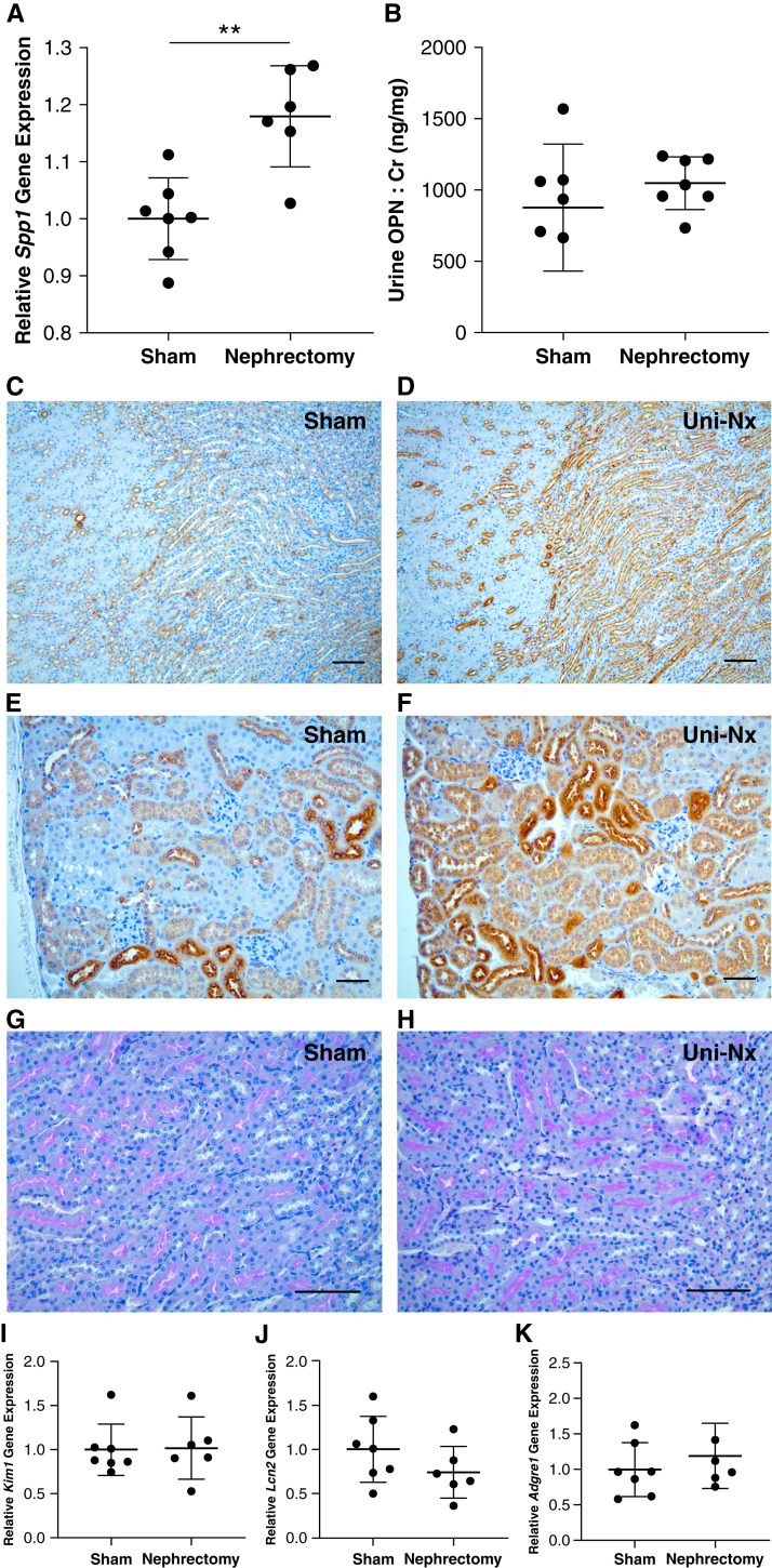
Figure 3.
Kidney OPN expression is increased by reduction of functional nephrons (unilateral nephrectomy; Uni-Nx). (A) Spp1 (OPN) gene expression by quantitative real-time PCR was increased in the remaining kidney at 3 weeks post unilateral nephrectomy compared with sham-operated littermates. (B) No difference was observed in urine OPN (normalized to Cr). (C) and (D) IHC staining of residual kidneys from nephrectomy mice demonstrated markedly increased OPN expression (brown staining) in medullary tubules near the corticomedullary junction compared with sham controls (×10 magnification; scale bar=100 μm). Moreover, nearly all tubular segments in the kidney cortex (E) and (F) exhibited increased OPN expression in the Uni-Nx group (×20 magnification; scale bar=50 μm). Importantly, no evidence of tubular injury was observed by periodic acid-Schiff staining of kidney sections (G) and (H) (×20 magnification; scale bar=100 μm) or gene expression for Kim1 or Lcn2 (NGAL) (I) and (J). Additionally, Adgre1 (F4/80) expression was comparable for the two groups (K), suggesting no difference in macrophage accumulation in response to changes in OPN. Representative images were selected from a minimum of three per group (**P<0.01 by Student’s t test; all data presented as mean±SD; gene expression normalized to HPRT).

